Biotinidase Deficiency is an inherited autosomal recessive disorder in which the enzyme biotinidase is deficient. The enzyme helps to recycle the vitamin biotin, and its deficiency leads to decreased levels of biotin in the body.
What is Biotinidase Deficiency?
Biotin is an important vitamin that must be provided through diet as it metabolizes the carbohydrates, fats, and amino acids present in food. The Biotinidase enzyme is mostly present in your liver and kidneys, and its main function is to cleave and liberate the biotin that you take from the diet. The constant recycling of biotin effectively helps to maintain ongoing cycles in the body, such as gluconeogenesis and fatty acid synthesis.
A deficiency of the Biotinidase enzyme influences the recycling of biotin, bringing out unfavorable symptoms since biotin-dependent carboxylation of certain enzymes does not take place. Partial deficiency of Biotinidase enzyme (10-30% of enzyme activity ) in your body doesn’t cause significant signs and symptoms, while patients with less than 10% activity of Biotinidase enzyme present with serious manifestations and complications in eyes, brain, and skin.1Wolf, B. (2000). Biotinidase Deficiency. In M. P. Adam (Eds.) et. al., GeneReviews®. University of Washington, Seattle.
Causes of Biotinidase Deficiency
This deficiency develops when a specific gene called BTD on chromosome 3p25 becomes defective due to inherited causes, and it transmits in an autosomal recessive pattern, which means that both of the parents must have the biotinidase deficiency to transmit it to their offspring. The defective gene BTD cannot recycle the biotin that you take from your diet, and the biotin becomes unavailable for carboxylase enzymes in your body. As a result, the molecular reactions in your body do not get catalyzed, causing the substrates to accumulate and develop toxicity in the body.2Pindolia, K., Jordan, M., & Wolf, B. (2010). Analysis of mutations causing this deficiency. Human mutation, 31(9), 983–991. https://doi.org/10.1002/humu.21303
How common is Biotinidase Deficiency?
The incidence of biotinidase deficiency is very low. There are only 1 out of 40000 to 1 out of 60000 cases, and the number of cases in different countries varies. Nowadays, newborn babies are screened for this deficiency in many countries worldwide.
The average age of developing this deficiency is the first six weeks of life. Therefore, doctors pay great attention to screening babies. In most patients, symptoms of this deficiency take time to develop, and it may take them ten years to manifest the first symptom. 3Canda, E., Kalkan Uçar, S., & Çoker, M. (2020). Biotinidase Deficiency: Prevalence, Impact And Management Strategies. Pediatric health, medicine and therapeutics, 11, 127–133. https://doi.org/10.2147/PHMT.S198656
Pathophysiology of Biotinidase Deficiency
The biotin vitamin is present in several foods, such as milk, egg yolk, green vegetables, and meat. In addition to exogenous biotin, your body also makes endogenous biotin in your large intestine. Biotin is essential for carboxylase enzymes in your body. When you take biotin through your diet, apocarboxylase converts into holocarboxylase in the presence of the holocarboxylase synthetase enzyme. This conversion needs the attachment of biotin; without biotin, the conversion is impossible. Then, the holocarboxylase enzyme undergoes several proteolytic reactions in your body, ultimately releasing biocytin and biotinyl-peptides. Here, biotinidase’s function occurs as it recycles the used biotin so that it is continuously available for carboxylases.
When the biotinidase enzyme is deficient in your body, biotin becomes unavailable for the following four enzymes:
- 3-methylcrotonyl coenzyme carboxylase
- Pyruvate carboxylase
- Acetyl-CoA carboxylase
- Propionyl-CoA carboxylase
Insufficient biotin in the body prevents carboxylases from catalyzing substrates, leading to their excessive accumulation. These substrates are lactic acid, alanine, methyl citrate, and acetyl-coA, and they cause several symptoms in the brain and skin.4Tomar, R., Vashisth, D., & Vasudevan, R. (2012). Biotinidase deficiency. Medical journal, Armed Forces India, 68(1), 81–83. https://doi.org/10.1016/S0377-1237(11)60118-4
Symptoms of Biotinidase Deficiency
Symptoms of this deficiency depend on the severity of its deficiency. In the case of partial biotinidase deficiency, symptoms are not serious and significant. However, with profound deficiency, mild to moderate symptoms in several parts of your body are present. These are:
Neurological Symptoms:
The main neurological symptom of this deficiency is seizures, which can be generalized, tonic-clonic, or myoclonic. Studies show that giving biotin to patients suffering from seizures significantly lessens the frequency and severity of seizures. Untreated cases can often lead to permanent brain damage due to several episodes of seizures.
Children suffering from this deficiency also experience developmental delays. Moreover, this deficiency affects the function of the auditory nerve, which causes sensorineural hearing loss. Further neurological presentations you may experience in severe biotinidase deficiency include ataxia (loss of balance and coordination of your body) and optic nerve atrophy.5Suchy, S. F., McVoy, J. S., & Wolf, B. (1985). Neurologic symptoms of biotinidase deficiency: possible explanation. Neurology, 35(10), 1510–1511. https://doi.org/10.1212/wnl.35.10.1510
Respiratory Symptoms:
This deficiency causes apnea, a sudden loss of breathing, especially during sleep. It also affects the laryngeal muscles, which can produce stridor (an abnormal sound made when you breathe air in). Biotinidase deficiency also causes difficulty swallowing by involving the bulbar muscles.
Ocular Symptoms:
The presenting sign could be a sudden loss of vision. Some patients also present with conjunctivitis, which involves eye itching, sneezing, and nasal discharge. Your ophthalmologist might find optic atrophy on your eye examination, which is a cause of vision loss.6Salbert, B. A., Astruc, J., & Wolf, B. (1993). Ophthalmologic findings in biotinidase deficiency. Ophthalmologica. Journal international d’ophtalmologie. International journal of ophthalmology. Zeitschrift fur Augenheilkunde, 206(4), 177–181. https://doi.org/10.1159/000310387
Hair Problems:
It affects hair in a way that you lose your hair, leading to its thinning. Besides losing hair, you also experience a change in hair color and texture.7Trüeb R. M. (2016). Serum Biotin Levels in Women Complaining of Hair Loss. International journal of trichology, 8(2), 73–77. https://doi.org/10.4103/0974-7753.188040

Skin Manifestations:
The facial rash is one manifestation of biotinidase deficiency. It typically appears around the lips and other orifices. Sometimes, a scaly appearance is dominant on the skin, along with the rash. Some physicians might confuse these skin manifestations with eczema or seborrheic dermatitis. Therefore, a detailed history and examination are essential to differentiate biotinidase deficiency from other skin problems.8Mock D. M. (1991). Skin manifestations of biotin deficiency. Seminars in dermatology, 10(4), 296–302.
Immunological Abnormalities:
If you are suffering from any viral or fungal infection along with a biotinidase deficiency, it suggests a deterioration of your cellular immunity. Abnormalities in your immunity make you more prone to infections. Taking sufficient biotin restores your natural immunity.
How to diagnose Biotinidase deficiency?
In addition to taking a history and performing examinations, your physician may advise you to undergo the following tests to confirm biotinidase deficiency.
Neonatal Screening:
Neonatal screening for biotinidase deficiency is a recommended part of early screening in several countries because it is as crucial as any other screening. This screening identifies the mutated BTD gene in the neonate, which helps take the necessary steps to treat biotinidase deficiency.9Heard, G. S., Secor McVoy, J. R., & Wolf, B. (1984). A screening method for biotinidase deficiency in newborns. Clinical chemistry, 30(1), 125–127.
Serum Tests:
Some important serum tests to carry out for the diagnosis of this deficiency are:
Arterial Blood Gas
Biotinidase deficiency disrupts the function of carboxylase enzymes, which fail to remove excessive lactic acid from the body. As a result of its accumulation, acidosis occurs, which is detected by an arterial blood gas test.
Ammonia
In some patients, biotinidase deficiency also leads to hyperammonemia in their body.
Serum Amino Acids & Biotinidase Profile
Checking the level of biotinidase enzyme in your body is also helpful in diagnosing its deficiency. This deficiency also affects the level of serum amino acids in your body.

Urine Tests
This deficiency results in ketones and organic acids forming in your urine. Therefore, physicians recommend urine tests.
Imaging Studies:
For the diagnosis, the following imaging tests are carried out:
MRI & MRS
Magnetic resonance imaging is beneficial in patients having neurological symptoms of biotinidase deficiency. Children with this deficiency show cerebral atrophy, edema, and even ventricular enlargement on the MRI report. Moreover, some physicians suggest magnetic resonance spectroscopy for patients with serious brain manifestations. MRS measures the biochemical changes in your brain by detecting signals emerging from molecules, and in this way, it easily detects any functional defect in your brain.
PET Scan
Severe biotinidase deficiency affects the cerebral metabolic activity in your brain. Positron emission tomography demonstrates these changes effectively. Furthermore, a PET scan is carried out after giving the patients a dose of biotin, which shows an obvious difference in metabolic activity.
CT Scan
MRI cannot easily detect findings such as calcifications in your brain that are formed due to this deficiency, and for this purpose, a CT scan plays a remarkable role.
EEG
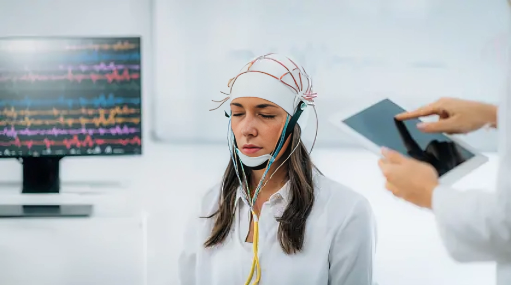
Electroencephalograms help detect electrical activity in the brain by placing electrodes on the scalp. In the case of biotinidase deficiency, they detect the abnormal morphology and spikes in sleep as well as the changes that occur during a myoclonic and tonic-clonic seizure.10Biswas, A., McNamara, C., Gowda, V. K., Gala, F., Sudhakar, S., Sidpra, J., Vari, M. S., Striano, P., Blaser, S., Severino, M., Batzios, S., & Mankad, K. (2023). Neuroimaging Features of Biotinidase Deficiency. AJNR. American journal of neuroradiology, 44(3), 328–333. https://doi.org/10.3174/ajnr.A7781

Ophthalmological Tests:
The effects of biotinidase deficiency on the eyes make ophthalmological tests crucial. Fundoscopic examination, visual field testing, and visual evoked potentials are recommended for this deficiency. All of the above tests help detect optic nerve injury and eye atrophy.
Hearing Tests:
It is mandatory to perform audiological tests in all patients with biotinidase deficiency, as hearing loss is a very common manifestation. Even after treatment, this deficiency may persist in some patients. Some physicians also advise brainstem auditory evoked response for patients, as it easily detects hearing abnormalities.11Strovel, E. T., Cowan, T. M., Scott, A. I., & Wolf, B. (2017). Laboratory diagnosis, 2017 update: a technical standard and guideline of the American College of Medical Genetics and Genomics. Genetics in medicine: Official Journal of the American College of Medical Genetics, 19(10), 10.1038/gim.2017.84. https://doi.org/10.1038/gim.2017.84
Treatment of Biotinidase Deficiency
The only best therapy is taking biotin in low or high doses.
Oral Free Biotin:
Physicians usually do not prescribe the bound biotin in different multivitamin tablets as it doesn’t help to subside the biotinidase deficiency. Taking oral-free biotin is The best way to treat this deficiency. Initially, the prescribed dose of biotin is less, which is 5-10 mg/d, and the physicians notice whether this dose helps treat the symptoms. If the symptoms don’t subside by taking low doses of biotin, increasing the dose by up to 40 mg/d is advisable. Patients with profound biotinidase deficiency should take free biotin their whole life. However, people with mild biotinidase deficiency only have to take biotin as long as they have symptoms.12Szymańska, E., Średzińska, M., Ługowska, A., Pajdowska, M., Rokicki, D., & Tylki-Szymańska, A. (2015). Outcomes of oral biotin treatment in patients with biotinidase deficiency – Twenty years follow-up. Molecular genetics and metabolism reports, 5, 33–35. https://doi.org/10.1016/j.ymgmr.2015.09.004
Symptomatic Treatment:
Besides taking biotin to treat biotinidase deficiency, treating the symptoms is also substantial. You should always stay in touch with a neurosurgeon if you experience seizures or any other neurological symptoms. Injection baclofen is considered to cure spasticity and dystonia (uncontrollable contraction of your muscles). For symptoms of respiratory and ocular origin, consult a pulmonologist and ophthalmologist.
In short, treatment is not done by a single doctor; a team of doctors should work together to get better results in less time.
Complications of Biotinidase Deficiency
Untreated biotinidase deficiency can result in the following complications if not treated in early childhood:13Taitz, L. S., Leonard, J. V., & Bartlett, K. (1985). Long-term auditory and visual complications of biotinidase deficiency. Early human development, 11(3-4), 325–331. https://doi.org/10.1016/0378-3782(85)90086-6
- Optic atrophy
- Developmental delay
- Acquired retinal dysplasia
- Metabolic abnormalities
- Deafness
- Organic aciduria
- Infections
- Coma
- Death
Prognosis & Life Expectancy
Better treatment and management at an early stage results in a better prognosis. Along with the early diagnosis of biotinidase deficiency, it is crucial to continue the treatment for longer to treat the deficiency completely. People with asymptomatic biotinidase deficiency can have a good prognosis if they treat the deficiency before the appearance of symptoms.
However, if the deficiency is too severe and it has already caused neurological symptoms, unfortunately, there is no going back, even after taking biotin therapy.
Conclusion
In conclusion, biotinidase deficiency is a deficiency of the enzyme biotinidase due to a mutated BTD gene in your body. The level of biotin is reduced in your body because biotinidase helps to recycle it. Several other carboxylase enzymes cannot function properly, too, and this results in problems in your brain, eyes, or skin. This deficiency mostly presents as seizures, permanent brain damage, loss of vision, respiratory problems, thinning of hair, and hearing loss. It would be great to consult a team of doctors, including a neurologist, dermatologist, physician, and ophthalmologist, for the its treatment. Take oral biotin as long as you have symptoms for better outcomes. To avoid complications like optic atrophy, hearing loss, or coma, early diagnosis and treatment of this deficiency are essential. Continuous and effective treatment promises a good prognosis.
Refrences
- 1Wolf, B. (2000). Biotinidase Deficiency. In M. P. Adam (Eds.) et. al., GeneReviews®. University of Washington, Seattle.
- 2Pindolia, K., Jordan, M., & Wolf, B. (2010). Analysis of mutations causing this deficiency. Human mutation, 31(9), 983–991. https://doi.org/10.1002/humu.21303
- 3Canda, E., Kalkan Uçar, S., & Çoker, M. (2020). Biotinidase Deficiency: Prevalence, Impact And Management Strategies. Pediatric health, medicine and therapeutics, 11, 127–133. https://doi.org/10.2147/PHMT.S198656
- 4Tomar, R., Vashisth, D., & Vasudevan, R. (2012). Biotinidase deficiency. Medical journal, Armed Forces India, 68(1), 81–83. https://doi.org/10.1016/S0377-1237(11)60118-4
- 5Suchy, S. F., McVoy, J. S., & Wolf, B. (1985). Neurologic symptoms of biotinidase deficiency: possible explanation. Neurology, 35(10), 1510–1511. https://doi.org/10.1212/wnl.35.10.1510
- 6Salbert, B. A., Astruc, J., & Wolf, B. (1993). Ophthalmologic findings in biotinidase deficiency. Ophthalmologica. Journal international d’ophtalmologie. International journal of ophthalmology. Zeitschrift fur Augenheilkunde, 206(4), 177–181. https://doi.org/10.1159/000310387
- 7Trüeb R. M. (2016). Serum Biotin Levels in Women Complaining of Hair Loss. International journal of trichology, 8(2), 73–77. https://doi.org/10.4103/0974-7753.188040
- 8Mock D. M. (1991). Skin manifestations of biotin deficiency. Seminars in dermatology, 10(4), 296–302.
- 9Heard, G. S., Secor McVoy, J. R., & Wolf, B. (1984). A screening method for biotinidase deficiency in newborns. Clinical chemistry, 30(1), 125–127.
- 10Biswas, A., McNamara, C., Gowda, V. K., Gala, F., Sudhakar, S., Sidpra, J., Vari, M. S., Striano, P., Blaser, S., Severino, M., Batzios, S., & Mankad, K. (2023). Neuroimaging Features of Biotinidase Deficiency. AJNR. American journal of neuroradiology, 44(3), 328–333. https://doi.org/10.3174/ajnr.A7781
- 11Strovel, E. T., Cowan, T. M., Scott, A. I., & Wolf, B. (2017). Laboratory diagnosis, 2017 update: a technical standard and guideline of the American College of Medical Genetics and Genomics. Genetics in medicine: Official Journal of the American College of Medical Genetics, 19(10), 10.1038/gim.2017.84. https://doi.org/10.1038/gim.2017.84
- 12Szymańska, E., Średzińska, M., Ługowska, A., Pajdowska, M., Rokicki, D., & Tylki-Szymańska, A. (2015). Outcomes of oral biotin treatment in patients with biotinidase deficiency – Twenty years follow-up. Molecular genetics and metabolism reports, 5, 33–35. https://doi.org/10.1016/j.ymgmr.2015.09.004
- 13Taitz, L. S., Leonard, J. V., & Bartlett, K. (1985). Long-term auditory and visual complications of biotinidase deficiency. Early human development, 11(3-4), 325–331. https://doi.org/10.1016/0378-3782(85)90086-6

