
Dermatomyositis is a rare illness that causes your immune system to attack your muscles, skin, and sometimes other organs. It results in debilitating muscle weakness and skin rashes. There is no cure for this illness, but your symptoms improve with treatment.
What is Dermatomyositis?
Dermatomyositis is a rare, chronic autoimmune condition characterized by inflammation of both the skin and skeletal muscles. It is classified under a broader group of disorders known as inflammatory myopathies.. The word “dermatomyositis” comes from the Greek language. “Derma” means skin, “myo” means muscle, and “itis” means inflammation, so the term means “inflammation of the skin and muscles”.
Dermatomyositis typically presents with muscle weakness (particularly in the proximal muscles such as the shoulders and hips) and distinctive skin rashes, including the heliotrope rash (a violet discoloration around the eyes) and Gottron’s papules (scaly eruptions over the knuckles).
Epidemiologically, it is a rare disorder, with an estimated incidence of 1 to 10 cases per million people per year. The condition can affect both adults and children (juvenile dermatomyositis), with a slight female predominance.
The disease was first clearly described in 1887 by German physician Heinrich Unverricht, and its classification and understanding have since evolved through continued clinical and pathological research.1Dermatomyositis – Symptoms, diagnosis and treatment | BMJ Best Practice. (2025). Bmj.com. https://bestpractice.bmj.com/topics/en-gb/595?locale=tr
What causes Dermatomyositis?
This illness is a complex disease with no single known cause. Researchers believe that a variety of factors may play a role.
Cancer:
In about 40% of cases, dermatomyositis can be a sign of an underlying cancer. Your symptoms might be related to the body’s reaction to a hidden tumor. The most common cancers linked to this illness include:
- Ovarian cancer
- Breast cancer
- Lung cancer
Genetic Factors:
You may have a genetic makeup that increases your risk for dermatomyositis. Although it rarely affects multiple people in the same family, certain immune-related genes, like:
- HLA-DR3
- HLA-DR5
- HLA-DR7
A specific gene variation called TNF-α-308A has also been linked to photosensitivity in adults and calcium buildup in children, especially in people of European descent.2Rider, L. G., Miller, F. W., & Lundberg, I. E. (2019). Genetic risk and pathogenesis in idiopathic inflammatory myopathies. Current Opinion in Rheumatology, 31(6), 571–578. https://doi.org/10.1097/BOR.0000000000000650
Immune System Dysfunction:
This illness can develop if you have certain antibodies in your blood called autoantibodies that signal an overactive immune response. These include antinuclear antibodies and others that target proteins inside your cells. If someone in your family has another autoimmune disease, it could suggest a shared immune background.
Infections:
Sometimes, infections can trigger the condition. Microbes like Toxoplasma or Borrelia (which causes Lyme disease) have also been considered. Viruses have also been linked to this rare illness, such as:
- Coxsackievirus
- Parvovirus
- HIV
- HTLV-1
Medical Treatments:
Some medications and medical procedures can cause or increase the symptoms of this illness, such as:
- Statins
- Interferons
- Chemotherapy drugs
- Penicillamine
- Tumor necrosis factor inhibitors
- Silicone implants3Dermatomyositis: MedlinePlus Medical Encyclopedia. (2016). Medlineplus.gov. https://medlineplus.gov/ency/article/000839.htm
What are the symptoms of Dermatomyositis?
Dermatomyositis mainly causes muscle weakness and skin changes. Many people notice the skin symptoms before any muscle problems begin.
Skin Symptoms:
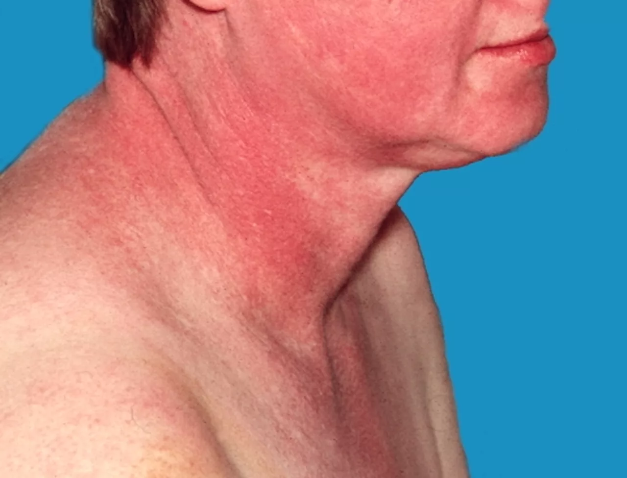
You might see a rash that is purplish or lilac-colored, called a heliotrope rash, often around your eyes and sometimes with swelling. This rash can also appear on your upper chest or back, especially around the neck (shawl sign) or above the breasts (V-sign). Other areas like your face, upper arms, thighs, or hands may also be affected.
Another common rash is called Gottron’s sign — these are red or violet, sometimes scaly, raised bumps that appear on your finger joints. Gottron’s papules can also show up on elbows, knees, or feet. These rashes tend to get worse when you’re in the sun and can be itchy, painful, or even bleed.
In amyopathic dermatomyositis, you will only have skin symptoms without muscle weakness or abnormal muscle tests.
Muscle Symptoms:
You may feel progressive weakness in muscles close to your body, such as your shoulders and thighs. This makes activities like climbing stairs, lifting objects, or standing up from a chair harder.
Respiratory Symptoms of Dermatomyositis:
In some cases, dermatomyositis affects your breathing by involving the diaphragm muscle. About 40% of people experience breathing difficulties, which may worsen over time.
Other Symptoms:
About 30% of people have mild joint pain and swelling. You may also develop trouble swallowing as the muscles in your throat weaken, and some people experience acid reflux.4Okogbaa, J., & Batiste, L. (2019). Dermatomyositis: An Acute Flare and Current Treatments. Clinical Medicine Insights Case Reports, 12.
What are the types of Dermatomyositis?
This illness presents in several distinct forms, each with unique clinical features that influence diagnosis and treatment.
Classic Dermatomyositis:
The most common type, classic dermatomyositis, involves:
- The heliotrope rash around the eyes
- Gottron’s papules on the knuckles
- Progressive muscle weakness, especially in the muscles closest to the trunk, like the shoulders and hips.
Patients typically develop skin symptoms before muscle involvement, and managing both skin and muscle inflammation is essential for treatment.
Amyopathic Dermatomyositis:
Amyopathic dermatomyositis is also known as dermatomyositis sine myositis. It is characterized by the classic skin manifestations without any muscle weakness for at least six months to two years.
In amyopathic dermatomyositis, your muscle strength stays normal, and blood tests for muscle enzymes are typically unremarkable. However, muscle symptoms can still develop over time. The condition also carries risks such as lung involvement and an increased likelihood of malignancy.
Juvenile Dermatomyositis:
Juvenile dermatomyositis occurs in children under the age of 16 and shares many features with adult dermatomyositis, including skin rash and muscle weakness. However, it is distinguished by a higher likelihood of developing calcinosis. Calcinosis involves calcium deposits forming in soft tissues and can cause significant complications. Moreover, the disease course in children can be more severe and often requires long-term treatment and follow-up.
Clinically Amyopathic Dermatomyositis:
Clinically amyopathic dermatomyositis is a broader term that overlaps with amyopathic dermatomyositis. It refers to patients who have prominent skin symptoms but minimal or no muscle involvement. Although muscle weakness and elevated muscle enzymes are absent or mild, these patients may still experience serious complications such as interstitial lung disease, which necessitates regular pulmonary monitoring.
Hypomyopathic Dermatomyositis:
Hypomyopathic dermatomyositis describes a condition in which patients exhibit the typical skin findings of dermatomyositis. However, muscle involvement is not that severe. There is only subclinical muscle involvement, which is detectable through elevated muscle enzymes or muscle biopsy. Obvious muscle weakness is usually absent. This form may remain mild or progress to classic dermatomyositis.
Paraneoplastic Dermatomyositis:
Paraneoplastic dermatomyositis occurs as a manifestation of an underlying cancer. Dermatomyositis symptoms are such cases that prompt a search for an undiagnosed cancer. Treatment of the cancer can lead to improvement in dermatomyositis symptoms.5Iaccarino, L., Ghirardello, A., Bettio, S., Zen, M., Gatto, M., Punzi, L., & Doria, A. (2014). The clinical features, diagnosis and classification of dermatomyositis. Journal of Autoimmunity, 48-49, 122–127. https://doi.org/10.1016/j.jaut.2013.11.005
How is Dermatomyositis diagnosis established?
Diagnosing dermatomyositis requires a thorough evaluation.
History & Physical Exam:
When you visit the doctor, they will take a detailed history to understand when your symptoms started and how they have changed over time. They’ll ask about any difficulties you have with daily activities that involve your muscles.
During your physical exam, the doctor will carefully check your muscle strength, especially in areas close to your trunk like your hips and shoulders. They will also examine your skin closely for any specific rashes that help identify dermatomyositis.

Laboratory Investigations:
Several blood tests are ordered by your doctor while establishing a dermatomyositis diagnosis.
- A CBC test can reveal anemia or signs of inflammation.
- ESR and CRP measure general inflammation in your body.
- A basic metabolic panel is done to check your kidney and liver function to monitor overall health and the effects of medications.
- Tests for thyroid function may be done since thyroid problems can cause or worsen muscle weakness.
- Your doctor will check antinuclear antibodies or ANA, which are positive in about 80% of dermatomyositis cases. This shows autoimmune activity.
- Myositis-specific autoantibodies like anti-Jo1 and anti-Mi-2 help classify the disease subtype and predict lung involvement.
- Creatine kinase, or CK, is the most sensitive enzyme indicating muscle damage and inflammation.
- Aldolase is another muscle enzyme that can be elevated, especially if CK is normal or borderline.
- Other muscle-related enzymes, such as LDH, can also increase due to muscle injury.
- Sometimes tests for infections or cancer markers are done if there’s suspicion of associated malignancy or infection.
Electromyography:
Electromyography, or EMG, measures the electrical activity of your muscles and their response to nerve stimulation. In dermatomyositis, EMG detects:
- Increased insertional activity and fibrillation potentials suggest ongoing muscle fiber injury.
- Short-duration, low-amplitude motor unit potentials reflect myopathic changes.
- Polyphasic discharges and repetitive high-frequency spikes indicate muscle irritability.
Muscle Biopsy:
A muscle biopsy is considered the gold standard investigation for diagnosing dermatomyositis. A small tissue sample is taken from your muscle, typically from a site showing abnormalities on imaging studies like MRI or electromyography (EMG). Under the microscope, doctors look for:
- Mononuclear inflammatory cells, primarily T cells and macrophages, infiltrating the perimysial and endomysial connective tissue surrounding the muscle fibers.
- Perifascicular atrophy, a hallmark finding in dermatomyositis, where muscle fibers near the edges of muscle bundles shrink due to ischemia caused by small vessel inflammation.
- Muscle fiber degeneration and regeneration, showing that your muscle tissue is being damaged and trying to heal.
- Phagocytosis of muscle fibers, where immune cells engulf and remove damaged or dying muscle fibers.
Imaging:
Several imaging modalities show changes in your tissues as a result of dermatomyositis:
- MRI detects muscle edema and inflammation noninvasively. It can check for changes in internal organs if needed. It can also guide biopsies to the most affected muscle areas.
- X-ray scans are used to identify calcifications in your soft tissues or joints. They can also find out if you’re developing interstitial lung disease. CT scans are ordered if any abnormality is seen on X-ray scans.6Zainab Qudsiya, & Waseem, M. (2023, August 7). Dermatomyositis. Nih.gov; StatPearls Publishing. https://www.ncbi.nlm.nih.gov/books/NBK558917/
Is there any treatment for Dermatomyositis?
There is currently no cure for dermatomyositis, but you can still manage the symptoms effectively with various dermatomyositis treatment options.
Medications:
Medications are the mainstay of treatment and help control both muscle and skin symptoms.
- You’ll most likely start with prednisolone, which quickly reduces inflammation. It can be taken as pills or given through an IV if symptoms are severe.
- If steroids alone don’t work for you, your doctor may add drugs like azathioprine or methotrexate to calm your immune system and lower side effects from steroids.
- Intravenous immunoglobulins or IVIG are used if your condition doesn’t improve with other treatments. IVIG helps act on your immune system and eases muscle weakness.
- If you have a persistent rash, antimalarial medications like hydroxychloroquine can help clear your skin and reduce flare-ups.
- If nothing else is working, your doctor may try biologic therapy with drugs like rituximab. It is a targeted treatment that helps control resistant cases.
Supportive Care:
Dermatomyositis is a chronic condition, so several measures can be taken to help you stop the progression of the disease or reduce the severity of its symptoms:
- A physical therapist can help you work out with gentle exercises that rebuild muscle strength and prevent stiffness.
- You might find relief from microwave or ultrasound therapy, which soothes tight or painful muscles.
- Braces or mobility aids can support you during recovery, especially if walking or daily tasks become difficult.
- Don’t forget your high-SPF sunscreen and protective clothing when going outdoors.
Surgery:
In some cases, calcium deposits are painful or lead to infections, your doctor may recommend surgical removal to protect your nerves and soft tissues.7Dermatomyositis – Diseases | Muscular Dystrophy Association. (2015, December 18). Muscular Dystrophy Association. https://www.mda.org/disease/dermatomyositis
Life Expectancy of Dermatomyositis
If you’re diagnosed with this rare illness and no associated cancer is found, your outlook is generally encouraging. Studies show that more than 95% of people with dermatomyositis are still alive five years after diagnosis. Overall, the five-year survival rate ranges between 70% and 93%. Your chances of living 10 years or more are also strong, with long-term survival rates around 70–80%. 8Vinit Deolikar, Raut, S. S., Saket Toshniwal, Gaidhane, S. A., & Acharya, S. (2024). Unveiling Dermatomyositis: A Tragic Tale of Mortality in a 23-Year-Old. Cureus. https://doi.org/10.7759/cureus.56058
Prognosis for Dermatomyositis
The prognosis varies, but many people see significant improvement with timely treatment. However, it’s important to understand that this condition can impact multiple organs. A variety of dermatomyositis complications can occur, such as:
- 20–25% of adults with dermatomyositis may develop a related cancer, often within the first three years.
- If your lungs are involved, you could develop interstitial lung disease, which can severely affect your breathing and oxygen levels.
- In rare cases, your heart can be affected. This can lead to arrhythmias or myocarditis, which may pose significant health risks.
- Esophageal muscle weakness increases the risk of aspiration pneumonia, a serious complication which is life-threatening.
- Severe infections can occur if you’re on long-term immunosuppressive therapy.9Tambe, S., & HemangiR Jerajani. (2013). Dermatomyositis associated with malignancy: A report of 3 cases. Indian Dermatology Online Journal, 4(4), 326–326. https://doi.org/10.4103/2229-5178.120664
Dermatomyositis Rash vs. Lupus Rash
If you have dermatomyositis, you might notice a purple or lilac rash around your eyes called a heliotrope rash. You may also see raised, scaly bumps on your knuckles, elbows, or knees called Gottron’s papules. On the other hand, if you have lupus, the rash usually looks like a red butterfly across your cheeks and nose, but does not cover the folds next to your nose. The lupus rash often gets worse when you are in the sun. These differences can help you and your doctor tell dermatomyositis and lupus apart.
Conclusion
Dermatomyositis is a rare and lifelong autoimmune condition. It affects your skin and muscles, and sometimes other organs. There is no cure, but symptoms can be managed through medication and supportive care.
Refrences
- 1Dermatomyositis – Symptoms, diagnosis and treatment | BMJ Best Practice. (2025). Bmj.com. https://bestpractice.bmj.com/topics/en-gb/595?locale=tr
- 2Rider, L. G., Miller, F. W., & Lundberg, I. E. (2019). Genetic risk and pathogenesis in idiopathic inflammatory myopathies. Current Opinion in Rheumatology, 31(6), 571–578. https://doi.org/10.1097/BOR.0000000000000650
- 3Dermatomyositis: MedlinePlus Medical Encyclopedia. (2016). Medlineplus.gov. https://medlineplus.gov/ency/article/000839.htm
- 4Okogbaa, J., & Batiste, L. (2019). Dermatomyositis: An Acute Flare and Current Treatments. Clinical Medicine Insights Case Reports, 12.
- 5Iaccarino, L., Ghirardello, A., Bettio, S., Zen, M., Gatto, M., Punzi, L., & Doria, A. (2014). The clinical features, diagnosis and classification of dermatomyositis. Journal of Autoimmunity, 48-49, 122–127. https://doi.org/10.1016/j.jaut.2013.11.
- 6Zainab Qudsiya, & Waseem, M. (2023, August 7). Dermatomyositis. Nih.gov; StatPearls Publishing. https://www.ncbi.nlm.nih.gov/books/NBK558917/
- 7Dermatomyositis – Diseases | Muscular Dystrophy Association. (2015, December 18). Muscular Dystrophy Association. https://www.mda.org/disease/dermatomyositis
- 8Vinit Deolikar, Raut, S. S., Saket Toshniwal, Gaidhane, S. A., & Acharya, S. (2024). Unveiling Dermatomyositis: A Tragic Tale of Mortality in a 23-Year-Old. Cureus. https://doi.org/10.7759/cureus.56058
- 9Tambe, S., & HemangiR Jerajani. (2013). Dermatomyositis associated with malignancy: A report of 3 cases. Indian Dermatology Online Journal, 4(4), 326–326. https://doi.org/10.4103/2229-5178.120664


