What is Hypophosphatasia?
Hypophosphatasia is a rare inborn metabolic disorder that affects the mineralization of bone and teeth.
Phosphate is one of the essential minerals necessary for the growth of your bone—hypophosphatasia results when you have a low alkaline phosphatase level in the body. Mineralization is a chemical process in which calcium and phosphorus are deposited in your bones and teeth. This process makes your bones firm and rigid. In hypophosphatasia, your bone becomes soft and fragile due to defective mineralization, resulting in bone fracture and deformity.
The prevalence of hypophosphatasia is not well understood; it varies from region to region. In the USA, hypophosphatasia affects 500-600 people each year, while in Canada, the prevalence of this disease is about 1:100000 per year.1Correa, R. R. (2023, June 5). Hypophosphatasia (HPP): Background, Pathophysiology, Epidemiology. Medscape.com; Medscape. https://emedicine.medscape.com/article/945375-overview#a6
Causes of Hypophosphatasia
Hypophosphatasia is a rare genetic disorder caused by a mutation in the ALPL gene. This gene carries genetic information to form an enzyme called tissue nonspecific alkaline phosphatase (TNSALP). This enzyme plays an essential role in bone mineralization. Mutation in the ALPL gene causes abnormal production of tissue nonspecific alkaline phosphatase, resulting in defective mineralization.2Nunes ME. Hypophosphatasia. 2007 Nov 20 [updated 2023 Mar 30]. In: Adam MP, Mirzaa GM, Pagon RA, Wallace SE, Bean LJH, Gripp KW, Amemiya A, editors. GeneReviews® [Internet]. Seattle (WA): University of Washington, Seattle; 1993–2023. PMID: 20301329.
The severity of hypophosphatasia depends on the type of mutation. Mutations in the ALPL gene that lead to complete elimination of TNSALP are the more severe forms of the disease, while mutations that reduce the activity of TNSALP are milder forms of hypophosphatasia.
Symptoms of Hypophosphatasia
The symptomatology of hypophosphatasia varies depending on the severity of the disease and the type of mutation. Symptoms can occur at any time before birth, after birth, or in adulthood.3Mornet, E. (2007). Hypophosphatasia. Orphanet Journal of Rare Diseases, 2(1). https://doi.org/10.1186/1750-1172-2-40
Clinical Forms of Hypophosphatasia:
This disease has about six clinical forms according to age and severity.
Prenatal Severe: (Autosomal Recessive Disease)
In the prenatal severe form, there is in-utero (inside the uterus) defective mineralization. The characteristic feature of this form is the protrusion of osteochondral (composed of bone and cartilage) spurs, also called bowdler’s spur, below the skin of arms or legs, and respiratory insufficiency with hypercalcemia. This results in the following symptoms:
- Breathing difficulty
- Rachitic chest deformity (ends of the ribs are swollen)
- Seizures
- Shortening of bone length
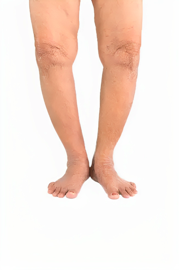
Prenatal Benign: (Autosomal Recessive & Autosomal Dominant )
In this form, despite prenatal (before birth) symptoms, skeletal bone defects improve progressively (during the third trimester). Symptoms of prenatal benign hypophosphatasia include the following:
- Shortening of long bones
- Bowing of legs
- The presence of dimples over the deformity
Infantile Form: (Autosomal Recessive)
Children with this form are normal at birth. Symptoms appear postnatally (after six months). These symptoms include:
- Premature craniosynostosis (a condition in which the skull bones fuse too early).
- Difficulty in breathing due to hypoplastic changes in the lungs
- Short stature
- Loss of teeth (deciduous)
Hypercalcemia is also present in this form. Hypercalcemia is characterized by irritability, loss of appetite, vomiting, constipation, polyuria, excessive thirst, and hypotonia (decreased tone).
Childhood Form: (Autosomal Recessive & Autosomal Dominant)
This form affects children up to 5 years of age. In this form, the signs and symptoms are:
- Intracranial hypertension
- Skeletal abnormalities
- Gait deformity (waddling gait)
- Short stature
- Bone pain
- Fracture of bones
- Focal bony defects
- Loss of teeth (incisors)
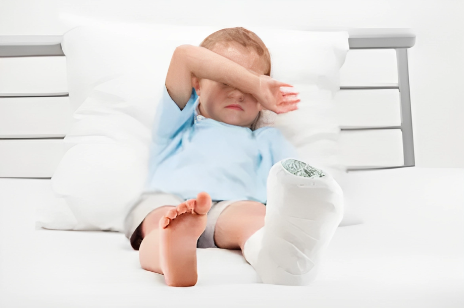
Adult Form: (Autosomal Dominant)
This form is more prevalent in the Middle Ages (13-18). In this form, the patient presents with4Whyte MP, Teitelbaum SL, Murphy WA, Bergfeld MA, Avioli LV. Adult hypophosphatasia. Clinical, laboratory, and genetic investigation of a large kindred with a review of the literature. Medicine (Baltimore) 1979;58:329–47.
- Pain in foot
- Bone fracture (usually metatarsals)
- Pseudo fracture of the femur
- Osteoarthritis is the late feature of this disease
- Tooth loss
Odontohypophosphatasia: (Autosomal Recessive & Autosomal Dominant)
This form mainly affects teeth, which is characterized by the loss of teeth before the average expected period. The anterior teeth are most commonly affected.5Beumer J, 3rd, Trowbridge HO, Silverman S, Jr, Eisenberg E. Childhood hypophosphatasia and the premature loss of teeth. A clinical and laboratory study of seven cases. Oral Surg Oral Med Oral Pathol. 1973;35:631–40. doi: 10.1016/0030-4220(73)90028-5.
How to Diagnose Hypophosphatasia?
Hypophosphatasia is diagnosed based on history, clinical symptoms, laboratory findings, radiological findings, and histopathology.6Nunes ME. Hypophosphatasia. 2007 Nov 20 [Updated 2023 Mar 30]. In: Adam MP, Mirzaa GM, Pagon RA, et al., editors. GeneReviews® [Internet]. Seattle (WA): University of Washington, Seattle; 1993-2023. Available from: https://www.ncbi.nlm.nih.gov/books/NBK1150/
History:
Knowledge about the background of the disease is necessary to make a diagnosis.7 Cole DE. Hypophosphatasia update: recent advances in diagnosis and treatment. Clin Genet. 2008;73(3):232–235. doi: 10.1111/j.1399-0004.2007.00958.x. Your doctor will ask some important questions. These questions include the following:
- Biodata
- Any previous positive history of this disease
- Family history
- Bone pain
- Any deformity in the bones?
- Tooth loss?
After a proper history, a physical examination is required to see clinical symptoms.
Laboratory Investigations:
In laboratory investigation, your doctor will prescribe baseline tests and total serum alkaline phosphatase. The activity of this enzyme is reduced in these patients with hypophosphatasia. However, the activity of alkaline phosphatase enzymes depends upon the severity of the disease. In general, when you have a more severe disease, the activity of the alkaline phosphatase enzyme will be lower.
In addition, low alkaline phosphatase activity is not only present in hypophosphatasia but also manifested in other diseases like celiac disease, early pregnancy, and hypothyroidism.
Some specific markers are used to diagnose hypophosphatasia. However, increasing pyridoxal phosphate levels (PLP) may be considered the most sensitive marker for HPP.8Whyte MP. Hypophosphatasia and the role of alkaline phosphatase in skeletal mineralization. Endocr Rev. 1994;15:439–61. doi: 10.1210/er.15.4.439.
Laboratory Features:
- Hypercalciuria (high levels of calcium in the urine during the first year of life)
- Increased vitamin B6 level
- Urine amino acid chromatogram, which shows increased urinary excretion of phosphoethanolamine and proline
- Increased inorganic pyrophosphate level (PPi)
Genetic Testing:
Genetic testing confirms the diagnosis of hypophosphatasia when other clinical and laboratory data do not support the diagnosis of hypophosphatasia. Additionally, at the molecular level, genetic screening indicates mutations in genes.
Single Gene Testing
In single gene testing, only ALPL gene analysis detects gene mutations that can be due to deletion or insertion.

Multiple Gene Testing
In Multiple gene testing, ALPL and other gene screenings are done to determine differential diagnoses.
Antenatal Diagnosis:
An antenatal diagnosis is required when there is a positive history of this disease in a previous child or pregnancy. Therefore, Chorionic villus DNA sampling is performed in the laboratories for the antenatal diagnosis of hypophosphatasia.
Radiological Investigations:
X-ray is the first line of investigation to diagnose hypophosphatasia. X-ray shows the following features.
- Bowing of long bones with osteochondral spur behind the skin
- Infantile rickets: Widened skull sutures, brachycephaly (a deformity in which there is a shorter anteroposterior diameter of the skull)
- Alveolar bone loss
- Pseudofracture
- Pathological bone fracture
- Rachitic deformity on chest
Histopathology:
Histopathology of bone shows rachitic abnormalities in the growth plate. Osteoclasts and osteoblasts appear normal with low alkaline phosphatase activity. In addition, Histopathology of the teeth shows a decrease in cementum (a hard calcified layer that covers your teeth).
How to treat Hypophosphatasia?
There is no cure for hypophosphatasia, but symptomatic relief is possible. Symptomatic treatment includes the following:9Facht, R. R. C. M. M. E. F. F. F. C. A. (n.d.). Hypophosphatasia (HPP): Background, pathophysiology, epidemiology. https://emedicine.medscape.com/article/945375-overview
- Enzyme replacement therapy
- Supplementation with zinc and magnesium
- Proper monitoring of calcium homeostasis
- Painkillers such as NSAIDs to relieve pain
- Pyridoxine to treat seizures
Supportive Treatment:
A multi-professional team gives supportive treatment to the patients. This team includes:
- Orthopedic surgeon to see bone-related complications and pain management.
- Neurosurgeon for the management of craniosynostosis (a condition in which a baby’s skull bones join earlier than normal age)
- Nephrologist to manage renal disease
- An endocrinologist to manage any hormonal abnormality.
- Pulmonologist for respiratory support
- Physiotherapist for proper physiotherapy
- Dental surgeon to deal with dental abnormality
Genetic Counseling:
As discussed earlier, this disease is linked to genetic mutation and can be transferred to the next generation. Genetic counseling is important to know how much your child is at risk of getting this disease if you have hypophosphatasia.
Autosomal Recessive Hypophosphatasia
In this disease pattern, if both parents are heterozygous for the ALPL gene, then 25% of the siblings are affected, 25% remain unaffected, and 50 % are heterozygous (carriers). Moreover, Heterozygous siblings may remain asymptomatic or show mild symptoms.
Autosomal Dominant Hypophosphatasia
In this form of the disease, if the parents are homozygous for the ALPL gene. All the siblings are affected. However, if the parents are heterozygous for the ALPL, it affects 50 percent of the siblings.
Prognosis of Hypophosphatasia
The perinatal form of hypophosphatasia is a lethal one, and the patient dies within days or weeks due to respiratory complications. However, patients with adult or odontohypophosphatasia have a normal life span.
Difference between Hypophosphatemia & Hypophosphatasia
Hypophosphatemia is an X-linked disorder with a low level of phosphate in the body ( <2.5 mg/dl). The normal phosphate level in the body is more than 7 mg/dl.
Hypophosphatasia is an autosomal recessive or autosomal dominant disorder. It is caused by a mutation in the ALPL gene, which results in a low level of alkaline phosphatase activity.
Conclusion
To conclude, hypophosphatasia is a genetic disorder related to the defective activity of TNSALP (tissue nonspecific alkaline phosphatase). However, There’s no standard treatment for this disease. A team of healthcare professionals with multiple specialties provides a symptomatic treatment approach. Genetic counseling plays an important role in preventing this disease.
Refrences
- 1Correa, R. R. (2023, June 5). Hypophosphatasia (HPP): Background, Pathophysiology, Epidemiology. Medscape.com; Medscape. https://emedicine.medscape.com/article/945375-overview#a6
- 2Nunes ME. Hypophosphatasia. 2007 Nov 20 [updated 2023 Mar 30]. In: Adam MP, Mirzaa GM, Pagon RA, Wallace SE, Bean LJH, Gripp KW, Amemiya A, editors. GeneReviews® [Internet]. Seattle (WA): University of Washington, Seattle; 1993–2023. PMID: 20301329.
- 3Mornet, E. (2007). Hypophosphatasia. Orphanet Journal of Rare Diseases, 2(1). https://doi.org/10.1186/1750-1172-2-40
- 4Whyte MP, Teitelbaum SL, Murphy WA, Bergfeld MA, Avioli LV. Adult hypophosphatasia. Clinical, laboratory, and genetic investigation of a large kindred with a review of the literature. Medicine (Baltimore) 1979;58:329–47.
- 5Beumer J, 3rd, Trowbridge HO, Silverman S, Jr, Eisenberg E. Childhood hypophosphatasia and the premature loss of teeth. A clinical and laboratory study of seven cases. Oral Surg Oral Med Oral Pathol. 1973;35:631–40. doi: 10.1016/0030-4220(73)90028-5.
- 6Nunes ME. Hypophosphatasia. 2007 Nov 20 [Updated 2023 Mar 30]. In: Adam MP, Mirzaa GM, Pagon RA, et al., editors. GeneReviews® [Internet]. Seattle (WA): University of Washington, Seattle; 1993-2023. Available from: https://www.ncbi.nlm.nih.gov/books/NBK1150/
- 7Cole DE. Hypophosphatasia update: recent advances in diagnosis and treatment. Clin Genet. 2008;73(3):232–235. doi: 10.1111/j.1399-0004.2007.00958.x.
- 8Whyte MP. Hypophosphatasia and the role of alkaline phosphatase in skeletal mineralization. Endocr Rev. 1994;15:439–61. doi: 10.1210/er.15.4.439.
- 9Facht, R. R. C. M. M. E. F. F. F. C. A. (n.d.). Hypophosphatasia (HPP): Background, pathophysiology, epidemiology. https://emedicine.medscape.com/article/945375-overview
