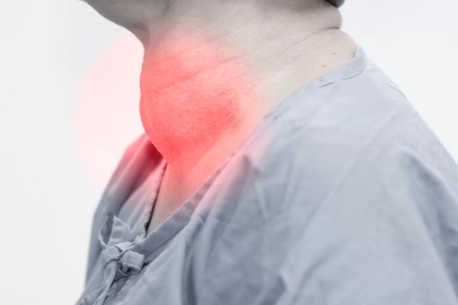
An enlarged thyroid is called a goiter, while the presence of nodules on a goiter is called multinodular goiter, which can either be toxic or non-toxic 1Unlu, M. T., Kostek, M., Aygun, N., Isgor, A., & Uludag, M. (2022). Non-Toxic Multinodular Goiter: From Etiopathogenesis to Treatment. Sisli Etfal Hastanesi tip bulteni, 56(1), 21–40. https://doi.org/10.14744/SEMB.2022.56514. This butterfly-shaped gland in our neck is responsible for releasing hormones that help regulate our sleep, heart, gut, skin, and weight.

While most multinodular goiters are asymptomatic and benign, toxic multinodular goiters still have a risk of becoming cancerous. Hence, if you feel like you have a lump or are diagnosed with it, your doctor will likely screen you for thyroid cancer as well.
Toxic vs. Non-toxic Multinodular Goiter: What are the Different Types?
Goiter is usually found during routine physical neck examinations or incidental ultrasounds.
Nontoxic Multinodular Goiter
Non-toxic multinodular goiter is a condition when the nodularity on the thyroid gland does not affect its functioning but can still cause:
- Visible lump around the neck
- Dysphagia
- Changes in voice
- Breathing problems
- Coughing
Toxic Multinodular Goiter
However, in a toxic multinodular goiter, excess thyroid hormone is produced, resulting in a condition called ‘hyperthyroidism,’ which can lead to:
- Abnormal weight loss
- Hand tremors
- Sweating
- Irregular heart rate
- Muscle weakness
- Heat intolerance
- Insomnia
- Increased hunger
- Diarrhea
If you notice any such symptoms, you need to consult a doctor immediately because a toxic multinodular goiter can become an emergency due to the risk of developing life-threatening hyperthyroidism called a ‘thyroid storm.’
What causes Multinodular Goiter?
While the cause remains ambiguous, certain risk factors increase the probability of developing multinodular goiter. Non-modifiable risk characteristics include female gender, age 55 and above, a positive family history (of goiter or thyroid problems), or autoimmune problems such as Grave’s disease or Hashimoto thyroiditis. Modifiable risk factors include smoking, exposure to estrogen drugs, and a lack of iodine.
Iodine Deficiency
Low iodine was previously one of the biggest risk factors for developing it. However, with the introduction of iodized salt in the 1920s, iodine deficiency is much less common now 2Hoang, T. D., Mai, V. Q., Clyde, P. W., Glister, B. C., & Shakir, M. K. (2011). Multinodular goiter as the initial presentation of systemic sarcoidosis: limitation of fine-needle biopsy. Respiratory care, 56(7), 1029–1032. https://doi.org/10.4187/respcare.01000. Hence, in regions where iodized salt was released late, the older generation is still at a higher risk of acquiring multinodular goiter.
How to Diagnose a Multinodular Goiter?

A physical examination is the first step in diagnosing a multinodular goiter. The doctor will have a look and feel of the gland to look for any nodularity. After a detailed history and examination, if your doctor suspects an abnormality, you will probably be offered a range of tests to exclude toxic multinodular goiter, non-toxic multinodular goiter, or even cancer.
Common tests
- Thyroid Profile: The thyroid profile (aka Thyroid Function Test) is a blood test to check the level of thyroid hormones (TSH, T3, and T4) and thyroid antibodies. A thyroid profile is one of the first tests offered during a goiter presentation, as it helps pick up toxic multinodular goiter early on. Toxic multinodular goiter has a hyperthyroid picture, increased T3 and T4, and a low TSH count.
- Imaging Tests: Your doctor may order ultrasound imaging that uses sound waves to visualize the thyroid gland’s internal structure. Ultrasound can help guide the doctor about the number, nature, and location of nodules, which can help develop a plan of action.
- Fine-Needle Aspiration (FNA) Biopsy: Biopsy is the next step after a positive ultrasound, especially if the nodule is greater than 1 cm. FNA biopsy uses a needle to take a chunk of thyroid cells from the nodules. A microscope then assesses whether the cells are cancerous or not.
Complications
Patients of multinodular goiter are at risk of developing thyroid cancer as 4-17% of glands after goiter removal are found to have remnants of carcinoma; more often than not, papillary carcinoma is the most common one 3Medeiros-Neto, G. (2016). Multinodular Goiter. In K. R. Feingold (Eds.) et. al., Endotext. MDText.com, Inc.. In fact, in a 2015 study on 134 patients with multinodular goiter, 46.3% were found to develop thyroid cancer 4Campbell, M. J., Seib, C. D., Candell, L., Gosnell, J. E., Duh, Q. Y., Clark, O. H., & Shen, W. T. (2015). The underestimated risk of cancer in patients with multinodular goiters after a benign fine needle aspiration. World journal of surgery, 39(3), 695–700. https://doi.org/10.1007/s00268-014-2854-y.
Treatment
Since most goiters are asymptomatic, they rarely require any treatment. However, goiters can present with varied types and problems that need treatment accordingly.
Observation
Non Toxic goiter with noncancerous nodules is often asymptomatic and does not even require much medical attention and treatment, and often goes undiagnosed.
Regardless, several treatments are available, the most common being observation. Your doctor would conduct routine examinations to ensure its dormancy as it is mostly benign.
Medication
When it comes to goiter, medicine rarely plays a huge role. In combination with goiter, medication is only used in hyperthyroid states. Carbimazole and propylthiouracil are the drugs of choice for hyperthyroidism because of their ability to cease the production of thyroid hormones. It is important to note that these medicines can take from 3 weeks to 2 months to show effects.
Need for Surgery
Thyroidectomy, the surgical goiter removal of all or part of the thyroid gland, is only opted for in extreme circumstances like excessive overgrowth, recurrence, or in the case of malignant nodules. For example, in a study in which out of 10-15% of patients with goiters require surgery, 12% have recurring states 5Moalem, J., Suh, I., & Duh, Q. Y. (2008). Treatment and prevention of recurrence of multinodular goiter: an evidence-based review of the literature. World journal of surgery, 32(7), 1301–1312. https://doi.org/10.1007/s00268-008-9477-0. However, even in the cases of benign nodules, we can opt for surgery if it is heavily impacting the quality of life.
Goiter surgery is essential for cancer-causing nodules commonly seen in a toxic multinodular goiter. According to a 2015 study, ‘Dunhill Thyroidectomy’ is considered a safer procedure for goiter removal as it poses lesser complications 6Mauriello, C., Marte, G., Canfora, A., Napolitano, S., Pezzolla, A., Gambardella, C., … & Candela, G. (2016). Bilateral benign multinodular goiter: what is the adequate surgical therapy? A review of the literature. International Journal of Surgery, 28, S7-S12.. However, one drawback of goiter surgery is the need for lifelong supplementation of exogenous thyroid hormones.
Radioactive Iodine Therapy
In toxic multinodular goiter, radioactive iodine therapy is the main choice, especially for smaller goiters. The radioactive iodine reduces hyperthyroidism by targeting the nodules and shrinking the goiter. Although radioactive iodine therapy is safe, it still comes with contraindications like pregnancy, lactation, cancer suspicion, and an age group of fewer than 15 years 7Gurgul, E., & Sowinski, J. (2011). Primary hyperthyroidism–diagnosis and treatment. Indications and contraindications for radioiodine therapy. Nuclear medicine review. Central & Eastern Europe, 14(1), 29–32.. Hence, it requires caution.
How to Shrink Multinodular Goiter Naturally?
If you have a multinodular goiter and want to avoid its seemingly daunting treatments, there are also home remedies on how to shrink goiter naturally.
An iodine-rich diet prevents goiters and shrinks them; milk, cheese, and tuna are good sources of iodine and can help fight iodine loss.
Selenium-rich foods like eggs and sunflower seeds help in the metabolism of thyroid hormones. While home remedies such as massaging olive oil on your neck or consuming lemon and garlic are great for their anti-inflammatory and detoxifying properties, which can help shrink your goiter.
Prevention
Prevention is better than cure. By tweaking your lifestyle, you can significantly lessen your chances of multinodular goiter by controlling modifiable risk factors like iodine deficiency, smoking, and alcohol consumption.
When it comes to prevention, iodine plays the most important role. By consuming 5g of iodized salt daily, you can significantly reduce your risk of goiter 8Gurgul, E., & Sowinski, J. (2011). Primary hyperthyroidism–diagnosis and treatment. Indications and contraindications for radioiodine therapy. Nuclear medicine review. Central & Eastern Europe, 14(1), 29–32..
Conclusion
Multinodular goiters might seem scary but are mostly asymptomatic and harmless. If they pose a medical issue, they can be easily cured by tried and tested medical treatments like radioactive iodine therapy or goiter surgery. However, they can sometimes lead to more serious conditions like hyperthyroidism and cancer. Hence, a cautious mind and safer life choices can help prevent this the first time.
Refrences
- 1Unlu, M. T., Kostek, M., Aygun, N., Isgor, A., & Uludag, M. (2022). Non-Toxic Multinodular Goiter: From Etiopathogenesis to Treatment. Sisli Etfal Hastanesi tip bulteni, 56(1), 21–40. https://doi.org/10.14744/SEMB.2022.56514
- 2Hoang, T. D., Mai, V. Q., Clyde, P. W., Glister, B. C., & Shakir, M. K. (2011). Multinodular goiter as the initial presentation of systemic sarcoidosis: limitation of fine-needle biopsy. Respiratory care, 56(7), 1029–1032. https://doi.org/10.4187/respcare.01000
- 3Medeiros-Neto, G. (2016). Multinodular Goiter. In K. R. Feingold (Eds.) et. al., Endotext. MDText.com, Inc.
- 4Campbell, M. J., Seib, C. D., Candell, L., Gosnell, J. E., Duh, Q. Y., Clark, O. H., & Shen, W. T. (2015). The underestimated risk of cancer in patients with multinodular goiters after a benign fine needle aspiration. World journal of surgery, 39(3), 695–700. https://doi.org/10.1007/s00268-014-2854-y
- 5Moalem, J., Suh, I., & Duh, Q. Y. (2008). Treatment and prevention of recurrence of multinodular goiter: an evidence-based review of the literature. World journal of surgery, 32(7), 1301–1312. https://doi.org/10.1007/s00268-008-9477-0
- 6Mauriello, C., Marte, G., Canfora, A., Napolitano, S., Pezzolla, A., Gambardella, C., … & Candela, G. (2016). Bilateral benign multinodular goiter: what is the adequate surgical therapy? A review of the literature. International Journal of Surgery, 28, S7-S12.
- 7Gurgul, E., & Sowinski, J. (2011). Primary hyperthyroidism–diagnosis and treatment. Indications and contraindications for radioiodine therapy. Nuclear medicine review. Central & Eastern Europe, 14(1), 29–32.
- 8Gurgul, E., & Sowinski, J. (2011). Primary hyperthyroidism–diagnosis and treatment. Indications and contraindications for radioiodine therapy. Nuclear medicine review. Central & Eastern Europe, 14(1), 29–32.


