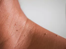
Myiasis is a serious parasitic infestation that occurs when certain species of flies lay eggs on or in human tissue. After hatching, these larvae or maggots feed on your tissue, which results in pain, swelling, and a foul odor. If untreated, myiasis results in severe tissue damage.
What is Myiasis?
Myiasis is the invasion of living tissue by fly larvae, which feed on human or animal flesh after hatching. The term myiasis comes from the Greek word “myia”, which means “fly.” Myiasis is not commonly found in the United States. However, it is more prevalent in the tropical and subtropical parts of the world, such as Central and South America, Africa, and the Caribbean.1CDC. (2024, September 13). About Myiasis. Myiasis. https://www.cdc.gov/myiasis/about/index.html
What is Myiasis caused by?
Myiasis occurs when fly larvae infest living tissue as a necessary part of their life cycle, opportunistically in wounds or decaying tissue, or by accidental entry into your body. Risk factors for myiasis include:
- Flies are more likely to lay eggs on your skin if you have poor hygiene, don’t bathe regularly, or wear dirty clothes.
- Open wounds give larvae a direct way in if you don’t keep them clean and covered.
- Weak immunity from illness or malnutrition slows your healing, giving larvae more time to grow.
- Having limited mobility can make it harder for you to maintain hygiene and wound care, which increases your risk of myiasis.
- Fly-infested environments, whether you live near livestock, in a tropical climate, or around unsanitary conditions, bring you into closer contact with egg-laying flies.2Andreatta, E., & Bonavina, L. (2021). Wound myiasis in Western Europe: prevalence and risk factors in a changing climate scenario. European Surgery, 54(6), 289–294. https://doi.org/10.1007/s10353-021-00730-y
Myiasis Transmission:
The transmission of myiasis depends on the type of fly and how they lay their eggs. Here’s how it usually spreads:
- Some flies lay their eggs on mosquitoes or other blood-feeding insects. When these insects bite a human, the warmth triggers the eggs to hatch, and the larvae burrow into the skin. This is common with the human botfly, Dermatobia hominis in South America, and the tumbu fly, Cordylobia anthropophaga in Africa.
- Screwworm flies, like Cochliomyia hominivora and Chrysomya bezziana, lay eggs directly in wounds, ulcers, or necrotic tissue. The larvae feed on living or dead tissue and cause wound myiasis. This is far more common in areas with poor hygiene or inadequate wound care.
- Some larvae enter the skin, move through tissues, and result in migratory myiasis. This can happen when people walk barefoot on contaminated soil or have contact with infected animals like cattle or horses. The Hypoderma species of warble flies and the Gasterophilus species of horse botflies are transmitted by this method.
- In rare cases, people can swallow fly eggs or larvae through contaminated food or water.
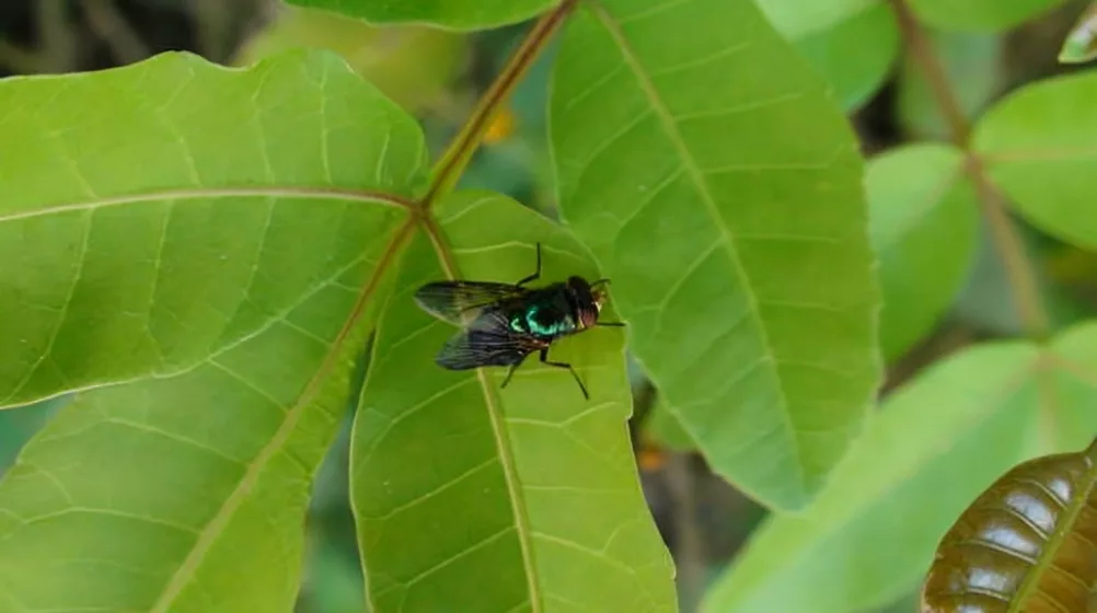
Types of Myiasis
Myiasis can be classified into several types, some of which are:
Sanguinivorous Myiasis:
Sanguinivorous myiasis happens when fly larvae feed on your blood instead of burrowing into your skin. Unlike other types, these larvae act more like ticks or fleas. They attach to your exposed skin and suck blood, usually at night. This type is mainly caused by Auchmeromyia senegalensis (Congo floor maggot) and is more common in rural Africa, especially where people sleep on bare floors.
Cutaneous Myiasis:
Cutaneous myiasis happens when fly larvae infest the skin. It has two main forms: furuncular and migratory.
- Furuncular myiasis happens when fly larvae enter unbroken skin. You may notice a boil-like lump that gets bigger over time. The area can feel swollen and painful. A small hole in the lump lets the larvae breathe. After several weeks, they grow and eventually come out of the skin. This type is more common if you have traveled to tropical regions. The main culprits are Dermatobia hominis (human botfly) in Central and South America and Cordylobia anthropophaga (tumbu fly) in sub-Saharan Africa.
- Migratory myiasis happens when fly larvae burrow under your skin and move through tissues. This creates red, winding tracks that spread over time. You may feel irritation and an intense crawling sensation as the larvae move. Unlike furuncular myiasis, where larvae stay in one spot, these keep migrating through the skin. The most common causes are Gasterophilus (horse botflies), which mainly affect horses but can infest humans, and Hypoderma (warble flies), which usually infect cattle but sometimes burrow under human skin.
Wound Myiasis:
Wound myiasis happens when fly larvae infest open wounds, ulcers, or dead tissue. If you have an untreated wound, flies may lay their eggs directly inside it. The larvae feed on both dead and living tissue, which can slow down healing and cause infections. Unlike other types, this does not require healthy skin but only an open wound. If you are in a tropical or rural area, especially in the Americas, Africa, or Asia, you should keep your wounds clean and covered. The most common culprits are Cochliomyia hominivorax (New World screwworm) and Chrysomya bezziana (Old World screwworm).
Cavitory Myiasis:
Cavitary myiasis occurs when fly larvae infest natural openings in your body, such as the nose, ears, eyes, mouth, or even deeper areas like the respiratory or gastrointestinal tract. Unlike cutaneous myiasis, where larvae burrow into the skin, these larvae enter existing cavities and feed on living or decaying tissue. This can cause pain, swelling, foul-smelling discharge, and even tissue destruction in severe cases.3Francesconi, F., & Lupi, O. (2012). Myiasis. Clinical Microbiology Reviews, 25(1), 79–105. https://doi.org/10.1128/cmr.00010-11
Signs & Symptoms of Myiasis
Myiasis presents differently depending on where the larvae are located. The most common sign is a painful or itchy skin lesion with a small central opening where the larvae breathe.4Blechman, A. B. (2025, February 6). Myiasis: Background, Pathophysiology, Etiology. Medscape.com; Medscape. https://emedicine.medscape.com/article/1491170-overview
| Type of Myiasis | Clinical Presentation |
| Furuncular Myiasis | Painful, boil-like swellings with a small central hole, Itchiness, Tenderness |
| Migratory Myiasis | Red, winding tracks, Intense creeping sensation and itching as larvae migrate |
| Wound Myiasis | Foul-smelling discharge Severe pain |
| Sanguinivorous Myiasis | Irritation Anemia |
| Nasal Myiasis | Bad-smelling nasal discharge Frequent nosebleeds Facial swelling Destruction of the nasal passages |
| Aural Myiasis | Foul-smelling ear discharge Itching and an earache Hearing loss Eardrum damage |
| Ophthalmomyiasis | Redness Swelling Tearing Blurred vision Corneal ulcers Vision loss |
| Oral Myiasis | Swelling and pain in the mouth Trouble eating or speaking Foul odor Soft tissue damage |
| Gastrointestinal Myiasis | Nausea Vomiting Diarrhea Stomach pain Blood in your stool Larvae in your vomit or feces |
| Respiratory Myiasis | Persistent cough Trouble breathing Chest pain Cough up blood Blocked airways |
How is a Myiasis diagnosis established?
Myiasis is usually diagnosed clinically, though tests are required to determine what type of fly larvae have been deposited in your body.
History & Physical Exam:
Your doctor will start by inquiring about your symptoms and any possible exposure to flies or larvae. A physical exam will focus on the affected areas. If needed, your doctor may use a dermatoscope or apply gentle pressure to encourage the larvae to emerge.
Lab Investigations:
Various lab tests can indicate or confirm myiasis in your body. These include:
- A CBC test can help identify signs of infection. You may have an increased white blood cell count in the form of leukocytosis and eosinophilia. This presentation is especially likely if your immune system is reacting strongly to the larvae.
- Inflammatory markers like ESR and CRP are elevated if there is significant inflammation or secondary infection.
- Microscopic examination of discharge or tissue from myiasis-affected sites can reveal larval structures. This confirms the presence of fly larvae. If there is any pus or exudate, your doctor may examine it under a microscope.
- If a biopsy is performed, histopathological testing can show inflammatory changes and larvae in cross-section. This is rarely needed but may be useful in deep or unclear cases.
- Stool examination is done if intestinal myiasis is suspected, because larvae or eggs might be present in stool samples.
- Bacterial culture and sensitivity tests of wound swabs, blood, and tissue samples help detect secondary bacterial infections.
Imaging:
For early and more serious myiasis cases, different imaging modalities are often employed.
- MRI helps your doctor see if larvae have invaded deeper tissues, like your brain, breast, face, or eyes. It gives a clear picture of how much damage has occurred and where the larvae are.
- CT scans may be needed if the infestation is deep inside your body. This helps check how far the larvae have spread.
- Ultrasound scans, especially high-resolution Doppler scans, can help confirm if larvae are present, especially in the early stages. It shows their size and exact location under your skin.5Dutto, M., Zavattero, C., Elio Vinai, Lauria, G., & Stefano Vanin. (2023). [Diagnostic atypique d’une myiase]. PubMed, 3(2). https://doi.org/10.48327/mtsi.v3i2.2023.370
What is the treatment for Myiasis?
In most cases, if left untreated, the larvae will eventually fall out within 5 to 7 weeks, but that’s not ideal. Proper treatment ensures faster recovery, less discomfort, and a lower risk of infection.
Maggot Removal:
One of the simplest ways to remove the larvae is by cutting off their air supply, making them crawl out on their own. Covering the wound with petroleum jelly, liquid paraffin, beeswax, or even bacon strips tricks the larvae into moving up to the surface, where they can be picked off with tweezers or forceps. This can take anywhere from a few hours to a full day.
For some types of myiasis, making the wound opening slightly bigger can help get them out faster. Once the larvae are removed, the wounds are thoroughly cleaned and dresed to avoid reinfestation or infection. If you have wound myiasis, doctors may also flush the wound with antiseptics and use special solutions like chloroform in light oil or ether to weaken the larvae before removal.

Surgery:
If the larvae are too deep or won’t come out with suffocation techniques, a minor surgical procedure might be needed. A doctor will numb the area with lidocaine before making a small incision or using a punch tool to open the wound for better access.
Another technique involves injecting lidocaine into the base of the wound to push the larvae out with pressure. Since larvae latch on tightly with tiny hooks, they must be removed carefully to avoid breaking them apart inside the wound, which could lead to inflammation or infection.
If myiasis affects sensitive areas like the eyes, brain, or deep tissues, more complex surgery may be required to prevent serious damage. After removal, the wound is cleaned, stitched if needed, and monitored closely for proper healing.
Medications:
A combination of antiparasitic and antibiotic medications is used to kill both the fly larvae and any bacteria they expose you to:
- Oral or topical ivermectin affects the larvae’s nervous system by increasing chloride levels, paralyzing and killing them. It works well for deeper infections and eye myiasis.6Patel, B. C., Ostwal, S., Sanghavi, P. R., Joshi, G., & Singh, R. (2018). Management of Malignant Wound Myiasis with Ivermectin, Albendazole, and Clindamycin (Triple Therapy) in Advanced Head-and-Neck Cancer Patients: A Prospective Observational Study. Indian Journal of Palliative Care, 24(4), 459. https://doi.org/10.4103/IJPC.IJPC_112_18
- Albendazole stops the larvae from using energy by blocking important cell structures. Without nutrition, they die. It helps when larvae are inside tissues. Thiabendazole also works similarly, which makes larvae weak and easier to remove. It is mainly used for larvae that move under your skin.
- Broad-spectrum antibiotics help prevent or treat bacterial infections that can develop in myiasis wounds. These include amoxicillin-clavulanate, cephalexin, and clindamycin.
Preventing Recurrence:
It is important to take certain safety precautions to reduce the risk of myiasis infestations, as well as other fly-borne infections:
- Wear protective clothing such as long-sleeved shirts, pants, and hats to minimize skin exposure. Use insect repellents on your exposed skin and clothing to deter flies.
- Sleep on raised beds, in screened rooms, or under a mosquito net at night to prevent fly contact. Avoid sleeping outside if you have open wounds, as this increases the risk of fly infestation.
- In regions where flies are common, clothing should be hot-ironed and thoroughly dried to eliminate any fly eggs.
- To prevent wound myiasis, maintain proper wound hygiene with regular cleaning, antiseptic application, and appropriate dressings.
- In hospitals or indoor settings, make sure that windows remain closed or are properly screened to prevent flies from entering.7Patel, B. C., Shrenik Ostwal, Sanghavi, P. R., Joshi, G., & Singh, R. (2018). Management of malignant wound myiasis with ivermectin, albendazole, and clindamycin (triple therapy) in advanced head-and-neck cancer patients: A prospective observational study. Indian Journal of Palliative Care, 24(4), 459–459. https://doi.org/10.4103/ijpc.ijpc_112_18
Complications of Myiasis
The complications often arise when the infestations remain untreated for too long, such as:
- If a larva isn’t fully removed, any remaining parts can trigger a strong foreign body reaction, causing swelling, pain, and delayed healing.
- Secondary bacterial infections are also common. If the wound turns red, swollen, or starts oozing pus, antibiotics may be needed.
- In severe cases, untreated secondary infections can lead to cellulitis or even sepsis.
- Myiasis can also serve as an entry point for Clostridium tetani, increasing the risk of tetanus.8Villwock, J. A., & Harris, T. M. (2014). Head and Neck Myiasis, Cutaneous Malignancy, and Infection: A Case Series and Review of the Literature. The Journal of Emergency Medicine, 47(2), e37–e41. https://doi.org/10.1016/j.jemermed.2014.04.024
Conclusion
It is a parasitic infection caused by fly larvae called maggots, which are deposited in your skin, body openings, and sometimes even organs. Common in regions with limited healthcare, the infestation can lead to severe complications if left untreated. Treatment involves the removal of the maggots through surgery, wound care, and antibiotics.
In regions with limited healthcare, the infestation can lead to severe complications if left untreated. Treatment involves the removal of the maggots through surgery, wound care, and antibiotics.
Refrences
- 1CDC. (2024, September 13). About Myiasis. Myiasis. https://www.cdc.gov/myiasis/about/index.html
- 2Andreatta, E., & Bonavina, L. (2021). Wound myiasis in Western Europe: prevalence and risk factors in a changing climate scenario. European Surgery, 54(6), 289–294. https://doi.org/10.1007/s10353-021-00730-y
- 3Francesconi, F., & Lupi, O. (2012). Myiasis. Clinical Microbiology Reviews, 25(1), 79–105. https://doi.org/10.1128/cmr.00010-11
- 4Blechman, A. B. (2025, February 6). Myiasis: Background, Pathophysiology, Etiology. Medscape.com; Medscape. https://emedicine.medscape.com/article/1491170-overview
- 5Dutto, M., Zavattero, C., Elio Vinai, Lauria, G., & Stefano Vanin. (2023). [Diagnostic atypique d’une myiase]. PubMed, 3(2). https://doi.org/10.48327/mtsi.v3i2.2023.370
- 6Patel, B. C., Ostwal, S., Sanghavi, P. R., Joshi, G., & Singh, R. (2018). Management of Malignant Wound Myiasis with Ivermectin, Albendazole, and Clindamycin (Triple Therapy) in Advanced Head-and-Neck Cancer Patients: A Prospective Observational Study. Indian Journal of Palliative Care, 24(4), 459. https://doi.org/10.4103/IJPC.IJPC_112_18
- 7Patel, B. C., Shrenik Ostwal, Sanghavi, P. R., Joshi, G., & Singh, R. (2018). Management of malignant wound myiasis with ivermectin, albendazole, and clindamycin (triple therapy) in advanced head-and-neck cancer patients: A prospective observational study. Indian Journal of Palliative Care, 24(4), 459–459. https://doi.org/10.4103/ijpc.ijpc_112_18
- 8Villwock, J. A., & Harris, T. M. (2014). Head and Neck Myiasis, Cutaneous Malignancy, and Infection: A Case Series and Review of the Literature. The Journal of Emergency Medicine, 47(2), e37–e41. https://doi.org/10.1016/j.jemermed.2014.04.024

