What is Presbycusis?
The term presbycusis means “old hearing”. It is defined as the symmetrical bilateral age-related sensorineural hearing loss that is irreversible. Presbycusis is the most common problem in society nowadays. It is a complex hearing disorder that becomes noticeable at or after the age of 60 years. It progresses slowly, but there are some stressors that speed up the deterioration process.
However, bilateral high-frequency hearing impairment with poor speech discrimination is the hallmark of this disease. The association between high tone deafness and advanced age was first discovered in 1899 by Zwaardemaker. Moreover, there is no treatment for this disease, but hearing aids can be used to overcome the symptoms.1Age-Related Hearing Loss (Presbycusis) — Causes and treatment. (2023, March 17). NIDCD. https://www.nidcd.nih.gov/health/age-related-hearing-loss2Cheslock, M. (2023, May 29). Presbycusis. StatPearls – NCBI Bookshelf. https://www.ncbi.nlm.nih.gov/books/NBK559220/3Saadi, R. A., MD. (n.d.). Presbycusis workUp: laboratory studies, imaging studies, other tests. https://reference.medscape.com/article/855989-workup#showall
Understanding of the normal hearing mechanism (Conduction of the sound waves)
There are three parts of your ear (external ear, middle ear, and inner ear). During this process, sounds from the outer side of the ear come in the form of air vibration that is captured by your external ear and transferred toward the tympanic membrane. When the external air strikes the tympanic membrane, it behaves like a drum. Air from the tympanic membrane enters the middle ear with a specific frequency and amplitude. In addition, your middle ear comprises three small bones (malleus, incus, and stapes). At this stage, the middle ear performs three functions:
- Transfer of acoustic vibration coming from the tympanic membrane toward the cochlea
- Matches impedance between the outer and inner ear, which helps in efficient sound transmission.
- Protects the inner ear through the acoustic reflex, which dampens loud sounds
After reaching the inner ear, the acoustic vibrations are transferred to the cochlea. The cochlea contains receptors that convert physical energy (vibrations) into electrical energy (nerve signal). The nerve signal then travels toward the auditory cortex through the cochlear nerve.
Epidemiology
Presbycusis is the most common cause of hearing impairment that is most commonly linked with aging. The prevalence of this disease is not well-known due to the differences between the investigators. According to some studies in the United States (USA), it affects about 25-30% of people between the ages of 65 and 74, and for people ages greater than 75, the incidence of this disease was about 40-50%.
So, according to the above discussion, presbycusis affects more than half of the people aged around 70-75 years and nearly all people over the age of 90.4Age-Related Hearing Loss (Presbycusis) — Causes and treatment. (2023, March 17). NIDCD. https://www.nidcd.nih.gov/health/age-related-hearing-loss5Cheslock, M. (2023, May 29). Presbycusis. StatPearls – NCBI Bookshelf. https://www.ncbi.nlm.nih.gov/books/NBK559220/6Saadi, R. A., MD. (n.d.). Presbycusis workUp: laboratory studies, imaging studies, other tests. https://reference.medscape.com/article/855989-workup#showall

Pathophysiology
Multiple factors contribute to presbycusis. These factors are intrinsic (present inside the body), such as genetics, and extrinsic (present outside), like noise, medications, smoking, and other comorbidities.
In addition, some age-related histological changes affect your auditory system (cochlea and auditory cortex). These changes are associated with different clinical outcomes depending upon the severity of the disease. Histologically, age-related hearing loss is a slowly progressive disorder that results in either cochlear degeneration or due to loss of auditory nerve fibers.7Cheslock, M. (2023, May 29). Presbycusis. StatPearls – NCBI Bookshelf. https://www.ncbi.nlm.nih.gov/books/NBK559220/
What are the Types of Presbycusis?
On temporal bone analysis and correlating them with hearing loss, there are four main types and two subtypes of presbycusis.
Sensory Presbycusis:
In this type, there is a loss of hair cells in the cochlea with epithelial atrophy. The process begins from the basal turn of the cochlea and processes slowly toward the apex. Moreover, these histological changes result in a decline in the high-frequency threshold. However, speech discrimination is not affected.
Neural Presbycusis (Metabolic Presbycusis):
Neural presbycusis is associated with the atrophy of cochlear nerve cells and the central neural pathway (spiral ganglions). According to research, about 2100 neurons out of 35000 are estimated to be lost every decade. In addition, the loss of these neurons is genetically predetermined. The effect is noticeable when more than 90% of the neurons are lost. Clinically, it is characterized by severe loss of speech and discrimination compared to others.
Strial Presbycusis:
Strial presbycusis involves degeneration of stria vascularis (supply blood to the inner ear). These cells are essential for maintaining the endocochlear potential of endolymph. However, this process most commonly occurs in the last three decades of life.
Mechanical Presbycusis:
This type has a conductive ear impairment due to age-related changes in the cochlear duct (thickening and stiffening of the basilar membrane). Moreover, this membrane is an essential part of the organ of Corti that converts physical signals into electrical signals.
Subtypes of Presbycusis
There are two subtypes of presbycusis. These are the following:
Mixed Presbycusis
In this type, more than one pathological change is involved.
Indeterminate Presbycusis
In this type, no pathology is present.8Cheslock, M. (2023, May 29). Presbycusis. StatPearls – NCBI Bookshelf. https://www.ncbi.nlm.nih.gov/books/NBK559220/9Saadi, R. A., MD. (n.d.). Presbycusis workUp: laboratory studies, imaging studies, other tests. https://reference.medscape.com/article/855989-workup#showall
Otosclerosis vs. Presbycusis
As we discussed above, the middle ear plays an important role in hearing. Otosclerosis most commonly affects the middle ear. The term “otosclerosis” combines “osteo,” meaning bone, and “sclerosis,” meaning abnormal hardening. This condition is a bony disease that causes abnormal hardening of the bones in the middle ear, particularly the stapes. It limits the ability of stapes to vibrate, resulting in hearing impairment because these vibratory movements of stapes are essential for hearing. It can occur at any age.
Presbycusis is an age-related problem characterized by bilateral sensorineural hearing loss. Additionally, poor speech discrimination and high-tone deafness with advanced age are hallmarks of the disease.
What are the Causes of Presbycusis?
Presbycusis is a multifactorial disease. There can be multiple causes of this disease.
Genetic Factors:
The most common and potential cause of presbycusis is gene mutation of the mitochondrial DNA complex. As age progresses, the perfusion capability of the cochlea is reduced, resulting in the formation of reactive oxygen metabolites.
These reactive metabolites damage your inner ear as well as the mitochondrial DNA complex. As mitochondria are involved in oxidative phosphorylation, damage to mitochondria results in reduced oxidative phosphorylation that interferes with the normal function of the inner ear. Some studies support the evidence of presbycusis due to damaged mitochondrial DNA.
According to Dai et al., the damaged mitochondrial DNA causes anatomical changes in the middle ear (narrowing of the vaso nervorum). According to Picks, the rate of apoptosis increases in people with damaged mitochondrial DNA complex.
About two specific mitochondrial gene deletions (mtDNA4834 and mtDNA 4977) have been linked with age-related hearing impairment.10Saadi, R. A., MD. (n.d.). Presbycusis workUp: laboratory studies, imaging studies, other tests. https://reference.medscape.com/article/855989-workup#showall
Hormonal Factors:
Some hormones are also linked with presbycusis. These hormones include sex hormones, glucocorticoids, and glutamate. An increase in glucocorticoid level for a prolonged duration and loss of Kappa B results in loss of spiral ganglion neurons.11Guimaraes, P., Frisina, S. T., Mapes, F., Tadros, S. F., Frisina, D. R., & Frisina, R. D. (2006). Progestin negatively affects hearing in aged women. Proceedings of the National Academy of Sciences of the United States of America, 103(38), 14246–14249. https://doi.org/10.1073/pnas.0606891103
Nutritional Factors:
There are some nutritional factors that are linked to the presbycusis. According to the research done by Berner, the deficiency of folic acid and vitamin B12 may have a relationship with age-related hearing loss, but the results are less significant. However, Martin Villares et al. found that high cholesterol levels are linked with hearing loss, and the results are positive.12Martín Villares, C., San Román Carbajo, J., Domínguez Calvo, J., Fernández Pello, M. E., Pomar Blanco, P., & Tapia Risueño, M. (2005). Perfil lipídico de la sordera ligada al envejecimiento [Lipid profile and hearing-loss aged-related]. Nutricion hospitalaria, 20(1), 52–57.
According to Gao et al., it was noted that there is a decline in the concentration of gamma-aminobutyric acid (GABA) in the brain in people with presbycusis compared to normal ones.13Gao, F., Wang, G., Ma, W., Ren, F., Li, M., Dong, Y., Liu, C., Liu, B., Bai, X., Zhao, B., & Edden, R. A. (2015). Decreased auditory GABA+ concentrations in presbycusis demonstrated by edited magnetic resonance spectroscopy. NeuroImage, 106, 311–316. https://doi.org/10.1016/j.neuroimage.2014.11.023
Ototoxic Factors:
Multiple ototoxic agents contribute to causing presbycusis. They can be medicine or chemicals. Medicines include:
- Loop diuretics
- Salicylates
- Aminoglycosides
Additionally, some environmental toxins are also involved in causing presbycusis. These are:
- Lead
- Carbon monoxide
- Mercury
- Toluene
Avoiding exposure to these toxic agents can prevent age-related hearing impairment.
Noise Exposure:
Several research studies indicate that people living in a noisy area or who have sustained noise-induced ear damage at a young age are more prone to develop severe presbycusis elderly. Histologically, persistent noise exposure results in the loss of spiral ganglion neurons.
Other Causes of Presbycusis:
There are some other causes14Cheslock, M. (2023, May 29). Presbycusis. StatPearls – NCBI Bookshelf. https://www.ncbi.nlm.nih.gov/books/NBK559220/15Saadi, R. A., MD. (n.d.). Presbycusis workUp: laboratory studies, imaging studies, other tests. https://reference.medscape.com/article/855989-workup#showall that contribute to presbycusis. These are:
- Diabetes mellitus
- High blood pressure
- White people
- Smoking
- Bacterial or viral infections
- Low socioeconomic status
Signs & Symptoms of Presbycusis
There can be multiple symptoms of presbycusis depending upon the type and severity of the disease. Symptoms of presbycusis vary from person to person and with age. The most common complaints of this disease are:
- Inability to hear high-pitched sounds
- Loss of speech discrimination, specifically in noisy places
- The affected person easily understands men’s voices than women
- Tinnitus (It is a clinical condition in which the person hears sounds from inside of the body rather than outside of the body), e.g., ringing, buzzing
- Other people’s speech sounds mumbled or muffled
Consult your family doctor when you have these symptoms, and book an appointment.16Cheslock, M. (2023, May 29). Presbycusis. StatPearls – NCBI Bookshelf. https://www.ncbi.nlm.nih.gov/books/NBK559220/
How to Diagnose Presbycusis?
The diagnosis of presbycusis is mainly based on history, physical examination, laboratory investigations, and radiological investigations.
History:
It is the first basic step of investigation to reach the diagnosis of every disease. There are multiple questions to be asked. These are:
- Do you have a hearing impairment? If yes, then how much? Onset and duration?
- Can you hear high-pitched words like S or th?
- Do you feel ringing or buzzing in your ear?
- Is there any discharge coming from your ear?
- Your occupation?
- Do your family members have this same problem?
- Any history of previous illness?
Physical Examination:
After taking a detailed history, your doctor does your physical examination to confirm or support the diagnosis. During the physical examination, your doctor does some tests to confirm his diagnosis. These tests are:
Otoscopy
It is the most widely used procedure in daily clinical settings. An otoscope is an instrument that helps visualize the structure of the ear, specifically the external auditory canal, tympanic membrane (ear drum), and middle ear.
Rinne Test
Rinne’s test diagnoses conductive hearing loss. To perform the test, the vibrating tuning fork is initially placed on the mastoid process behind the ear. The patient is asked to indicate when they can no longer hear the sound while the other ear is covered to prevent external noise interference. Once the patient reports that they can no longer hear the sound through bone conduction, the tuning fork is quickly moved to a position near the ear canal (about 3-4 cm away) to test air conduction. The patient is then asked to indicate when they can no longer hear the sound through air conduction. Normally, air conduction is greater than bone conduction, meaning the patient should hear the tuning fork sound longer in front of the ear than when it was placed on the mastoid process.
Weber’s Test
Weber’s test is specifically used to differentiate between conductive and sensory hearing loss. This test is performed as follows:
- Strike a tuning fork on an elastic object or clinician’s elbow that produces a pure tune.
- After striking, place the tuning fork on the forehead, bridge of the nose, or chin.
- Ask the patient in which ear they are hearing a loud voice.
- Normally, sound is equal on both sides. In sensorineural hearing loss, sound is louder in the normal ear, while in conductive hearing loss, sound is more clear and louder in the affected ear.
Audiometry Testing
It is the examination of choice to diagnose the presbycusis. It tests your ability to hear the sounds of varying intensities and frequencies. Moreover, there are about three parts of audiometry.
- Mechanical sound transmission
- Sound transmission via nerve impulse
- To check the ability of speech discrimination
There are a few tests that are part of audiometry. These are:
Speech Audiometry
In this test, your doctor checks how loud speech or words you can hear or how clearly you can differentiate between the words.
Pure Tone Testing
In pure tone testing, patients listen to voices of varying frequencies through headphones and indicate when they hear the sound. Audiometry generates results in the form of an audiogram. Presbycusis initially impacts high tones, with low tones being affected as the condition advances. Moreover, on audiogram, presbycusis is characterized by symmetrical bilateral hearing loss of more than 2000 Hertz.
Tympanometry
This test measures the function of the middle ear by assessing the mobility of the tympanic membrane (eardrum) in response to changes in air pressure, helping to detect issues like fluid in the middle ear or eardrum perforations.
Laboratory Investigations:
Laboratory investigations are usually not required in this case but are advised if there are some comorbidities. However, these investigations are:
- Complete blood count (CBC) to check for anemia
- White blood cell count to check for acute or chronic infections
- HBA1C to check the progression of Diabetes Mellitus
Radiological Investigations:
Radiological investigations are necessary when there is a suspicion of a tumor or to see the anatomical structure of the ear. These investigations include17Screening for handicapping hearing loss in the elderly. (2003, January 1). PubMed. https://pubmed.ncbi.nlm.nih.gov/12540314/18Saadi, R. A., MD. (n.d.). Presbycusis workUp: laboratory studies, imaging studies, other tests. https://reference.medscape.com/article/855989-workup#showall
- CT scan
- MRI
How to Treat Presbycusis?
This disease has no specific treatment because age-related hearing loss is irreversible. However, hearing aids are used to help relieve your symptoms. These devices positively impact the quality of life and communication of the affected person. These devices are the following:
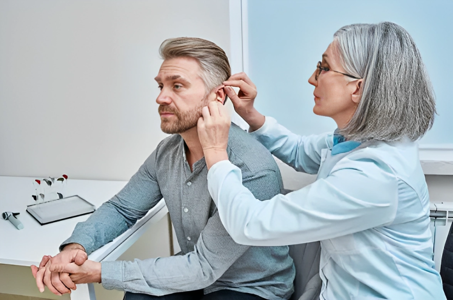
Amplification Devices:
These devices do not repair normal hearing mechanisms but help to amplify sounds. Moreover, they are electrical devices that can stay inside or behind the ear.

Cochlear Implants:
These are tiny devices that doctors surgically implant into the ears of people with severe hearing loss.
Assistive Listening Devices:
It includes telephone and cellphone devices that send signals to the patients with the help of a headset he wears. Moreover, it helps to amplify the specific sound you want to hear when there is noise in the background.
Lip Reading:
Lip reading is a technique that helps people with loss of speech discrimination and also helps people wearing hearing aids in noisy environments.19Saadi, R. A., MD. (n.d.). Presbycusis workUp: laboratory studies, imaging studies, other tests. https://reference.medscape.com/article/855989-workup#showall
Pharmacological Intervention
There is some ongoing research proposing the medical treatment of presbycusis. However, clinical trials on some medicines (antioxidants and anti-inflammatory) show that medications also slow down the process of age-related hearing loss.20Cheslock, M. (2023, May 29). Presbycusis. StatPearls – NCBI Bookshelf. https://www.ncbi.nlm.nih.gov/books/NBK559220/
Living with Presbycusis
If you or your family member suffers from this disease, your family doctor can refer you to a doctor with expertise in hearing loss treatment. Such as:
- Audiologist
- Otolaryngologist (ENT)
In addition to this, talking with your family and friends also helps you overcome the disease.
- Telling friends and family members about the problem and educating them can help.
- If you have a problem with speech discrimination, look at the faces and lip movements. Watching facial expressions and lip movements helps you understand the conversation.
- Ask people to speak out loud.
- Be aware of your environment because it can affect your hearing.
Conclusion
To conclude, presbycusis is an age-related sensorineural hearing loss that is irreversible. Symptoms of presbycusis involve hearing loss in both ears, loss of speech discrimination, and ringing or ticking sound in the ears. While it is an irreversible change, doctors prescribe hearing devices that assist with hearing by amplifying sounds. However, anti-inflammatory medications and antioxidants may also help. There is no definitive treatment approach. Further clinical trials are needed to prove the efficacy of drugs in presbycusis.
Refrences
- 1Age-Related Hearing Loss (Presbycusis) — Causes and treatment. (2023, March 17). NIDCD. https://www.nidcd.nih.gov/health/age-related-hearing-loss
- 2Cheslock, M. (2023, May 29). Presbycusis. StatPearls – NCBI Bookshelf. https://www.ncbi.nlm.nih.gov/books/NBK559220/
- 3Saadi, R. A., MD. (n.d.). Presbycusis workUp: laboratory studies, imaging studies, other tests. https://reference.medscape.com/article/855989-workup#showall
- 4Age-Related Hearing Loss (Presbycusis) — Causes and treatment. (2023, March 17). NIDCD. https://www.nidcd.nih.gov/health/age-related-hearing-loss
- 5Cheslock, M. (2023, May 29). Presbycusis. StatPearls – NCBI Bookshelf. https://www.ncbi.nlm.nih.gov/books/NBK559220/
- 6Saadi, R. A., MD. (n.d.). Presbycusis workUp: laboratory studies, imaging studies, other tests. https://reference.medscape.com/article/855989-workup#showall
- 7Cheslock, M. (2023, May 29). Presbycusis. StatPearls – NCBI Bookshelf. https://www.ncbi.nlm.nih.gov/books/NBK559220/
- 8Cheslock, M. (2023, May 29). Presbycusis. StatPearls – NCBI Bookshelf. https://www.ncbi.nlm.nih.gov/books/NBK559220/
- 9Saadi, R. A., MD. (n.d.). Presbycusis workUp: laboratory studies, imaging studies, other tests. https://reference.medscape.com/article/855989-workup#showall
- 10Saadi, R. A., MD. (n.d.). Presbycusis workUp: laboratory studies, imaging studies, other tests. https://reference.medscape.com/article/855989-workup#showall
- 11Guimaraes, P., Frisina, S. T., Mapes, F., Tadros, S. F., Frisina, D. R., & Frisina, R. D. (2006). Progestin negatively affects hearing in aged women. Proceedings of the National Academy of Sciences of the United States of America, 103(38), 14246–14249. https://doi.org/10.1073/pnas.0606891103
- 12Martín Villares, C., San Román Carbajo, J., Domínguez Calvo, J., Fernández Pello, M. E., Pomar Blanco, P., & Tapia Risueño, M. (2005). Perfil lipídico de la sordera ligada al envejecimiento [Lipid profile and hearing-loss aged-related]. Nutricion hospitalaria, 20(1), 52–57.
- 13Gao, F., Wang, G., Ma, W., Ren, F., Li, M., Dong, Y., Liu, C., Liu, B., Bai, X., Zhao, B., & Edden, R. A. (2015). Decreased auditory GABA+ concentrations in presbycusis demonstrated by edited magnetic resonance spectroscopy. NeuroImage, 106, 311–316. https://doi.org/10.1016/j.neuroimage.2014.11.023
- 14Cheslock, M. (2023, May 29). Presbycusis. StatPearls – NCBI Bookshelf. https://www.ncbi.nlm.nih.gov/books/NBK559220/
- 15Saadi, R. A., MD. (n.d.). Presbycusis workUp: laboratory studies, imaging studies, other tests. https://reference.medscape.com/article/855989-workup#showall
- 16Cheslock, M. (2023, May 29). Presbycusis. StatPearls – NCBI Bookshelf. https://www.ncbi.nlm.nih.gov/books/NBK559220/
- 17Screening for handicapping hearing loss in the elderly. (2003, January 1). PubMed. https://pubmed.ncbi.nlm.nih.gov/12540314/
- 18Saadi, R. A., MD. (n.d.). Presbycusis workUp: laboratory studies, imaging studies, other tests. https://reference.medscape.com/article/855989-workup#showall
- 19Saadi, R. A., MD. (n.d.). Presbycusis workUp: laboratory studies, imaging studies, other tests. https://reference.medscape.com/article/855989-workup#showall
- 20Cheslock, M. (2023, May 29). Presbycusis. StatPearls – NCBI Bookshelf. https://www.ncbi.nlm.nih.gov/books/NBK559220/
