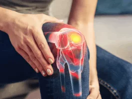
Osteomalacia, commonly referred to as the softening of the bones, is a metabolic bone disease. It is caused by suboptimal bone mineralization. When the same condition occurs during infancy and childhood, it is referred to as rickets osteomalacia or simply rickets.
This condition can result from a deficiency of calcium, phosphorous, and/or vitamin D. Subsequently, the larger weight-bearing bones of the body become soft, lose their strength, deform, and fracture easily when insulted.
What Causes Osteomalacia?
It can be caused by several factors:
Vitamin D Deficiency
The leading cause of osteomalacia is a severe vitamin D deficiency that hinders the absorption of calcium and phosphorus in the bones. Lack of sunlight exposure and malnourishment are the most common reasons for vitamin D deficiency, which can lead to a gradual and persistent depletion of bone mineral reserves. The result of such a decrease may cause the bones to become more pliable and more prone to fracture.
Kidney Disease
Faulty functioning of the kidneys can cause osteomalacia. The kidneys are the sites where the active form of vitamin D is formed. In chronic kidney diseases, such as nephrotic syndrome and acidosis, not only is vitamin D metabolism impaired, but there is an unchecked removal of calcium and phosphorus from the body.
GIT Disease
Osteomalacia can also result from absorption disorders 1Thieler S, Schölmerich J. Gastrointestinale Erkrankungen und Osteomalazie [Gastrointestinal diseases and osteomalacia]. Internist (Berl). 2008 Oct;49(10):1197-8, 1200, 1202-5. German. doi: 10.1007/s00108-008-2117-9. PMID: 18719873. such as celiac disease and IBS, which prevent patients from absorbing vitamin D adequately from their diets. At times, patients may have a familial history of osteomalacia, in which case the cause is usually a metabolic disorder arising from a lack of normal enzymes.
Tumor-Induced Osteomalacia (TIO)
In a rare event, a mesenchymal tumor may cause osteomalacia. The culprit elevates the level of a signaling molecule (FGF 23), which acts on the kidney tubules to increase phosphate excretion from the body. This reduces the phosphate available for bone matrix formation and even causes a wasting of the bones. The most distinctive feature of this type of osteomalacia is the extremely high levels of phosphate and vitamin D in the blood. While in all other cases of osteomalacia, calcium, vitamin D, and phosphate levels are low.
What are the Most Common Osteomalacia Risk Factors?
Risk factors associated with osteomalacia include:
Gender
Women are more likely to suffer from osteomalacia. Pregnant and lactating women are at an increased risk.
Decreased Sunlight Exposure
Individuals who stay indoors or live in areas with a lesser number of sunny days are prone to osteomalacia.
Age
Increased age alters vitamin D metabolism and leads to bone softening.
GIT disease/surgery:
Malabsorption syndromes and stomach injury impair the absorption of essential minerals and vitamin D from food.
Kidney Disease
Kidney failure or suboptimal kidney functioning can lead to unabated phosphate excretion from the body. A diseased kidney is unable to metabolize vitamin D.
Familial History
If anyone in your immediate family has osteomalacia, you should get yourself assessed too. At times, vitamin D and calcium deficiency are a result of inheritable genetic mutations.
What are the Classic Osteomalacia Symptoms?
The symptoms may vary from patient to patient and will ultimately depend on the severity of the disease.
The most common symptoms include:
- Increased bouts of exhaustion
- Lethargy
- Pain in the arms, legs, and other bones
- Abnormal gait. Side-to-side treading in severe cases
- Increased frequency of fractures
- Muscle weakness
- Bowing of the weight-bearing bones
Osteomalacia vs. Osteoporosis
Osteomalacia leads to a general lack of mineralization, which decreases the overall bone mineral content and decreases the bone matrix. There is no effect on the rate of bone remodeling.
On the other hand, osteoporosis is the loss of minerals from the bone. It is more common in menopausal women. Osteoporotic patients will also exhibit accelerated rates of bone resorption. The vitamin D and serum calcium levels in osteoporosis patients may or may not be altered.2NORDIN, B. E. C. M.D., M.R.C.P., PH.D.†. Osteomalacia, Osteoporosis, and Calcium Deficiency. Clinical Orthopaedics 17():p 235-258,
How is Osteomalacia Diagnosed?
The investigations needed to diagnose the disease are based on your medical history. Consistent symptoms include a history of frequent fractures, bone pain, and muscle weakness. The most common tests include:
Blood Tests
Common blood tests include complete blood counts and serum levels of vitamin D, calcium, phosphate, and alkaline phosphatase. These form the first line of investigation in most cases. Your physician may also ask for renal and liver function tests.
Imaging Tests
These include X-rays and bone scans. Imaging allows for fracture identification and the quantification of the mineral content and may also reveal a decrease in bone thickness, which is consistent with a diagnosis of osteomalacia.
Bone Biopsy
A bone biopsy is an invasive testing modality that involves procuring a small segment of the affected bone. It allows for assessing the pathological changes that take place within the bone. A doctor will ask for it only if other tests are inconclusive or to rule out TIO.
What are the Different Treatment Options for Osteomalacia?
Treatment for osteomalacia primarily focuses on addressing the underlying cause and improving bone health. Here are different treatment options for osteomalacia:
A Healthy Diet
A balanced diet is preventive and therapeutic for osteomalacia patients. Foods that are rich in vitamin D and calcium include poultry, dairy, green vegetables, oily fish, fortified drinks, and cereals.
Exogenous Supplements
If you suffer from renal or liver insufficiency, taking vitamin D supplements can treat osteomalacia. This is true for everyone suffering from osteomalacia. A doctor can prescribe the right dosage for you based on the severity of your disease because some patients might need an intramuscular depot injection of vitamin D to sustain optimum levels.
Supplementation with vitamin D and calcium is essential if you have any of the risk factors for osteomalacia.
Increased Sunlight Exposure
Getting enough sunlight boosts your vitamin D levels. Consult your healthcare provider for advice on using suitable sunscreens for optimal vitamin D production.
Safe Exercises
Numerous studies have shown that walking for 30-45 minutes 5 days a week can help maintain muscle mass and lead to accelerated bone deposition.3Pinheiro MB, Oliveira J, Bauman A, Fairhall N, Kwok W, Sherrington C. Evidence on physical activity and osteoporosis prevention for people aged 65+ years: a systematic review to inform the WHO guidelines on physical activity and sedentary behaviour. Int J Behav Nutr Phys Act. 2020 Nov 26;17(1):150. doi: 10.1186/s12966-020-01040-4. PMID: 33239014; PMCID: PMC7690138. Improving muscle tone can alleviate the pain and weakness associated with the condition.
Is Osteomalacia Preventable?
Yes!
Maintaining a healthy lifestyle and vitamin D supplementation can prevent it. Seeking the help of a rheumatologist or an endocrinologist can prevent the early onset of the disease if you have a familial history of osteomalacia.
If you or your loved ones are experiencing any of the aforementioned osteomalacia symptoms, it is best to get in touch with a reliable physician. An accurate diagnosis of the disease can help you get the treatment you need.
Refrences
- 1Thieler S, Schölmerich J. Gastrointestinale Erkrankungen und Osteomalazie [Gastrointestinal diseases and osteomalacia]. Internist (Berl). 2008 Oct;49(10):1197-8, 1200, 1202-5. German. doi: 10.1007/s00108-008-2117-9. PMID: 18719873.
- 2NORDIN, B. E. C. M.D., M.R.C.P., PH.D.†. Osteomalacia, Osteoporosis, and Calcium Deficiency. Clinical Orthopaedics 17():p 235-258,
- 3Pinheiro MB, Oliveira J, Bauman A, Fairhall N, Kwok W, Sherrington C. Evidence on physical activity and osteoporosis prevention for people aged 65+ years: a systematic review to inform the WHO guidelines on physical activity and sedentary behaviour. Int J Behav Nutr Phys Act. 2020 Nov 26;17(1):150. doi: 10.1186/s12966-020-01040-4. PMID: 33239014; PMCID: PMC7690138.


