
 Spider angioma
Spider Angioma is a vascular lesion characterized by dilation of end vasculature under the skin surface. It is mostly associated with chronic liver disease and cirrhosis. The prognosis of this disease is good because it responds well to treatment and disappears after the cause is treated.
What Is Spider Angioma?
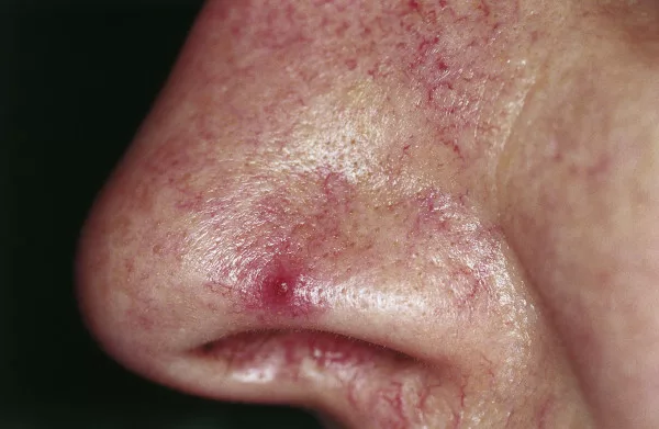
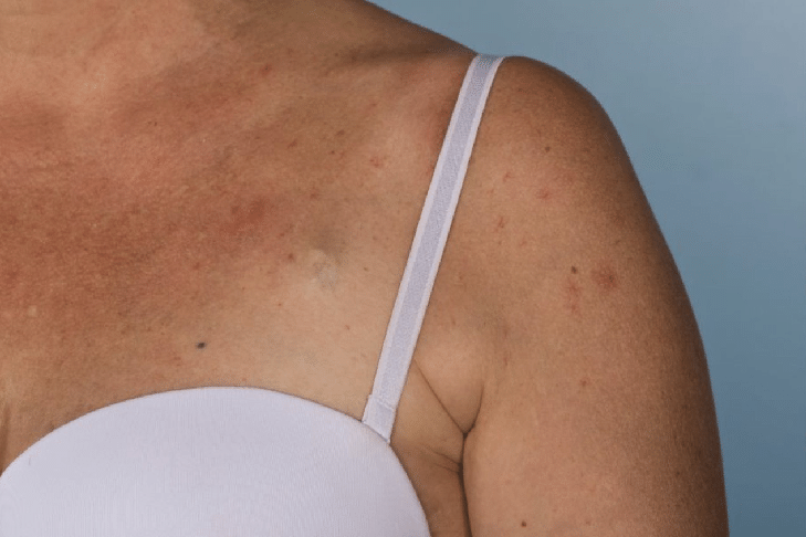
Spider angioma is a type of Chronic Vascular lesion in which there is dilatation of the end vasculature; it is also known as “Spider Nevus” or “Spider Telangiectasia’. ‘ It is a red or cherry-colored lesion that is most common in alcoholics, cirrhotic patients, patients with rheumatoid arthritis, and women who use contraceptive pills. The Lesion has three parts: body, legs, and erythema around the body. The body is a one to ten-millimeter dilatation of the vessel, while the legs are extensions from the body of the lesion, and they surround the area around it. Its legs radiate in a spider pattern around the body, so it is called “Spider Angioma.” 1Samant, H., & Kothadia, J. P. (2023). Spider Angioma. In StatPearls. StatPearls Publishing. Spider nevi most commonly appear on exposed areas such as the face, upper trunk, arms, hands, and fingers.
Pathophysiology Of Spider Angioma
In spider angioma, the muscles of arteries fail to contract. It results in the dilatation of the end arteries that form spider angioma.
Moreover, vasodilation of arteries, increased estrogen levels, hormonal imbalance, and the production of Substance P also dilate end arteries and form spider angiomas in the body.2 Vascular disorders of infancy and childhood. In: Hurwitz Clinical Pediatric Dermatology: A Textbook of Skin Disorders of Childhood and Adolescence, 3rd, Paller AS, Mancini AJ (Eds), WB Saunders, Philadelphia 2006. p.307.
How Common is Spider Angioma?
Spider Angioma is a benign condition that affects 10 -15 percent of the healthy population. In such cases, there are less than three lesions. However, more than three lesions indicate any underlying pathology. Multiple spider angiomas indicate chronic liver disease in 95 percent of the population.
In cirrhotic patients, the prevalence of spider angioma is 33 percent. However, its prevalence in pregnant women is 60 percent. 38 percent of the children have at least one spider angioma.3Gonzalez Ballerga, E., Pozo, M. O., Rubatto Birri, P. N., Kanoore Edul, V. S., Sorda, J. A., Daruich, J., & Dubin, A. (2018). Sublingual microcirculatory alterations in cirrhotic patients. Microcirculation (New York, N.Y.: 1994), 25(4), e12448. https://doi.org/10.1111/micc.12448
Causes of Spider Angiomas
There are various causes of spider angiomas, a few of them are listed below:
Chronic Liver Disease
One of the main causes of spider angiomas is chronic liver disease. Patients having cirrhosis and end-stage liver disease are more prone to develop spider angiomas. Doctors think that spider angiomas are a high-risk factor for mortality in such patients.
They have more than three spider angiomas on their body, and the cause behind them is an increase in vascular endothelial growth factor (VEGF) levels. 4Terp, K., & Izquierdo-Pretel, G. (2023). Spider Angioma Number and Location as Potential Prognostic Indicators in Chronic Liver Disease: A Case Report. Cureus, 15(1), e34193. https://doi.org/10.7759/cureus.34193
Thyrotoxicosis
Thyrotoxicosis is an increase in thyroid hormone levels in the body. Increased thyroid hormone increases estrogen levels, which can cause spider angiomas. Moreover, if you have thyrotoxicosis, blood flow in the blood vessels of your skin increases, resulting in the formation of Spider Angiomas.
Additionally, an autoimmune response in thyrotoxicosis is also a reason for developing spider angioma. Seventy percent of patients who have thyroid disease develop spider angiomas.
Rheumatoid Arthritis
Patients with rheumatoid arthritis can also develop spider angiomas. The primary reason is the use of hormone replacement therapy, which increases estrogen levels. Elevated estrogen levels can lead to the formation of spider angiomas. Additionally, rheumatoid arthritis is a chronic inflammatory condition, and the resulting inflammatory changes can increase blood flow to the skin, contributing to the development of spider angiomas.
Alcohol Intake
People who consume large amounts of alcohol in their lives develop liver disease with age. Liver diseases are a risk factor for developing spider angiomas. Chronic liver disease is often fatal.
Pregnancy
Spider Angiomas are common in pregnant women and affect almost two-thirds of pregnancies. They appear due to an increase in estrogen levels and blood flow.
Malnutrition
Malnourished people can also develop spider angioma.
Use Of Contraceptive Drugs
If you are using oestrogen-containing contraceptive pills, you can develop spider angiomas.
Genetic Factors
Genetic factors also play an important role in the development of spider angiomas. People with a family history of spider angiomas are at risk of developing them.
Symptoms Of Spider Angioma
Spider angioma may appear as a small reddish dot on your body that appears red or purplish in color with radiating legs. They can appear on your arms, face, legs, torso, fingers, neck and nasal mucosa.5Ralli, M., Candelori, F., Di Stadio, A., & de Vincentiis, M. (2022). Spider Angioma of the Nasal Mucosa. Ear, nose, & throat journal, 101(10), NP445–NP446. https://doi.org/10.1177/0145561320980202
In children, they form in the upper extremities. You can also notice them in the mucosa of your child’s oral cavity. Spider angiomas can also occur in the mucosa of GIT.6Singh, S., Sahoo, A. K., Ramam, M., & Bhari, N. (2018). Mucocutaneous spider angiomas in an adolescent with chronic liver disease. Archives of disease in childhood, 103(12), 1145. https://doi.org/10.1136/archdischild-2017-314406
When you press a spider angioma, it disappears and reappears after a few seconds. It is painless and does not bleed. However, if you injure it, it can bleed.
Grades Of Spider Angioma
Spider Angiomas are divided into four grades based on the number and size of lesions.
Grade 1
They are merely recognizable lesions with a small body, legs, and a small area of erythema surrounding the body. They are pink or light red.
Grade 2
These spider angiomas are medium (1-3 millimeters), with a small body and small legs radiating from it. They also have moderate-sized blood vessels radiating from their body.
Grade 3
The Grade 3 spider angiomas appear large, dark red, and purple, with a diameter of 3-6 millimeters and large radiating blood vessels.
Grade 4
Grade 4 spider angiomas are large lesions having large (greater than 6 millimeters) prominent bodies. The mortality rate of spider angioma is maximum in alcoholics and people having chronic liver disease. Such patients can also develop esophageal varices.
How to Diagnose Spider Angiomas?
Spider Angioma is a clinical diagnosis based on the characteristic spider-shaped appearance of the lesion. Proper evaluation, which includes a complete history, physical examination, and laboratory investigations, is necessary for the diagnosis.
History
If your doctor suspects spider angiomas on your body, he will ask the following questions related to the Spider angioma:
- When did it start, and what was the pattern by which it spread all over the body?
- Do you have any history of liver disease, thyroid disease, or rheumatoid arthritis?
- Your doctor may also inquire about alcohol intake and use of any drugs or pregnancy.
- Any history of confusion, shifting dullness, jaundice, itching of skin, or cirrhosis?
Examination
Examination of spider angioma is mainly clinical. However, dermatoscopy may help your doctor with diagnosis.
First, your doctor will examine the size, shape, spread, and pattern of distribution of the lesion. He will also check whether it fades away when pressure is applied. Secondly, the pressure over the area of the lesion is checked, which is usually greater than that of other body areas. Moreover, the skin temperature surrounding the lesion can also be higher than normal.
Diascopy
Your doctor will also perform a diascopy, using a glass slide to apply pressure to the lesion. The lesion will become pale, and when the pressure is removed, it will refill and appear red. This is characteristic of spider angioma; no other lesion shows this characteristic.
Skin Biopsy
A skin biopsy is important to rule out cancerous lesions like Basal cell carcinoma.
Systemic Investigations
Systemic investigations are important in the workup of spider angioma. Investigations like Liver function tests (LFTs), thyroid function tests (TFTs), and the tests for anticitrulinated proteins antibodies (ACPA) and rheumatoid factors are important to rule out the systemic causes of Spider angioma. There is generally no need for investigations in pregnant ladies because the spider angiomas in them fade out after childbirth. In children less than three years, they are physiological, and investigations are not needed.
Treatment Of Spider Angioma
Spider angiomas are divided into two main groups in relation to treatment
Benign Spider Angiomas or Physiological Spider Angiomas
They are benign if they are less than three and common in children and young adults. Benign spider angiomas resolve without any treatment.
In pregnant ladies, they appear due to pregnancy and resolve without any treatment.
Treatment Of Spider Angiomas Related To Diseases:
-
Liver Disease
Spider angiomas related to liver disease indicate chronic liver disease and usually appear in the end stage of liver disease. Spider angiomas can be treated for cosmetic concerns. The procedures generally yield good results, though there is a small risk of scarring. It is important to note that spider angiomas may recur after treatment. There are the following removal options for these spider angiomas:
Electrocauterization
In electro-cauterization, your doctor uses an electric current to burn the blood vessels, and lesions disappear.
Electro Dissection
In electro-dissection, your doctor uses an electric needle to remove the spider angiomas. This procedure burns out the lesion.
Fine Needle Cauterization
In fine needle cauterization, your doctor uses a fine, 595-nanometer needle to burn out the lesion.7Yang, B., Li, L., Zhang, L. X., Sun, Y. J., & Ma, L. (2015). Clinical Characteristics and Treatment Options of Infantile Vascular Anomalies. Medicine, 94(40), e1717. https://doi.org/10.1097/MD.0000000000001717
Potassium Titanyl Laser:
In this procedure, your doctor uses a Potassium Titanyl Laser (532-nanometer laser) to treat the lesion.8Becher, G. L., Cameron, H., & Moseley, H. (2014). Treatment of superficial vascular lesions with the KTP 532-nm laser: experience with 647 patients. Lasers in medical science, 29(1), 267–271. https://doi.org/10.1007/s10103-013-1330-5
Sclerotherapy
In sclerotherapy, doctors inject a colloidal solution into the spider angioma. This solution shrinks the blood vessels, and the spider angiomas disappear.
Cryotherapy
In cryotherapy, they freeze the spider angiomas with cold, liquid nitrogen. The results of these treatments are good. They can only leave a scar. However, there is a risk of recurrence.
In cirrhotic patients, spider angiomas only fade away after the liver transplantation.
-
Thyroid Disease
In patients with underlying thyroid disease who have spider angiomas, the treatment of thyroid disease also treats spider angiomas.
Can Spider Angiomas Reoccur After Treatment?
Yes, spider angiomas can reoccur after removal by electro-dissection, electrocauterization, or laser. To prevent this reoccurrence, you must follow your doctor’s recommendations and precautions.
Complications Of Spider Angiomas
Spider angiomas are mostly benign, painless, and do not bleed. However, they may have some compilations like:
- They can bleed if they are injured.
- They can also become inflamed or infected.
- They can form big scars.
- They can bleed if you injure them.
Spider Angioma Vs. Cherry Angioma
Cherry angioma is usually age-related; it is more common in young adults and increases with aging, while spider angiomas are common in children, young adults, and pregnant ladies and are genetically influenced. Cherry angioma appears as a reddish spot that is uniform in shape, while spider angiomas have legs radiating from their body, giving them the characteristic appearance of a spider.
On applying pressure, spider angiomas show complete blanching; however, if you have a cherry angioma, it shows partial blanching, and its edges are white.
Spider Angiomas have a body, legs, and an area of erythema surrounding them, while cherry angiomas only have a trunk and extremities.
Cherry angiomas are common on the extremities, while spider angiomas occur on the trunk, fingers, face, and torso.
Effect Of Oestrogen in the Formation of Spider Angioma
Increases in the levels of estrogen stimulate the formation of Spider angiomas. That’s why they are common in young adults, females in their reproductive age, people using hormonal contraceptives that contain estrogen in them, and pregnant ladies.
Additionally, spider angiomas are more common in women as compared to men due to high estrogen levels.
How Can I Prevent Spider Angiomas
You can prevent spider angiomas on your body by treating the underlying cause. For this, your doctor will evaluate and treat other diseases like thyroid problems, liver disease, and rheumatoid arthritis. Moreover, if you are a woman and you are taking estrogen-containing contraceptive pills, your doctor will advise you to use non-hormonal contraceptive methods or non-estrogen contraceptive pills.
Does Sun Exposure Cause Spider Angiomas?
Sun exposure for a long duration can also lead to spider angiomas on the face. Fair-skinned people are at a higher risk of developing spider angiomas.
Conclusion
Spider Angioma is a skin condition that causes spider-shaped lesions due to blood vessel dilation. Its causes include liver disease, thyrotoxicosis, pregnancy, and oestrogen-containing contraceptive pills. They are physiological in pregnant women and do not need any medical treatment because they disappear in the postpartum period. Your doctor diagnoses it clinically. It is treatable and has a good prognosis, except in cirrhotic patients who have higher mortality rates.
Refrences
- 1Samant, H., & Kothadia, J. P. (2023). Spider Angioma. In StatPearls. StatPearls Publishing.
- 2Vascular disorders of infancy and childhood. In: Hurwitz Clinical Pediatric Dermatology: A Textbook of Skin Disorders of Childhood and Adolescence, 3rd, Paller AS, Mancini AJ (Eds), WB Saunders, Philadelphia 2006. p.307.
- 3Gonzalez Ballerga, E., Pozo, M. O., Rubatto Birri, P. N., Kanoore Edul, V. S., Sorda, J. A., Daruich, J., & Dubin, A. (2018). Sublingual microcirculatory alterations in cirrhotic patients. Microcirculation (New York, N.Y.: 1994), 25(4), e12448. https://doi.org/10.1111/micc.12448
- 4Terp, K., & Izquierdo-Pretel, G. (2023). Spider Angioma Number and Location as Potential Prognostic Indicators in Chronic Liver Disease: A Case Report. Cureus, 15(1), e34193. https://doi.org/10.7759/cureus.34193
- 5Ralli, M., Candelori, F., Di Stadio, A., & de Vincentiis, M. (2022). Spider Angioma of the Nasal Mucosa. Ear, nose, & throat journal, 101(10), NP445–NP446. https://doi.org/10.1177/0145561320980202
- 6Singh, S., Sahoo, A. K., Ramam, M., & Bhari, N. (2018). Mucocutaneous spider angiomas in an adolescent with chronic liver disease. Archives of disease in childhood, 103(12), 1145. https://doi.org/10.1136/archdischild-2017-314406
- 7Yang, B., Li, L., Zhang, L. X., Sun, Y. J., & Ma, L. (2015). Clinical Characteristics and Treatment Options of Infantile Vascular Anomalies. Medicine, 94(40), e1717. https://doi.org/10.1097/MD.0000000000001717
- 8Becher, G. L., Cameron, H., & Moseley, H. (2014). Treatment of superficial vascular lesions with the KTP 532-nm laser: experience with 647 patients. Lasers in medical science, 29(1), 267–271. https://doi.org/10.1007/s10103-013-1330-5


