
What Is an Apicoectomy?
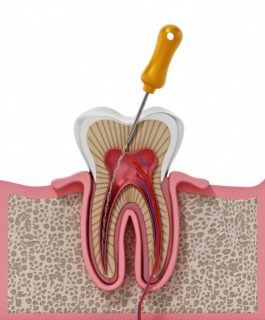
An apicectomy, also known as root-end surgery, is a minor surgical procedure performed to remove the tip of a tooth’s root (apex) and the surrounding infected tissue. It is typically recommended when a standard root canal treatment has failed to fully eliminate an infection or when inflammation persists around the root tip. If an infection at the root tip is not treated, it can progress into a chronic abscess or cyst, leading to persistent pain and bone loss around the tooth’s root. Over time, this can result in tooth mobility.
Inside every tooth, there is a soft tissue called dental pulp, which contains blood vessels and nerves extending from the crown to the root. When bacteria reach the root tip and cause infection, an apicectomy helps prevent further complications while preserving the natural tooth. This procedure is often the last resort before considering tooth extraction, making it an essential option in restorative dentistry.1von Arx, Thomas. (2011). Apical surgery: A review of current techniques and outcome. The Saudi Dental Journal. 23. 9-15. 10.1016/j.sdentj.2010.10.004.
Apicoectomy vs. Root Canal Treatment
Understanding the differences between an apicoectomy and a root canal can help you make an informed decision about your dental care. A root canal is a common procedure for treating infected or inflamed dental pulp. During this non-surgical treatment, the dentist removes the infected tissue, cleans the root canals, and seals them to prevent reinfection. It is typically the first choice for treating dental infections and preserving the natural tooth.
While both procedures address dental infections, they take different approaches. A root canal focuses on cleaning and sealing the inside of the tooth to remove infection. In contrast, an apicoectomy is a surgical procedure that removes the root tip and surrounding infected tissue when a previous root canal has failed. This option is considered when an infection persists or spreads despite prior treatment.
Why is an Apicectomy Recommended?
Dentists generally prefer non-invasive treatments for managing dental infections, with root canal therapy being the first choice. However, in some cases, a root canal may not be sufficient, making an apicoectomy necessary.
One common reason for an apicoectomy is persistent infection or cyst formation around the root tip despite previous root canal treatment. Additionally, some teeth have complex root structures, such as curved or blocked canals, making it difficult to fully clean and seal the root, leading to reinfection.2Soesilo, Diana & Pangabdian, Fani & Wijaya, Yongki & Puspita, Sinta. (2023). Apicoectomy for Periapical Lesion Treatment after Post Endodontic Failure on First Maxillary Premolar (Case Study). Asian Journal of Dental and Health Sciences. 3. 1-4. 10.22270/ajdhs.v3i1.38.
An apicoectomy may also be recommended when anatomical concerns arise after a root canal, such as root crowding, which can affect multiple teeth. In cases where a root canal contains obstructions like a metal post or calcified tissue, making retreatment impossible, surgically removing the root tip allows for effective infection control.
Other indications include pulp chamber perforation or damage that cannot be repaired with conventional treatment. In rare cases, even a previous apicoectomy may fail, requiring a repeat procedure. If an apicoectomy is suggested, it’s often because the only alternative is tooth extraction, which may require replacement options like implants, bridges, or removable dentures to maintain proper tooth alignment.
Contraindications for Apicectomy
An apicoectomy may not be recommended in cases where the affected tooth lacks an opposing tooth for functional support or does not play a significant role in supporting a fixed prosthesis. Additionally, if the tooth has severe periodontal disease leading to inadequate bone support or has sustained an irreparable vertical root fracture, restoration becomes unfeasible.3Agarwal, Twinkle & Govind, Shashirekha. (2024). Comprehensive Overview of the Apicoectomy Procedure: Indications, Technique, and Postoperative Care.. Journal of Biomedical and Pharmaceutical Research. 13. 80-85. 10.32553/jbpr.v13i5.1172.
Other contraindications include patients with medical conditions that pose a high surgical risk or those who may not tolerate an oral surgical procedure. Non-cooperative patients, particularly those unable to maintain proper post-surgical care, may also require alternative treatment approaches.
Preparation For Apicoectomy
Before undergoing an apicoectomy, several preparatory steps are taken:
- Consultation: To have a discussion about your condition with your dental professional.
- X-rays: To take images of the affected tooth and the surrounding bone.
- Pre-procedure Care Recommendations: Your dentist will recommend some antimicrobial mouthrinse, inflammation-reducing medicine, or antibiotics to reduce the infection at the root canal and prevent any post-procedure complications.
- Medical History Review: Provide details on your past and current medical problems, including a list of medications to find the best treatment solution for your condition.
Step-By-Step Procedure
Here are sequential steps involved in the apicoectomy procedure:
Local Anesthetic
Before the procedure begins, your dentist or endodontist will administer a local anesthetic to numb the area around the affected tooth. Long-acting anesthetic agents like bupivacaine may be used to ensure prolonged numbness beyond the procedure.
Gum Incision and Flap Design
Once anesthesia takes effect, a small incision is made in the gum near the affected tooth to expose the underlying bone. The choice of flap design depends on the location and complexity of the infection. Commonly used flaps include the full buccal/palatal mucoperiosteal flap, the sub-marginal mucoperiosteal flap (Luebke-Oschenbein flap), and the papilla-based incision flap.4Alfotawi, Randa. (2020). Flap Techniques in Dentoalveolar Surgery. 10.5772/intechopen.91165. These designs allow better access while minimizing scarring and healing complications. The semilunar flap is rarely used due to its unpredictable healing and scarring concerns.
Root Tip Access and Osteotomy
After lifting the gum tissue, the dentist carefully removes a small portion of bone to access the root tip, a process known as an osteotomy. If the cortical bone is thin, the root may be easily located, but in denser areas, a slow-speed handpiece with a round bur and water irrigation is used to prevent surgical emphysema. The osteotomy opening is kept minimal—around 4–5 mm—to promote faster healing. Smaller lesions heal within 6.4 months, while larger ones may take up to 11 months.5Agarwal, Twinkle & Govind, Shashirekha. (2024). Comprehensive Overview of the Apicoectomy Procedure: Indications, Technique, and Postoperative Care.. Journal of Biomedical and Pharmaceutical Research. 13. 80-85. 10.32553/jbpr.v13i5.1172.
Root Tip Removal and Cleaning
Once the root tip is exposed, a few millimeters of the root and surrounding infected tissue are removed. Curettage is performed to clear soft tissue from the periradicular area, ensuring complete removal of infected material. If the apicoectomy does not involve a retrograde filling, the natural healing process will seal the root end over time.
Root Canal Sealing
In cases where additional protection is needed to prevent reinfection, a biocompatible filling, such as mineral trioxide aggregate (MTA), is placed at the root tip. This ensures a secure seal, reducing the risk of bacterial invasion. Additional X-rays may be taken to confirm the proper sealing of the root and the surrounding bone structure.
Suturing and Healing
The gum tissue is then repositioned and sutured back in place, allowing it to heal naturally. Over time, the surrounding bone regenerates, reinforcing the treated area. The entire procedure typically lasts between 30 to 90 minutes, depending on the tooth’s location and complexity.
Recovery and Follow-Up
Post-apicoectomy surgery, patients may experience some pain, and recovery times can vary. Most patients can resume normal activities the day after apicoectomy surgery. It is important to note that bone around the root may take several months.
Applying cold compresses and following up with the dental professional for stitch removal, if necessary, are integral to the recovery process.
Tips for Apicoectomy Recovery
After completing an apicoectomy, patients can promote a smoother recovery process by following these simple steps:
- Gentle Dental Care: Conduct your dental routine with gentleness to prevent irritation to sensitive areas.
- Avoid Harmful Substances: Steer clear of smoking, crunchy foods, or actions that may harm stitches.
- Medication Compliance: Take prescribed medications as directed and adhere to the provided aftercare instructions.
- Over-the-Counter Painkillers: Alleviate discomfort or swelling by using over-the-counter pain relievers or anti-inflammatories.
- Follow-Up with Dental Professional: Schedule a follow-up appointment with your dental professional if stitches require removal. Note that many stitches dissolve on their own.
- Cold Compress Application: Apply a cold compress or ice pack to the affected area for twenty minutes on, twenty minutes off.
Is Apicoectomy Painful?
The invasiveness of an apicoectomy compared to a standard root canal surgery may result in a more extended recovery period with increased pain.
During the apicoectomy procedure, local anesthesia is given to manage the pain while removing the infected tissue. However, it is very common to have pain and swelling after the procedure is done. Your dentist will give you some painkillers and anti-inflammatory medicines to help manage the pain and swelling.
Studies say that post-procedure apicoectomy typically lasts for a few days.6Christiansen, R., Kirkevang, L. L., Hørsted-Bindslev, P., & Wenzel, A. (2008). Patient discomfort following periapical surgery. Oral surgery, oral medicine, oral pathology, oral radiology, and endodontics, 105(2), 245–250. https://doi.org/10.1016/j.tripleo.2007.08.023
Complications & Risks
Although apicoectomy is a safe surgical procedure, rarely it can lead to complications like nerve damage, or further root canal infection if potential spaces are left while suturing. These complications are not exclusive to apicoectomies and may arise in various dental procedures.
Apicoectomy Failure Signs
Considered rare, apicoectomies are deemed failures if they do not alleviate symptoms or heal properly. Apicoectomy infection is a rare but possible sign of the procedure failure.
However, instances of failure are uncommon, particularly when performed by experienced dental professionals.
Success Rate
The success rate of apicoectomies varies depending on whether a root-end filling is used. In a study analyzing treatment outcomes at a 12-month follow-up, apicoectomies without retrograde fillings had an 88.5% success rate, while those with retrograde fillings had a slightly lower success rate of 64.3%. However, this difference was not statistically significant.7Ajayi, J., Abiodun-Solanke, I., Olusile, O., Oginni, A., & Esan, T. (2018). COMPARATIVE STUDY OF TREATMENT OUTCOME IN APICECTOMIES WITH OR WITHOUT ROOT-END FILLING. Annals of Ibadan Postgraduate Medicine, 16(2), 109. https://pmc.ncbi.nlm.nih.gov/articles/PMC6580414/
Apicoectomies demonstrate a high success rate, with studies indicating excellent results up to 5 years later.8Truschnegg, A., Rugani, P., Kirnbauer, B., Kqiku, L., Jakse, N., & Kirmeier, R. (2020). Long-term Follow-up for Apical Microsurgery of Teeth with Core and Post Restorations. Journal of endodontics, 46(2), 178–183. https://doi.org/10.1016/j.joen.2019.11.002 They prove reliable in preserving teeth affected by infection or root problems.
Take-Away
An apicoectomy emerges as a valuable dental procedure, playing a pivotal role in saving a tooth when standard root canal therapy falls short. With a high success rate and a relatively low risk of complications, it provides a targeted solution for persistent infections or anatomical concerns. Understanding the procedure, recovery process, and potential complications empowers patients to make informed decisions about their dental health. Timely intervention and adherence to aftercare instructions significantly contribute to the success of an apicoectomy. If recommended by your dental professional, serious consideration is warranted, given its potential to preserve your natural tooth and prevent more extensive dental interventions. Dental health holds a crucial role in overall well-being, and addressing dental issues promptly prevents problems from escalating. Whether understanding various dental procedures’ nuances or recognizing signs of potential complications, staying informed is key to maintaining a healthy and vibrant smile.
Refrences
- 1von Arx, Thomas. (2011). Apical surgery: A review of current techniques and outcome. The Saudi Dental Journal. 23. 9-15. 10.1016/j.sdentj.2010.10.004.
- 2Soesilo, Diana & Pangabdian, Fani & Wijaya, Yongki & Puspita, Sinta. (2023). Apicoectomy for Periapical Lesion Treatment after Post Endodontic Failure on First Maxillary Premolar (Case Study). Asian Journal of Dental and Health Sciences. 3. 1-4. 10.22270/ajdhs.v3i1.38.
- 3Agarwal, Twinkle & Govind, Shashirekha. (2024). Comprehensive Overview of the Apicoectomy Procedure: Indications, Technique, and Postoperative Care.. Journal of Biomedical and Pharmaceutical Research. 13. 80-85. 10.32553/jbpr.v13i5.1172.
- 4Alfotawi, Randa. (2020). Flap Techniques in Dentoalveolar Surgery. 10.5772/intechopen.91165.
- 5Agarwal, Twinkle & Govind, Shashirekha. (2024). Comprehensive Overview of the Apicoectomy Procedure: Indications, Technique, and Postoperative Care.. Journal of Biomedical and Pharmaceutical Research. 13. 80-85. 10.32553/jbpr.v13i5.1172.
- 6Christiansen, R., Kirkevang, L. L., Hørsted-Bindslev, P., & Wenzel, A. (2008). Patient discomfort following periapical surgery. Oral surgery, oral medicine, oral pathology, oral radiology, and endodontics, 105(2), 245–250. https://doi.org/10.1016/j.tripleo.2007.08.023
- 7Ajayi, J., Abiodun-Solanke, I., Olusile, O., Oginni, A., & Esan, T. (2018). COMPARATIVE STUDY OF TREATMENT OUTCOME IN APICECTOMIES WITH OR WITHOUT ROOT-END FILLING. Annals of Ibadan Postgraduate Medicine, 16(2), 109. https://pmc.ncbi.nlm.nih.gov/articles/PMC6580414/
- 8Truschnegg, A., Rugani, P., Kirnbauer, B., Kqiku, L., Jakse, N., & Kirmeier, R. (2020). Long-term Follow-up for Apical Microsurgery of Teeth with Core and Post Restorations. Journal of endodontics, 46(2), 178–183. https://doi.org/10.1016/j.joen.2019.11.002


