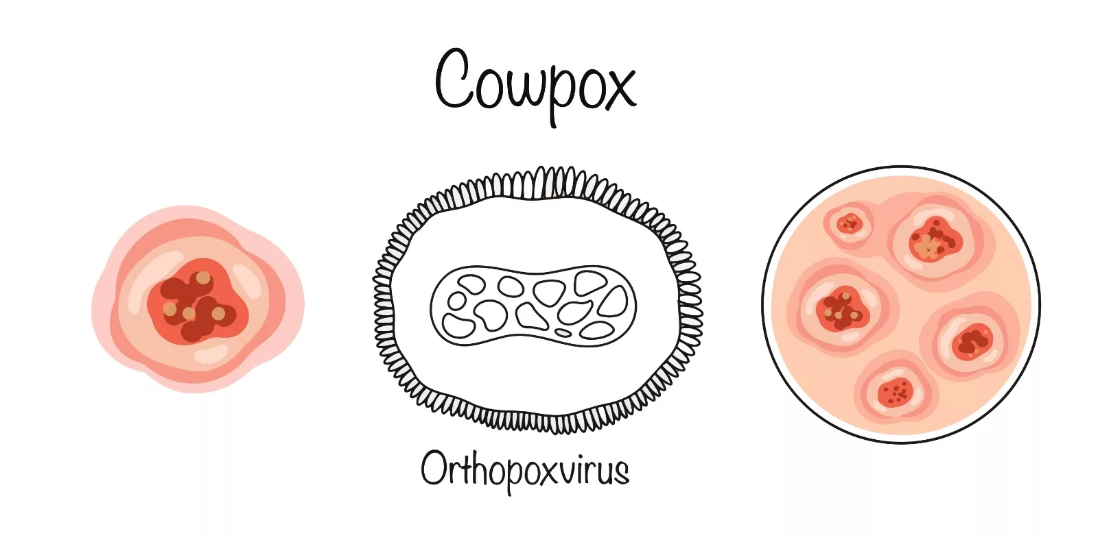
Cowpox, sometimes referred to as catpox, is a rare zoonotic viral infection that primarily affects the skin, causing large blisters and swollen lymph nodes. The condition is caused by the cowpox virus (CPXV), a member of the Orthopoxvirus genus in the Poxviridae family. CPXV is closely related to variola virus, the pathogen responsible for smallpox, and vaccinia virus, used in smallpox vaccines.
Cowpox was first identified in cows but is more commonly observed in domestic cats today. Transmission to humans typically occurs through direct contact with infected animals, leading to localized lesions. Interestingly, exposure to CPXV in healthy humans can induce cross-immunity to smallpox.1Bruneau RC, Tazi L, Rothenburg S. Cowpox viruses: a zoo full of viral diversity and lurking threats. Biomolecules. 2023;13(2):325.
The term “vaccinia” is sometimes used interchangeably with cowpox to describe human disease, though technically, vaccinia refers to the virus used in smallpox vaccination. It is important to distinguish cowpox from “cow pack” (milker’s nodule), a condition caused by the parapoxvirus.
Cowpox remains rare, and human cases are usually self-limiting, resolving without significant complications in otherwise healthy individuals. However, those with compromised immune systems may experience more severe symptoms.2Vanessa Ngan SW. Cowpox 2008 [Available from: https://dermnetnz.org/topics/cowpox.
Structure of CPXV
CPXV possesses a complex structure that is crucial for its function and pathogenicity.
Genome Structure:
CPXV is a linear double-stranded DNA virus. This virus consists of around 222,499 nucleotides, among the largest genomes of orthopoxviruses. This virus encodes about 223 proteins. There are no structural RNAs.
Virion Structure:
CPXV is approximately 250 to 350 nm in size. Its viral envelope contains unique glycoproteins. The virus also possesses inclusion bodies.3Cowpox [Available from: https://microbewiki.kenyon.edu/index.php/Cowpox.

Causes & Transmission
Cowpox virus usually has a low infectivity rate in humans. The transmission requires direct contact with the mucous membranes or skin lesions during the milking and handling of infected cows. Skin appears to be the most common entry point, but oronasal infection can also be possible. Cats are the primary source of this infectious disease. Wild animals such as voles, mice, and rodents can cause cat infections. Because of their close contact with humans, domestic cats play a key role in transmitting disease to human beings. Cats often contract the infection from wild animals such as voles, mice, or other rodents. Infected cats may develop lesions at the site of a bite or scratch, indicating transmission from an infected rodent.
Life Cycle & Pathogenesis of CPXV
After the incubation period, a red spot (macule) appears on the skin. This spot changes into a raised bump (papule), then a fluid-filled blister (vesicle), and eventually a pus-filled sore (pustule). The sore may burst and form a black scab surrounded by swelling and redness.4FRANK FENNER PAB, E. PAUL J. GIBBS, FREDERICK A. MURPHY, MICHAEL J. STUDDERT, DAVID O. WHITE. CHAPTER 21 – Poxviridae. Veterinary Virology. 1987:387-405.
The life cycle of CPXV is as follows:
Entry & Uncoating:
The cowpox virus enters host cells by direct fusion with the cell membrane, likely mimicking apoptotic mechanisms. Once inside, the viral core is released into the cytoplasm. Host and viral factors facilitate uncoating, allowing the release of viral DNA.5McFadden G. Poxvirus tropism. Nature Reviews Microbiology. 2005;3(3):201-13.
Transcription & Gene Expression:
The viral DNA acts as a template for transcription. Viral enzymes synthesize early mRNAs. This step also involves the necessary gene expression for viral replication and modulation of host defenses.6Moss B. Poxvirus DNA replication. Cold Spring Harbor perspectives in biology. 2013;5(9):a010199.
DNA Replication:
The virus takes over the cell’s machinery to make copies of itself. This happens in special areas of the cell called “viral factories.”7Rampersad S, Tennant P. Replication and expression strategies of viruses. Viruses. 2018:55.
Formation of New Viruses:
The virus assembles new copies using proteins and genetic material it produces.
Release of the Virus:
The new viruses leave the cell in two ways:
- Cell Lysis: The infected cell bursts, releasing viruses to infect nearby cells.
- Budding: The virus wraps itself in a layer from the cell membrane and exits, ready to spread.
After release, poxvirus employs various technology to escape the host’s immune system. It reduces the markers (MHC-I molecules) on infected cells that help the immune system identify and destroy them. Additionally, it releases special proteins to stop the signals (cytokines) that alert the immune system about the infection. It also interferes with the body’s natural antiviral defenses, allowing the virus to survive longer.8Pal M, Singh R, Parmar BC, Paulos K, Lema GAG. Human cowpox: A viral zoonosis that poses an emerging health threat. Journal of Advances in Microbiology Research. 2022;3(1):22-6.
The hallmark of cowpox infection is the development of painful pustular lesions on the skin. Swollen lymph nodes and fever often accompany these lesions. In severe immunocompromised individuals, it can lead to systemic diseases and potentially fatal outcomes.
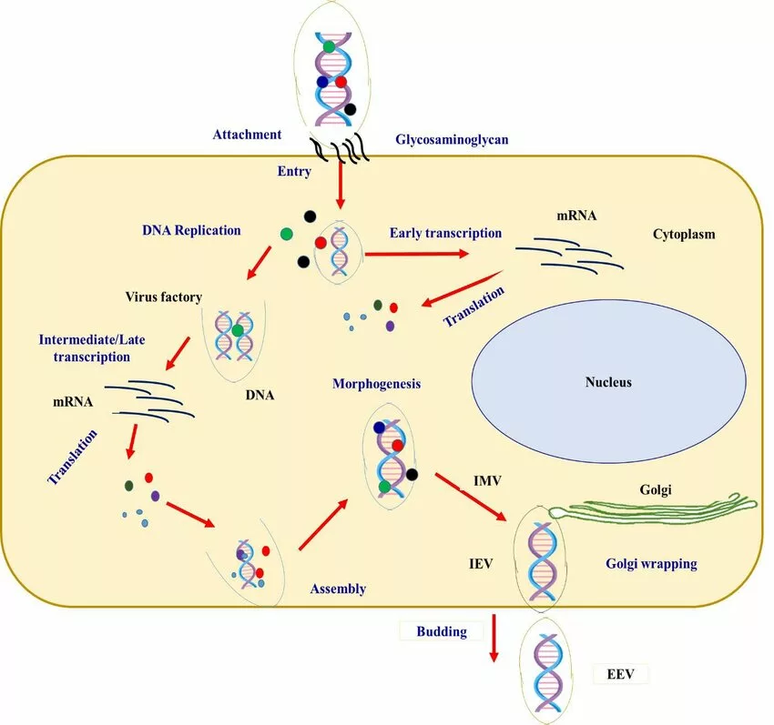
https://doi.org/10.18502/ijm.v14i6.11252, available via NCBI.”
Symptoms
The following are the symptoms of cowpox in animals and humans:
Symptoms in Animals:
- Cats: Cowpox often presents as skin lesions on the face, neck, paws, and forelimbs. Other symptoms include fever, swollen lymph nodes (lymphadenopathy), and secondary rashes. Cats often contract the virus from infected rodents.
- Cattle: Infected cows develop lesions on their teats and udder, typically observed in animals used for milking.
- Other Animals: Wild rodents act as natural reservoirs of the virus, and symptoms in other animals depend on their immune status and the severity of infection.9Pal M, Singh R, Parmar BC, Paulos K, Lema GAG. Human cowpox: A viral zoonosis that poses an emerging health threat. Journal of Advances in Microbiology Research. 2022;3(1):22-6.
Symptoms in Humans:
In most cases, infections are self-limiting. Immunocompromised individuals are at high risk of developing severe illnesses. The symptoms include:
- High fever
- Severe headache
- Back pain
- Abdominal pain
- Vomiting and flu-like symptoms
- Skin lesions, primarily on the hands and fingers, which may ulcerate and form black scabs
- Lesions on mouth
- Enlarged local lymph nodes
- Eczema
- Pneumonia
- Eye complaints such as conjunctivitis
- Corneal involvement
- Periorbital swelling10Pal M, Singh R, Parmar BC, Paulos K, Lema GAG. Human cowpox: A viral zoonosis that poses emerging health threat. Journal of Advances in Microbiology Research. 2022;3(1):22-6.
Diagnosis of Cowpox
The diagnosis of cowpox involves a combination of clinical and laboratory evaluations.
Clinical Evaluations:
Clinicians can diagnose the disease through characteristic symptoms, particularly skin lesions. A history of contact with infected animals, such as domestic cats or cattle, is significant.
Laboratory Diagnosis:
Polymerase chain reaction (PCR) electron microscopy, histopathology, and serology are the main diagnostic measures for cowpox.
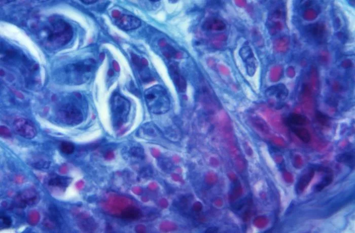
Histopathology
Pathologists examine tissue samples histologically to recognize the characteristic changes associated with the infection. These changes can include degeneration of epithelial cells or the presence of giant cells.
Electron Microscopy
Healthcare providers use electron microscopy for direct visualisation. Electron microscopy helps visualise the characteristic brick-shaped virus particles directly from the lesion material. While helpful, it cannot distinguish cowpox from other orthopoxviruses like smallpox or vaccinia.
PCR
PCR is the primary method for identifying CPXV infection. PCR detects viral DNA in the clinical samples. Samples can include tissue biopsies, exudates, and lesions. This method provides rapid and accurate diagnosis due to its high sensitivity and specificity.
Serology
Serology can help in detecting antibodies against CPXV. Clinicians do not use this method frequently for acute diagnosis.11Pelkonen PM, Tarvainen K, Hynninen A, Kallio ER, Henttonen H, Palva A, et al. Cowpox with severe generalized eruption, Finland. Emerging infectious diseases. 2003;9(11):1458.
Treatment for Cow Pox
Currently, there are no specific antiviral medications available for treating cowpox infection. Treatment focuses on supportive care for most cases, especially in moderate to severe instances.12Weese JS, Fulford MB. Companion Animal Zoonoses. 2011.
Supportive Therapy:
- Proper care of skin lesions involves keeping the affected area clean and protected to prevent secondary bacterial infections.
- Analgesics can help alleviate discomfort caused by lesions.
Antiviral Treatment:
- Cidofovir: This antiviral is used for orthopoxvirus infections, including cowpox, in severe cases. It has been shown to reduce mortality in experimental animal models of CPXV infection, though it is typically reserved for more severe instances.13Smee DF, Bailey KW, Sidwell RW. Comparative effects of cidofovir and cyclic HPMPC on lethal cowpox and vaccinia virus respiratory infections in mice. Chemotherapy. 2003;49(3):126-31.
- Vaccinia Immune Globulin (VIG): VIG can also be used to treat severe cases of cowpox, especially for individuals with systemic symptoms. It provides passive immunity that helps reduce the severity of the infection.14Petersen BW, Karem KL, Damon IK. Orthopoxviruses: Variola, vaccinia, cowpox, and monkeypox. Viral infections of humans: Epidemiology and control. 2014:501-17.
Prevention & Control
- Follow good hygiene procedures.
- Proper hand washing and avoiding bites from infected animals can help decrease cowpox transmission from animals.
- Avoid contact with cats or other animals showing symptoms of cowpox. Cats are the primary source of human infections. If a cat has skin lesions, especially in endemic areas, it should be evaluated by a veterinarian.
- One should wear gloves while handling animals with skin lesions.
- Clean your hands immediately after removing the gloves.
- Do not trap and keep wild rodents as pets.
- Pet keepers must be aware of the proper handling of pets to avoid bites and properly clean the bites.
- Monitoring cowpox cases in animals and humans is necessary for early detection and control.
- If exposure occurs, properly clean the affected area. If healthcare providers administer the vaccinia vaccine within three days of exposure to the virus, it can prevent the severity of the disease.
Cowpox Vaccine
The cowpox vaccine has a historical significance as it was instrumental in developing the first smallpox vaccine. Edward Jenner originated it from his experiment in the late 18th century. While there is no cowpox-specific vaccine currently in use, the historical cowpox vaccine helped pave the way for the development of the smallpox vaccine. The Cowpox virus was used to inoculate individuals against smallpox, as the cowpox virus is much less virulent. Vaccination with the vaccinia virus, derived from cowpox, is cross-protective against smallpox and cowpox.
Cowpox versus Smallpox
Smallpox and cowpox are both caused by viruses belonging to the same genus and family. Though they share some similarities, they differ significantly in some characteristics. Table 1 provides a complete comparison between the two.
| Features | Smallpox | Cowpox |
| Causative virus | Variola virus | Cowpox virus |
| Mode of Transmission | Transmission occurs during direct contact with the contaminated surfaces.
Human-to-human transmission (through respiratory droplets) |
Zoonotic mode of transmission
Infected animals, primarily domestic cats, rodents, and cattle, transmit this virus to humans directly. |
| The severity of the condition | Researchers find it to be severe in most cases.
Significant morbidity is associated with the disease. |
Generally mild.
Severe in immunocompromised patients Mortality rates are low. |
| Signs and Symptoms | High fever
Malaise Rash that progresses from macules to pustules. Lesions that occur during smallpox are more severe and extensive than those of cowpox.
|
Pustular lesions appear on the hands and face.
Systemic symptoms include fever and lymphadenopathy. Lesions usually heal within six to eight weeks. |
Final Remarks
Cowpox is an important subject of study within public health. Despite its rarity, it has the potential to pose risks as a zoonotic disease. The pathogenesis of cowpox demonstrates its ability to evade host immune defenses through various mechanisms. Understanding these mechanisms is crucial for developing therapeutic strategies against poxvirus infections. Additionally, keeping in mind the causes and transmission of this viral infection can help in avoiding the infection.
Refrences
- 1Bruneau RC, Tazi L, Rothenburg S. Cowpox viruses: a zoo full of viral diversity and lurking threats. Biomolecules. 2023;13(2):325.
- 2Vanessa Ngan SW. Cowpox 2008 [Available from: https://dermnetnz.org/topics/cowpox.
- 3Cowpox [Available from: https://microbewiki.kenyon.edu/index.php/Cowpox.
- 4FRANK FENNER PAB, E. PAUL J. GIBBS, FREDERICK A. MURPHY, MICHAEL J. STUDDERT, DAVID O. WHITE. CHAPTER 21 – Poxviridae. Veterinary Virology. 1987:387-405.
- 5McFadden G. Poxvirus tropism. Nature Reviews Microbiology. 2005;3(3):201-13.
- 6Moss B. Poxvirus DNA replication. Cold Spring Harbor perspectives in biology. 2013;5(9):a010199.
- 7Rampersad S, Tennant P. Replication and expression strategies of viruses. Viruses. 2018:55.
- 8Pal M, Singh R, Parmar BC, Paulos K, Lema GAG. Human cowpox: A viral zoonosis that poses an emerging health threat. Journal of Advances in Microbiology Research. 2022;3(1):22-6.
- 9Pal M, Singh R, Parmar BC, Paulos K, Lema GAG. Human cowpox: A viral zoonosis that poses an emerging health threat. Journal of Advances in Microbiology Research. 2022;3(1):22-6.
- 10Pal M, Singh R, Parmar BC, Paulos K, Lema GAG. Human cowpox: A viral zoonosis that poses emerging health threat. Journal of Advances in Microbiology Research. 2022;3(1):22-6.
- 11Pelkonen PM, Tarvainen K, Hynninen A, Kallio ER, Henttonen H, Palva A, et al. Cowpox with severe generalized eruption, Finland. Emerging infectious diseases. 2003;9(11):1458.
- 12Weese JS, Fulford MB. Companion Animal Zoonoses. 2011.
- 13Smee DF, Bailey KW, Sidwell RW. Comparative effects of cidofovir and cyclic HPMPC on lethal cowpox and vaccinia virus respiratory infections in mice. Chemotherapy. 2003;49(3):126-31.
- 14Petersen BW, Karem KL, Damon IK. Orthopoxviruses: Variola, vaccinia, cowpox, and monkeypox. Viral infections of humans: Epidemiology and control. 2014:501-17.

