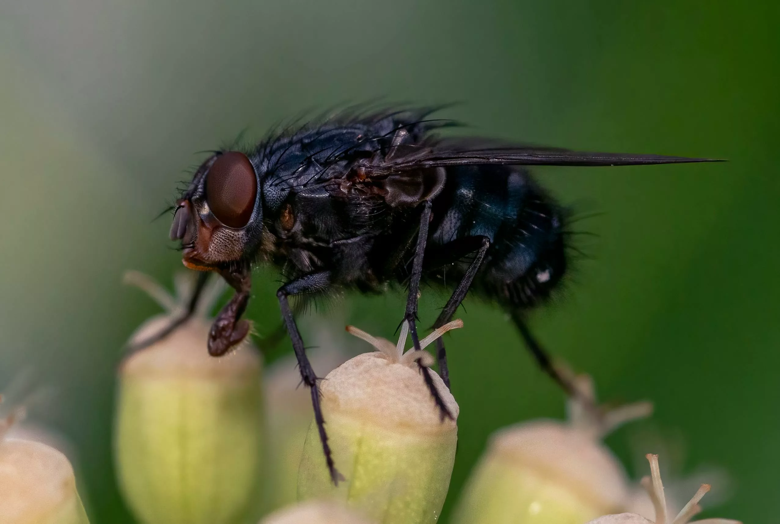
River blindness or Onchocerciasis is a parasitic infection caused by the worm Onchocera volvulus that causes skin and vision problems. The parasite spreads to humans through an infected blackfly’s bite. Onchocerciasis is prevalent in Africa (Sub-Saharan region), Brazil, and South America. Geospatial analyses reveal that control steps have led to a recent decline in onchocerciasis prevalence; however, many areas of western and central Africa still have infection prevalence above 5%.1Schmidt, C. A., Cromwell, E. A., Hill, E., Donkers, K. M., Schipp, M. F., Johnson, K. B., … & Hay, S. I. (2022).The prevalence of onchocerciasis in Africa and Yemen, 2000–2018: a geospatial analysis. BMC medicine, 20(1), 293. The infectious condition can cause severe skin rash and irreversible blindness in some cases. Thus, timely diagnosis and treatment is crucial. Doctors usually treat the disease with anti-parasitic drugs like ivermectin.
Onchocerciasis Symptoms
This parasitic infection is notorious for causing severe skin and eye symptoms. The intensity of the symptoms can cause debilitation and gravely hamper life. Symptoms appear sometime after you have been infected with the parasite larvae. The disease only develops after the larvae have produced a significantly large number of parasitic agents. Therefore, it generally takes 12 to 18 months for the first symptoms to appear.

Skin Presentations:
Onchodermatitis
Onchocerciasis-induced dermatitis (skin inflammation) i.e., onchodermatitis is a common presentation. This symptom presents as multiple papules that form along with skin rash (which resembles eczema). Generally, these lesions are hyperpigmented. The most common sites of dermatitis are:
- Face
- Trunk
- Extremities
Patients experience debilitating pruritus and skin rashes. Onchodermatitis is characterized by skin changes (sclerosis and edema) accompanying enlargement and lymph node softening.
Itching
Itch is one of the most common symptoms of tropical parasitic infections including onchocerciasis, schistosomiasis, scabies, African trypanosomiasis, etc.2Ju, T., Vander Does, A., Ingrasci, G., Norton, S. A., & Yosipovitch, G. (2022). Tropical parasitic itch in returned travellers and immigrants from endemic areas. Journal of the European Academy of Dermatology and Venereology, 36(12), 2279-2290. Pruritis caused by onchocerciasis is very intense and people are greatly affected by the severe itch. According to research data, the most frequent skin manifestation of onchocerciasis was itching, followed by swelling and rash, respectively. Many patients are irritated by the troublesome itching and eye problems.3Enk, C. D. (2006). Onchocerciasis—river blindness. Clinics in dermatology, 24(3), 176-180.
Types Of Skin Conditions:
The dermatological patterns are categorized into different types.4Murdoch, M. E. (2018). Onchodermatitis: where are we now?. Tropical medicine and infectious disease, 3(3), 94. A patient can have one or more patterns of skin presentation at one time.
Acute Papular Onchodermatitis (APOD)
There is widespread skin rash and itchy papules (on the face, trunk, and extremities) that convert into vesicles/pustules.
Chronic Papular Onchodermatitis (CPOD)
The itch intensifies and papules have flat-tops+hyperpigmentation. This is the most common skin presentation type mainly affecting the shoulders and buttocks.
Lichenified Onchodermatitis (LOD)
Itchy plaques form on the skin that is thickened, scaly, and hyperpigmented. Lymph node enlargement is also present in this type.
Onchocercal Atrophy
Weak, dry, and wrinkled skin forms on the lower back (and buttocks) region. The inelastic skin breaks down. The dry and scaly skin may appear like “lizard skin.”
Onchocercal Depigmentation
In this type, there is the formation of what experts call “leopard skin” characterized by areas of pigment loss surrounded by areas of normally colored skin. Studies show that leopard skin was the most common skin feature of the disease.5Egejuru, J., Nwokeji, C. M., Iwunze, J. I., & Amaechi, A. A. (2021). Onchocercal Skin Disease Profiles Following Ivermectin Treatment in the Middle Imo River Baisn, Nigeria. International Journal of TROPICAL DISEASE & Health, 42(23), 23-32.
Palpable Onchocercal Nodules
Nodules form in the subcutaneous layer (beneath the skin). The subcutaneous lumps are usually present over bony prominences and can stretch to 80 cm in length. These nodules contain adult worms. The bumpy nodules are called onchoceroma.6Ogbonna, E. C., & Ikani, O. R. (2020). Systematic review on onchocerciasis infection in Nigeria in the past five decades. It is a classic clinical feature of onchocerciasis. Thus, it can aid in diagnosing the parasitic infection.
Hanging Groin
Another classical presentation of the disease is the formation of atrophic skin folds in the groin region that accompanies lymph node enlargement. This pattern can help in diagnosis and is called hanging groin.
Eye Symptoms:
Reduced Visual Acuity
Ocular onchocerciasis begins with changes in the cornea (clear layer of the eye). However, other eye structures may also be infected simultaneously. Parasite infestation triggers a cascade of inflammatory responses within the body, increasing corneal opacity. Experts notice “snow-flake” opacities due to pinpoint inflammation of the cornea i.e., punctate keratitis.7Kwa, B. H., & Kwa, B. H. (2017). River Blindness: And It Was About Time. The Parasite Chronicles: My Lifelong Odyssey Among the Parasites that Cause Human Disease, 75-87. These spots of keratitis can coalesce later and cause hyperpigmentation i.e., sclerosing keratitis. Onchocercal keratitis is characterized by:
- Corneal opacification i.e., scarring of the cornea and subsequent loss of transparency
- Neovascularization i.e., formation of new blood vessels

The opacification of the cornea reduces the amount of light passing through the eye. Therefore, onchocercal keratitis leads to reduced visual acuity in patients.8Ament, C. S., & Young, L. H. (2006). Ocular manifestations of helminthic infections: onchocersiasis, cysticercosis, toxocariasis, and diffuse unilateral subacute neuroretinitis. International ophthalmology clinics, 46(2), 1-10.
Blindness
In addition to vision impairments, onchocerciasis can cause permanent, complete blindness (if left untreated). According to the WHO, onchocerciasis is the second leading cause of permanent blindness (after trachoma).9https://www.who.int/news-room/fact-sheets/detail/onchocerciasis The entry of the pathogen inside the body triggers multiple eye complications )in addition to the sclerosing keratitis) including secondary glaucoma, chorioretinitis, cataracts, and atrophy of the optic nerve. Thus, combining these conditions leads to a reduced field of vision, fading of color, and eventually blindness.
Neurological Complications:
Onchocerciasis can induce epileptic seizures (especially in pediatric patients and young patients). Researchers have found a strong association between the onset of tonic-clonic seizures and onchocerciasis. A 2023 study concluded that the parasitic infection directly or indirectly induces onchocerciasis-associated epilepsy (OAE). Clinicians believe that the parasites cross the blood-brain barrier and cause neurological complications. Therefore, there is a need to eliminate this parasite.10Hadermann, A., Amaral, L. J., Van Cutsem, G., Fodjo, J. N. S., & Colebunders, R. (2023). Onchocerciasis-associated epilepsy: an update and future perspectives. Trends in Parasitology, 39(2), 126-138.
Onchocerciasis Causes
It is caused by the worm Oncocera volvulus which is transmitted to humans by the African blackfly belonging to the Simulium species.
Onchocerciasis Risk Groups:
Blackflies commonly reside in fast-flowing rivers and streams. Therefore, tropical and agricultural areas have the highest number of cases. According to a study, the highest rate of infection was seen in farmers.11Nyagang, S. M., Cumber, S. N., Cho, J. F., Keka, E. I., Nkfusai, C. N., Wepngong, E., … & Fokam, E. B. (2020). Prevalence of onchocerciasis, attitudes and practices and the treatment coverage after 15 years of mass drug administration with ivermectin in the Tombel Health District, Cameroon. Pan African Medical Journal, 35(1).

Blackflies thrive mostly in countries with poor sanitation and a lack of clean drinking water. Therefore, the greatest number of cases are seen in the following regions:
- Brazil
- Republic of Congo
- Sub-Saharan Africa
- Yemen
- Venezuela
Transmission of Onchocerciasis:
As mentioned, the disease develops after the worm Onchocera volvulus enters the human body. The worm survives inside the blackflies after it ingests the worm larvae i.e., microfilariae. Once inside the fly, the larvae grow and when an infected fly bites a human, it transmits the parasites (in larval form).
The microfilariae mature into adults inside their new host. The mature worms reproduce and soon there are large numbers of worms that attack the skin, eyes, and other organs (such as the brain). However, the symptoms humans experience are due to the body’s own inflammatory response to the larvae population.
Is Onchocerciasis Contagious?
Onchocerciasis isn’t contagious and does not spread to another person by contact. However, it can be spread to a healthy individual from an infected one via a vector (blackfly bite).
Onchocerciasis Diagnosis
Diagnosing river blindness is a little tricky because significant symptoms appear only when there are very large numbers of worms circulating in the body.
History:
Healthcare providers begin with a complete history of the disease including travel history (to areas like Brazil, sub-Saharan Africa, etc.). Travel history is a critical question if there is the presence of simultaneous skin and eye symptoms.
Physical Examination:
Eye
For diagnosis, your doctor may use additional equipment to observe your eye lesions. A slit lamp (with a microscope) is conventionally used to better observe eye complications like keratitis, etc.
Skin
Your doctor might perform a skin biopsy to diagnose what is causing the skin symptoms. In the biopsy, the doctor will remove skin snips (small pieces of skin) from the affected part and observe it under a microscope to check for microfilariae. This is the most common method of onchocerciasis diagnosis. The healthcare provider may also surgically excise a nodule (if needed) from your skin for examination.
Blood Tests
In addition to the skin and eye tests, your doctor may also order blood tests to check antibodies against the pathogenic agent. However, this test only determines whether a person has been infected (ever) and does not provide details of any active infection.
Onchocerciasis Treatment
In most cases, oral anti-parasitic medications do a decent job of managing symptoms. Ivermectin is the first choice of many physicians. However, the drug is unable to kill the adult worm and only kills the larvae. Therefore, it reduces the worm load but does not eliminate it. So, patients have to take the pill at least once for 10-15 years.
Another drug (under trial) is moxidectin. In a blind phase 3 trial, single-dose moxidectin showed a greater reduction in skin microfilarial load than single-dose ivermectin.12Opoku, N. O., Bakajika, D. K., Kanza, E. M., Howard, H., Mambandu, G. L., Nyathirombo, A., … & Kuesel, A. C. (2018). Single dose moxidectin versus ivermectin for Onchocerca volvulus infection in Ghana, Liberia, and the Democratic Republic of the Congo: a randomised, controlled, double-blind phase 3 trial. The Lancet, 392(10154), 1207-1216. Moreover, this drug has stronger and longer microfilaria suppression as compared to ivermectin.13Milton, P., Hamley, J. I., Walker, M., & Basáñez, M. G. (2020). Moxidectin: an oral treatment for human onchocerciasis. Expert Review of Anti-Infective Therapy, 18(11), 1067-1081.
Onchocerciasis Prevention
You can prevent onchocerciasis by avoiding visits to blackfly pandemic areas. However, if unavoidable, you can adopt the following steps to stay safe:
- Cover your arms and legs to prevent fly bites. Use long sleeves/pants and ensure no exposed area when moving out.
- Spray diethyltoluamide (DEET) insecticides like permethrin on your clothes to repel insects (and flies).
Onchocerciasis Control Programs
The World Health Organization looks over different vector control programs such as the African Programme for Onchocerciasis Control’s (APOC) strategy where mass administration of ivermectin is carried out in the African populations. The organization aims to eliminate onchocerciasis in most endemic countries by 2025. Another program is the Onchocerciasis Elimination Programme for the Americans (OEPA).14https://www.who.int/teams/control-of-neglected-tropical-diseases/onchocerciasis/prevention-control-and-elimination
Final Word
Onchocerciasis is a parasitic infection caused by the Onchocera volvulus worm. The worm transmits to humans from the bite of an infected Simulium blackfly. The worm thrives as microfilaria (larva form) in the fly host and once inside the human body, it replicates, producing large numbers of worms that cause symptoms. Onchocerciasis causes severely itchy skin rash, depigmented lesions, and raised skin nodules. It also attacks the eyes and leads to corneal opacification, sclerosing keratitis, and even irreversible blindness. Doctors diagnose it by checking skin snips under a microscope for microfilariae. Treatment involves oral administration of the antiparasitic drug ivermectin. Newer drugs like moxidectin are under trial and are expected to show results superior to ivermectin therapy.
Refrences
- 1Schmidt, C. A., Cromwell, E. A., Hill, E., Donkers, K. M., Schipp, M. F., Johnson, K. B., … & Hay, S. I. (2022).The prevalence of onchocerciasis in Africa and Yemen, 2000–2018: a geospatial analysis. BMC medicine, 20(1), 293.
- 2Ju, T., Vander Does, A., Ingrasci, G., Norton, S. A., & Yosipovitch, G. (2022). Tropical parasitic itch in returned travellers and immigrants from endemic areas. Journal of the European Academy of Dermatology and Venereology, 36(12), 2279-2290.
- 3Enk, C. D. (2006). Onchocerciasis—river blindness. Clinics in dermatology, 24(3), 176-180.
- 4Murdoch, M. E. (2018). Onchodermatitis: where are we now?. Tropical medicine and infectious disease, 3(3), 94.
- 5Egejuru, J., Nwokeji, C. M., Iwunze, J. I., & Amaechi, A. A. (2021). Onchocercal Skin Disease Profiles Following Ivermectin Treatment in the Middle Imo River Baisn, Nigeria. International Journal of TROPICAL DISEASE & Health, 42(23), 23-32.
- 6Ogbonna, E. C., & Ikani, O. R. (2020). Systematic review on onchocerciasis infection in Nigeria in the past five decades.
- 7Kwa, B. H., & Kwa, B. H. (2017). River Blindness: And It Was About Time. The Parasite Chronicles: My Lifelong Odyssey Among the Parasites that Cause Human Disease, 75-87.
- 8Ament, C. S., & Young, L. H. (2006). Ocular manifestations of helminthic infections: onchocersiasis, cysticercosis, toxocariasis, and diffuse unilateral subacute neuroretinitis. International ophthalmology clinics, 46(2), 1-10.
- 9https://www.who.int/news-room/fact-sheets/detail/onchocerciasis
- 10Hadermann, A., Amaral, L. J., Van Cutsem, G., Fodjo, J. N. S., & Colebunders, R. (2023). Onchocerciasis-associated epilepsy: an update and future perspectives. Trends in Parasitology, 39(2), 126-138.
- 11Nyagang, S. M., Cumber, S. N., Cho, J. F., Keka, E. I., Nkfusai, C. N., Wepngong, E., … & Fokam, E. B. (2020). Prevalence of onchocerciasis, attitudes and practices and the treatment coverage after 15 years of mass drug administration with ivermectin in the Tombel Health District, Cameroon. Pan African Medical Journal, 35(1).
- 12Opoku, N. O., Bakajika, D. K., Kanza, E. M., Howard, H., Mambandu, G. L., Nyathirombo, A., … & Kuesel, A. C. (2018). Single dose moxidectin versus ivermectin for Onchocerca volvulus infection in Ghana, Liberia, and the Democratic Republic of the Congo: a randomised, controlled, double-blind phase 3 trial. The Lancet, 392(10154), 1207-1216.
- 13Milton, P., Hamley, J. I., Walker, M., & Basáñez, M. G. (2020). Moxidectin: an oral treatment for human onchocerciasis. Expert Review of Anti-Infective Therapy, 18(11), 1067-1081.
- 14https://www.who.int/teams/control-of-neglected-tropical-diseases/onchocerciasis/prevention-control-and-elimination


