Cutaneous T-cell lymphoma (a type of non-Hodgkin lymphoma) is an uncommon skin pathology. It results from the mutation of your T lymphocytes (white blood cells) that are part of your immune system. These malignant T-cells proliferate and migrate to your skin and are usually characterized by rash and itching followed by patches or plaque formation.
What is Cutaneous T-cell Lymphoma?
As the word suggests, cutaneous means skin, T-cell stands for T-lymphocyte (white blood cells), and lymphoma means cancer. Normally, the T-lymphocytes protect you against various pathogens. This cancer affects your body’s immune system and forms immature T-cells (cancerous). These immature T-cells migrate from blood to your skin and cause itching and rash, resulting in lesion formation. However, the appearance of the lesion changes as the disease progresses. The first presentation of this disease is severe pruritus (itching) followed by plaques or patch formation.1Schukow C, Ahmed A. Dermatopathology, Cutaneous Lymphomas. [Updated 2023 Feb 16]. In: StatPearls [Internet]. Treasure Island (FL): StatPearls Publishing; 2024 Jan-. Available from: https://www.ncbi.nlm.nih.gov/books/NBK589703/
Cutaneous T-cell lymphoma constitutes only 2% of all T-cell lymphoma. The overall incidence of CTCL in the United States of America (USA) is about 6% of 1 million people. The frequency is higher in males than females, with a ratio of 2:1.2Pinter-Brown, L. C., MD. (n.d.). Cutaneous T-cell lymphoma: practice essentials, background, pathophysiology. https://emedicine.medscape.com/article/2139720-overview#a7?form=fpf
Types of Cutaneous T-cell Lymphoma
The clinical picture of the patient depends upon the type of cancer. There are multiple types of this lymphoma; these are low-grade and high-grade tumors. The low-grade tumors are:
Mycosis Fungoides:
It is the most common type of CTCL. About 50% of the cases of CTCL are mycosis fungoides. It progresses very slowly and most commonly involves your skin. In rare cases (about 10), this tumor spreads to your lymph and can affect your body organs, such as the liver and spleen. It usually affects older people and is not life-threatening. Additionally, there are some variants of cutaneous T-cell lymphomas. These are:
- Folliculotropic mycosis fungoides
- Pagetoid reticulosis
- Granulomatous slack skin disease
Other low-grade tumors include:
- Primary cutaneous CD30+
- Lymphomatoid papulosis
- Subcutaneous panniculitis
High-Grade Tumors:
High-grade tumors include the following:
Sezary Syndrome
It is less common than mycosis fungoides and constitutes only 15 percent of the cases of CTCL. This syndrome not only affects your skin but also affects your bloodstream. Moreover, the clinical picture of this syndrome is generalized redness all over the body, also called erythroderma. Unlike mycosis fungoides, it spreads faster. Other high-grade tumors are:
- Adult T-cell lymphoma
- Extranodal NK/T cell lymphoma
- Primary cutaneous aggressive CD8+ lymphoma3Cutaneous T-cell lymphoma: Overview. (n.d.). https://www.aad.org/public/diseases/a-z/ctcl-overview
Causes of Cutaneous T-cell Lymphoma
The cause of cutaneous T-cell lymphoma is unknown. However, some triggering factors can promote the disease. These include:
Gene Mutations:
Some studies show some genetic mutations are responsible for CTCL.
Drugs:
Some drugs trigger the risk of cutaneous T-cell lymphoma. These drugs include immunosuppressive medications and hydrochlorothiazide diuretics.
Infections:
Some infections are involved in promoting this disease. These can be viral, bacterial, and fungal.4Varghese MT, Alsubait S. T-Cell Lymphoma. [Updated 2022 Nov 7]. In: StatPearls [Internet]. Treasure Island (FL): StatPearls Publishing; 2024 Jan-. Available from: https://www.ncbi.nlm.nih.gov/books/NBK564354/
Viral Infections
- Cytomegalovirus infection (CMV)
- Epstein barr virus infection (EBV)
- AIDS
- Polyomavirus
Bacterial & Fungal Infections
- Staphylococcus aureus
- Chlamydophila pneumonia
- Mycobacterium leprae
- Dermatophytes
Environment Factors:
Several physical and chemical factors, including industrial toxins and microbial agents, enhance the risk of cutaneous T-cell lymphoma.5Cutaneous T-Cell lymphoma. (2020, February 27). Johns Hopkins Medicine. https://www.hopkinsmedicine.org/health/conditions-and-diseases/lymphoma/cutaneous-tcell-lymphoma
Symptoms of Cutaneous T-cell Lymphoma
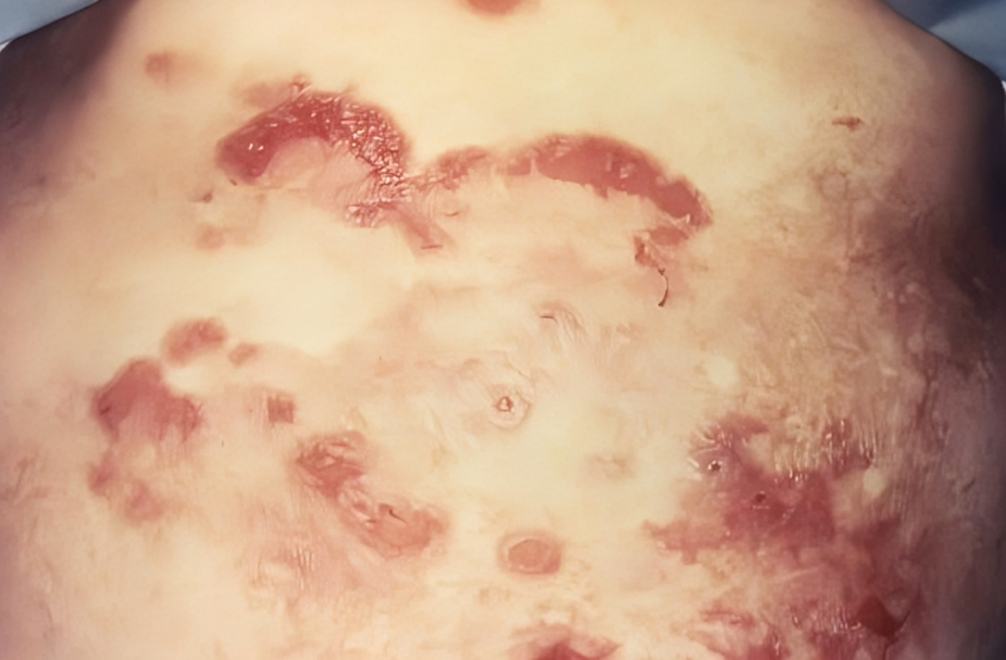
The clinical picture of this syndrome is similar to other skin disorders. However, the initial presentation of CTCL is scaly patches and hypo or hyperpigmented skin, specifically on the buttocks and body torso.6Cutaneous T-Cell lymphoma. (2020, February 27). Johns Hopkins Medicine. https://www.hopkinsmedicine.org/health/conditions-and-diseases/lymphoma/cutaneous-tcell-lymphoma The symptoms include:
- Hypopigmented or hyperpigmented patches
- Mottled skin appearance
- Red skin (erythroderma) that is itchy and dry
- Plaque formation (hard skin)
- Hard papules
- Swelling in the skin
- Cracked skin
- Lymph node swelling (most commonly involving the neck, axilla, and groin region)
How to diagnose Cutaneous T-cell Lymphoma?
Your doctor will diagnose based on history, clinical examination, lab investigations, imaging, and histopathology.
History:
During history, your doctor will ask you about your medical illness, presenting complaints, disease onset, drug history, and itching. If you have any drug allergies or medical illnesses, tell your doctor.
Clinical Examination:
Your doctor will examine your lesion and skin color, erythema, scaling, and area of skin involvement. He will also check your lymph nodes to assess the spread of the disease. Moreover, ocular involvement can also occur in the later stages of cutaneous T-cell lymphoma. Multiple biopsies can also be taken to check the aggressiveness of the tumor.
Laboratory Investigations:
Laboratory investigations that your doctor advises are :
Complete Blood Count
A complete blood count helps to see the blood picture. Patients with chronic skin disease may have anemia.
Serum Electrolytes
People with this syndrome may have an electrolyte imbalance. Therefore, serum electrolytes are advised.
Liver Function Test
This test is advised to check the hepatic involvement of the disease.
Lactate dehydrogenase level (LDH) and uric acid level:
LDH and Uric acid levels are markers of aggressive patterns of the disease. They are usually raised.
HIV Testing
As this disease affects people with compromised immune status. Therefore HIV screening is necessary to rule out the disease.
Imaging Studies:
Imaging studies include:
Chest Radiography
Chest radiography is advised to check the spread of disease in the lungs.
CT Scan
CT scans of the abdomen and pelvis help to stage the tumor and spread the disease.
PET Scan
This test is advised when there is a suspicion of visceral involvement.
Skin Biopsy
A biopsy involves your doctor taking a piece of your skin or lymph nodes and sending it to the pathology lab for evaluation to confirm the diagnosis. In mycosis fungoides, multiple skin biopsies are taken to check the grade of the lesion. Moreover, a skin biopsy is a confirmatory test to diagnose CTCL lesions.

Histopathology:
The histopathological criteria of the cutaneous T-cell lymphoma are:
- Band-like infiltrate in the upper dermal layer of the skin
- Infiltration of lymphocytes with tropism (affinity)
- Spongiosis of epidermis
- Hypochromic or cribriform nuclei
Staging of Cutaneous T-cell Lymphoma
Staging of CTL with Mycosis Fungoides & Sezary Syndrome:
The TNM staging of cutaneous T-cell lymphoma, according to the American Joint Committee, is:7Duhovic, C., Child, F., & Wain, E. M. (2012). Management of cutaneous T-cell lymphoma. Clinical Medicine, 12(2), 160–164. https://doi.org/10.7861/clinmedicine.12-2-160
Skin:
According to skin involvement, the staging is:
T1: A limited number of patches and plaques covering less than 10% of the skin area
T2: Multiple patches, hard papules, and plaques covering more than 10 percent of the skin area
T3: One or more tumors having a size of more than 1 cm diameter
T4: Redness (erythema) covering more than 80% of the body
Nodal Involvement:
N0: No abnormal lymph nodes are assessed
N1: Abnormal peripheral lymph nodes (Dutch grade 1) and can clone negative (N1a) or clone positive (N1B)
N2: Abnormal peripheral lymph nodes (Dutch grade 2)
N3: Abnormal peripheral lymph nodes (Dutch grade 3)
Nx: Abnormal lymph nodes with no histological confirmation
Organ Involvement:
M0: No organ involvement
M1: Organs are involved
Vessels Involvement:
B0: No significant vessels are involved (less than 5% peripheral blood lymphocytes involvement)
B1: Low blood tumor burden having more than 5% peripheral blood lymphocyte involvement
B2: High blood tumor burden contains more than 1000 microliter sezary cells
Staging of CTL without Mycosis Fungoides & Sezary Syndrome:
Skin:
T1: solitary skin lesions less than 5 cm (T1a) and more than 5 cm (T1b)
T2: regional skin involvement
T2a: disease involving less than 15 cm of circular skin area
T2b: disease involving more than 15 cm and less than 30cm of skin area
T2c: disease involving more than 30% of skin area
T3: Generalized skin involvement
Nodal involvement:
N0: No lymph node involvement
N1: One lymph node is involved
N2: Two or more peripheral lymph node involvement
N3: Central lymph node involvement
Metastasis:
M0: No evidence of extracutaneous metastasis
M1: Extracutaneous metastasis is present
Stage Grouping of CTL:
Stage IA: T1, N0, M0, B0 or B1
The skin lesions cover more than 10 % of the skin surface (T1). There are no tumors, lymph node involvement, or metastatic cells. The sezary cell count is low.8Lymphoma of the Skin Stages | Staging Skin Lymphoma. (2024). Cancer.org. https://www.cancer.org/cancer/types/skin-lymphoma/detection-diagnosis-staging/staging.html
Stage IB: T2, N0, M0, B0 or B1
The skin lesions extend over more than 10% of the skin surface (T2). There are no tumors, lymph node involvement, or metastatic cells present. The presence of Sezary cells is minimal. Furthermore, there is no dissemination of lymphoma cells to other organs.
Stage IIA: T1 or T2, N1 or N2, M0, B0 or B1
Only skin lesions which cover more than 80 % of the skin surface (T2 or T2). Lymph nodes are generally enlarged, but the pattern does not look abnormal. There is a low number of sezary cells and no spread of lymphoma cells to other organs.
Stage IIB: T3, N0 to N2, M0, B0 or B1
One skin lesion is a tumor 1cm or more in size (T3). There is a low number of sezary cells, and lymphoma cells have not spread to other organs. Lymph nodes may be normal or enlarged, but the pattern does not look abnormal on microscopy.
Stage IIIA: T4, N0 to N2, M0, B0
Skin lesions cover 80 percent of the skin (T4). There is a low number of sezary cells; lymphoma cells have not spread to other organs and are less than 5 percent. Lymph nodes may be normal or enlarged, but the pattern does not look abnormal on microscopy.
Stage IIIB: T4, N0 to N2, M0, B1
Skin lesions cover 80 percent of the skin (T4). There is a low number of sezary cells, and lymphoma cells have not spread to other organs. Lymph nodes may be normal or enlarged, but the pattern does not look abnormal on microscopy.
Stage IVA1: Any T, N0 to N2, M0, B2
Skin lesions that cover most of the surface or any amount of the skin surface. There are many sezary cells, and lymphoma cells have not spread to other organs. Lymph nodes may be normal or enlarged, but the pattern looks significantly abnormal on microscopy.
Stage IVA2: Any T, N3, M0, any B
Skin lesions that cover most of the surface or any amount of the skin surface. There may be a low or high number of sezary cells, and lymphoma cells have not spread to other organs. Lymph nodes may be normal or enlarged, but the pattern looks significantly abnormal on microscopy.
Stage IVB: Any T, any N, M1, any B
Skin lesions that cover most of the surface or any amount of the skin surface. There may be a low or high number of sezary cells, and lymphoma cells have spread to other organs (liver and spleen). Lymph nodes may be normal or enlarged, but the pattern looks significantly abnormal on microscopy.
How to treat Cutaneous T-cell Lymphoma?
The treatment of cutaneous T-cell lymphoma is based on the stage of the disease. Topical treatment is usually preferred in stage 1 carcinoma, and systemic therapy is indicated in stage 2 or greater. While there is no cure for this disease, symptomatic treatment is given to improve the patient’s quality of life.
Skin directed therapy:
In early-stage cancer, skin-directed therapy is given to alleviate the symptoms. It includes:
Topical therapy
- High-potency corticosteroids are applied over the plaques
- The use of emollients may benefit itching by improving skin condition
- Topical retinoids (vitamin A and its derivatives)
- Topical chemotherapeutic agents (kill cancer cells) that are applied to skin
- UVB radiation therapy under a specific strength of energy
- Topical electron beam radiation therapy
Systemic therapy:
Systemic therapy is for stage 2 or more. In this therapy, your doctor will treat your disease systemically. However, it includes the following:
Low-dose interferon alpha (IFN-a)
CTCL is an autoimmune disease. IFN-a alters your body’s immune system and is an oncogene (cancer cells) suppressor. A subcutaneous dose of 3-9 MU three times a week. Therefore, it can cause flu-like symptoms.
Extracorporeal photopheresis
It is the most accepted and popular therapy to treat mycosis fungoides (stage 3). Therefore, it is an immunomodulatory therapy. In this therapy, a photosensitizer is injected into the body, followed by UVA radiation.
Bexarotene
A retinoic acid derivative binds with the PXR receptor and interferes with cell proliferation, differentiation, and apoptosis. This therapy is taken orally once a day. The side effects of this drug are hyperlipidemia, neutropenia, and hypothyroidism. Therefore, thyroxine therapy and lipid-lowering drugs are indicated in some patients.
Methotrexate
It is an immunosuppressant drug and less frequently used to treat CTCL due to its side effects (hepatotoxicity and leukopenia).
Chemotherapy:
In advanced-stage carcinoma, chemotherapy is the only option to limit the disease progression. However, it is considered a palliative treatment option. Chemotherapeutic agents include:
- Gemcitabine
- Chlorambucil
- Doxorubicin
- Cyclophosphamide
Targeted therapy:
It targets specific molecules on the surface of the skin. It targets those cells, halts their activity, and stops the spreading of the tumor. Targeted therapy includes:
- Rituximab
- Romidepsin
- Brentuximab
Transplantation:
Allogeneic transplantation is indicated when the disease is refractory. It is associated with high morbidity and mortality.9Hristov, A. C., Tejasvi, T., & A Wilcox, R. (2021). Cutaneous T-cell lymphomas: 2021 update on diagnosis, risk-stratification, and management. American journal of hematology, 96(10), 1313–1328. https://doi.org/10.1002/ajh.26299
Prevention of Cutaneous T-cell Lymphoma
You can not prevent cutaneous T-cell lymphoma. As this is an autoimmune disease. Therefore, there are some ways to boost your immune system. These are:
- Vaccination
- Healthy diet plan
- Stop smoking
- Stop drinking alcohol
- Maintain your sleep pattern
- Healthy exercise
- Avoid having stress
Does it cause any Complications?
Cutaneous T-cell lymphoma can cause many complications that can be fatal. These include:
- Infection at the biopsy site
- Heart failure
- Anemia due to chronic illness
- Fluid collection (edema)
- Malignancies
How do I take care of my skin with Cutaneous T-cell Lymphoma?
It makes your skin red, dry, and itchy. Therefore, you have to take care of your skin by following:
- Use emollients and take baths to make your skin moist
- Avoid going into the sun and wear clothes that help you protect yourself from the sun
- Try to avoid scratching your skin. Additionally, cold compresses or antihistamines can be used to treat itching.
Survival Rate of Cutaneous T-cell Lymphoma
CTCL is an incurable disease that is incurable in some patients who are in advanced stages. The prognosis of CTCL is as follows:
- Patients who have stage 1 CTCL (mycosis fungoides) have a survival rate of 10 years (98-99%)
- Patients having stage 2 disease have a survival rate of 3.2 percent (10-year survival rate is about 42%)
- Patients with stage 3 disease have a survival rate of 4-8 years, and a 10-year survival rate is 83%
- Patients with stage 4 disease (extracutaneous manifestation) have a survival rate of 1.5 years, and a 10-year survival date is about 20%
Cutaneous T-cell Lymphoma Vs. Eczema
Eczema is derived from the Greek word meaning “to boil.” It is the most common skin disorder characterized by severe pruritus that disturbs sleep and quality of life. The eczematous lesions are erythematous, vesicular, and oedematous. Moreover, the inflammatory process in eczema is progressive and spreads all over the body from its origin. Unlike cutaneous T-cell lymphoma, T-cells are not involved in causing eczema.
However, cutaneous T-cell lymphoma is a rare skin disease that is slow and progressive and takes years to develop. It is a lymphoproliferative disorder characterized by generalized erythroderma and plaque formation.
Conclusion
To conclude, cutaneous T-cell lymphoma results from a mutation in T lymphocytes. It presents with severe itching, redness, mottled skin appearance, cracked skin, and lymph node swelling. Biopsy is a confirmatory test for CTCL. Skin-directed therapy and systemic therapy are treatment options, depending on the stage of the disease. Allogeneic transplant is suitable for refractory CTCL. The prognosis and survival rate depend on the stage of CTCL.
Refrences
- 1Schukow C, Ahmed A. Dermatopathology, Cutaneous Lymphomas. [Updated 2023 Feb 16]. In: StatPearls [Internet]. Treasure Island (FL): StatPearls Publishing; 2024 Jan-. Available from: https://www.ncbi.nlm.nih.gov/books/NBK589703/
- 2Pinter-Brown, L. C., MD. (n.d.). Cutaneous T-cell lymphoma: practice essentials, background, pathophysiology. https://emedicine.medscape.com/article/2139720-overview#a7?form=fpf
- 3Cutaneous T-cell lymphoma: Overview. (n.d.). https://www.aad.org/public/diseases/a-z/ctcl-overview
- 4Varghese MT, Alsubait S. T-Cell Lymphoma. [Updated 2022 Nov 7]. In: StatPearls [Internet]. Treasure Island (FL): StatPearls Publishing; 2024 Jan-. Available from: https://www.ncbi.nlm.nih.gov/books/NBK564354/
- 5Cutaneous T-Cell lymphoma. (2020, February 27). Johns Hopkins Medicine. https://www.hopkinsmedicine.org/health/conditions-and-diseases/lymphoma/cutaneous-tcell-lymphoma
- 6Cutaneous T-Cell lymphoma. (2020, February 27). Johns Hopkins Medicine. https://www.hopkinsmedicine.org/health/conditions-and-diseases/lymphoma/cutaneous-tcell-lymphoma
- 7Duhovic, C., Child, F., & Wain, E. M. (2012). Management of cutaneous T-cell lymphoma. Clinical Medicine, 12(2), 160–164. https://doi.org/10.7861/clinmedicine.12-2-160
- 8Lymphoma of the Skin Stages | Staging Skin Lymphoma. (2024). Cancer.org. https://www.cancer.org/cancer/types/skin-lymphoma/detection-diagnosis-staging/staging.html
- 9Hristov, A. C., Tejasvi, T., & A Wilcox, R. (2021). Cutaneous T-cell lymphomas: 2021 update on diagnosis, risk-stratification, and management. American journal of hematology, 96(10), 1313–1328. https://doi.org/10.1002/ajh.26299
