What is DEXA Scan?
DEXA stands for dual-energy X-ray absorptiometry scan. This scan is also called a bone densitometry scan or, more specifically, a DXA scan. It is a speedy, painless method to check the health of your bones. The scan uses low-dose X-rays to measure the mineral content, density, and strength of bones, making it more accurate than regular X-rays at detecting low bone density. This scan takes 10 to 20 minutes.
There are two types of DEXA scans: central and peripheral. Central DEXA scans measure bone density in areas like the spine and hips, while peripheral DEXA scans assess bones in the wrist, heel, or finger. DEXA scans commonly diagnose conditions like bone thinning (osteopenia) and osteoporosis. Bones with more minerals and calcium are denser, stronger, and less likely to break. Bones become thinner as you get older or you develop some medical condition.
Osteopenia is the term used to describe the bones that are thinner than usual. It puts you at high risk for a more severe condition termed osteoporosis. It is a disorder that causes bones to become very brittle and thin. The disorder gets worse over time and usually affects older people. It is most common in females over the age of 65. Such people are at more risk for fractures, predominantly in their wrists, spine, and hips.1El Maghraoui, A., & Roux, C. (2008). DXA scanning in clinical practice. QJM: An International Journal of Medicine, 101(8), 605-617.2Truscott, J. G., Devlin, J., & Emery, P. (1996). DXA scanning. Baillière’s clinical rheumatology, 10(4), 679-698.
DEXA Vs. Standard X-Ray
A DEXA scanner also emits X-ray energy, just like a standard X-ray scanner. But, it differs from the X-ray scanner in several ways. The standard X-ray scanner aims to produce a high-quality picture of the bones and other parts of the body. It can not provide information on the density of the tissue. In contrast, a DEXA scan specifically assesses bone mineral density (BMD) but does not produce high-resolution images.
Both of these scanners have different ways of delivering X-ray energy. The length of time an individual is exposed to X-rays also differs. Standard X-rays use a more focused radiation beam with a longer exposure time. DEXA scanners use two X-ray energies: one absorbed by bone and the other by soft tissues. These energies are delivered via a fan-shaped beam, which allows the DEXA scan to cover an area quickly, reducing the time and overall radiation exposure.3Hoyer-Kuhn, H., Knoop, K., Semler, O., Kuhr, K., Hellmich, M., Schoenau, E., & Koerber, F. (2016). Comparison of DXA scans and conventional X-rays for spine morphometry and bone age determination in children. Journal of Clinical Densitometry, 19(2), 208-215.
Why are DEXA Scans used?
Physicians may use a DEXA scan to inspect your bones in some situations, such as:
- To check your bone fracture risk
- To diagnose osteopenia
- To diagnose osteoporosis
- To predict the risk for future fractures
The healthcare provider uses a bone density test to examine how your bones change over time.
- This scan measures the natural extent of bone loss and tracks your bone loss over time.
- Check your bone’s response to treatment for osteopenia and osteoporosis.
- Test bone density before starting a medication or treatment that can have a side effect.
Who Needs a DEXA Scan?
Some individuals have a greater risk of developing osteoporosis. While some might have other disorders that can affect their bone density. Individuals who need a regular DEXA scan include:
- Males older than 70
- Females older than 65
- People with broken bones older than 50
- People with biological osteoporotic grandparents
- People at increased risk of bone loss
People with some health conditions, such as:
- Autoimmune diseases
- Vitamin deficiency
- Excessive use of tobacco products or smoking
- Excessive intake of alcohol
Certain medications also increase the risk of bone density issues, such as:
- Hormone suppressants
- Corticosteroids
- Immunosuppressants
- Seizure medications
- Some kinds of medications used for treating cancer (such as aromatase inhibitors and methotrexate)4Panday, K., Gona, A., & Humphrey, M. B. (2014). Medication-induced osteoporosis: screening and treatment strategies. Therapeutic advances in musculoskeletal disease, 6(5), 185-202.
- Heparin
- Proton pump inhibitors
Some other risk factors for losing bone density include:
- A very low body weight
- Lack of physical activity
- Lack of calcium in your diet
Preparations for DEXA Scan
There are no exceptional preparations for this test. You would not have to change your routine before this test. You can drink and eat as usual and can take medications. Speak to your provider about the supplements you take. The provider will let you distinguish which ones you can not take before this test. Stop taking calcium add-ons 24 to 48 hours before this scan.
Wear comfortable clothes. Do not wear anything that has metal in it. If you are expecting or think you could be pregnant, inform the department. Clear it out before your scan appointment. This is because X-rays from the DEXA scan might harm the developing fetus.
In some healthcare centers, you might need to fill out a questionnaire beforehand. Some of the queries in this questionnaire include:
- Do you drink alcohol?
- Do you smoke?
- Are you still having periods?
- Are you on any bone-strengthening treatment?
- Are you on some medications that are thinning your bones, like steroids?
This questionnaire will be either sent to your home or the department for fill-in.
The Procedure For DEXA Scan
If you are found to be at risk of bone fractures, a central DEXA scan is recommended.
Procedure of Central DEXA Scan
You have to lay on your back on a comfortable table. You might need to lay with your legs straight and rest them on that padded table. The arm of the scanning machine will pass over your hips and spine. Another scanning machine will pass under you at the same time. Images from these two scanners will be combined. The healthcare provider will interpret the images on the computer screen. When the gears are scanning, you must stay still. Also, you can be requested to hold your inhalation to keep the images from blurring. The Central DEXA scan usually takes 10 to 30 minutes. The healthcare provider will get the results in a few days.
Procedure of Peripheral DEXA Scan
Sometimes, a bone density scan is only done on your hand, foot bones, and wrist. This scan is known as a peripheral DEXA scan (p-DEXA scan). The portable device for this scan has a box-like structure. The scan might be performed in a mobile health van, drug store, and health store office.
In this process, you will be asked to place your hand, finger, foot, or forearm in the space for imaging. This test takes some minutes. The provider receives the results shortly after the scan. This test can not give as many details as the central DEXA scan. But it can provide good information about your fracture risk. The duration of the scan varies depending on the size and number of bones undergoing scanning.
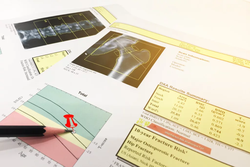
DEXA Scan Results
The bone density scan compares your bone density with the anticipated bone density. It will also compare your bone density with that of a healthy individual, who should be of your age, ethnicity, and gender. This difference in bone density is measured as a standard deviation (SD) score.
The providers give bone density results as T and Z scores. A high number of scores means that you have a high bone density. A lower score means a higher risk of bone fracture.
Your healthcare provider will use a T Score if you are a postmenopausal woman or man above or older than 50. This measurement compares your bone density with that of a healthy person of the same sex and age.
Providers use the Z score if you are a child, a male under 50, and a premenopausal woman. This measurement compares your bone density with that of other healthy people. These people should be of your weight, sex, age, racial or ethnic origin.5Carey, J. J., Delaney, M. F., Love, T. E., Richmond, B. J., Cromer, B. A., Miller, P. D., … & Licata, A. A. (2007). DXA-generated Z-scores and T-scores may differ substantially and significantly in young adults. Journal of Clinical Densitometry, 10(4), 351-358.
Interpretations of DEXA Scan Results
A radiologist will analyze the images. He will then send a signed report to your physicians, who will address the results with you. Other physicians, such as endocrinologists and rheumatologists, can also predict DEXA scans. The clinician will review your scan. He will consider the occurrence of clinical risk factors, including:
- Chronic renal and liver diseases
- Inflammatory bowel disease
- Rheumatoid arthritis
- Respiratory diseases
Your T score will show one of the following:
- A T score of -1 or above is a normal bone density.
- A T score that is between -1 and -2.4 shows low bone density (osteopenia). You may be at likelihood of developing osteoporosis.
- A T score of -2.5 indicates your possibility of having osteoporosis6MaryAnn De Pietro, C. (2022). DEXA (DXA) scan: Measuring bone density. Retrieved from https://www.medicalnewstoday.com/articles/324553
If your Z score is under -2, your bone density is low. Z scores are generally used for people under 30 and children who are still growing.
Bone density test results offer a good signal of bone strength. However, the results of the scan will not basically predict whether you will get a fracture or not. For instance, a person with low bone density might never break a bone. In contrast, a person with average bone density might have many breakages. This is due to other factors like sex, age, or previous falls. The healthcare provider will consider all your possible factors before determining the treatment.7El Maghraoui, A. (2011). Interpreting a DXA scan in clinical practice. Dual Energy X-Ray Absorptiometry, 1.
Preventive Measures
The provider will endorse some steps to avoid further bone loss. In case your scan results show that you have a low density, follow these recommendations:
- Addition of vitamin D and calcium to your diet.
- Increasing physical activity such as walking, using weight machines, jogging, etc.
- Taking prescribed medications to increase bone density.
The healthcare provider may recommend how often you need to have scans. Your provider will base these recommendations on your risk of bone fractures.
The provider recommends having a DEXA scan:
- Every two years for high-risk.
- Every three to five years for moderate risk
- Every 10 to 15 years for low-risk
Safety Profile
Bone density tests are very safe as they use a much lower level of radiation than typical X-rays. This means that the radiographer can stay in the screening room throughout this test. The amount of radiation you receive from this scan is so small and does not make you feel sick. Also, the risk of this radioactivity causing any harm in the future is surprizingly small. Despite being so safe, this scan is not recommended for pregnant women.
Impacts of DEXA Scan
DEXA scans are different from other imaging tests. The providers use this scan to screen a specific condition. The beneficial and harmful impacts of this scan are:
Benefits of DEXA Scan
DEXA scan can detect weak and brittle bones to aid in predicting the chances of a future fracture.
- It determines whether the bone density is improving, waning, or staying the same.
- This scan helps you and your provider to come up with plans to enhance your bone strength.
- The results of this scan help to prevent worsening conditions.
- DEXA is a quick, simple, and non-invasive technique.
- You do not need any anesthesia to undergo the scan.
- No radiation stays in your body after the scan.
- The equipment used for DEXA is convenient for both the patient and the provider.
Side Effects of DEXA Scan
There are a few risks to having a DEXA scan. You would not feel any pain during and after the scan. They are usually harmless for everyone, but:
- These small amounts of radiation can affect the development of unborn babies.
- There is a very slight increase in the possibility of future cancer. This is comparable to the side effects of X-rays. Every person is exposed to radiation in their daily life. However, any extra exposure (i.e., imaging test) can somewhat upsurge the risk of developing cancer.8Bisaccia, M., Rinonapoli, G., Meccariello, L., Ripani, U., Pace, V., Rollo, G., … & De Masi De Luca, A. (2019). Osteoporosis in male patients: epidemiology, clinical aspects, and DEXA Scan assessment. Clinical Cases in Mineral & Bone Metabolism, 16(1).
Limitations of the DEXA Scan
Despite the effectiveness of the DEXA scan, it still has some limitations.
- DEXA scan is of restricted use in people with spinal abnormality. Surgeons do not perform this scan on people who have had previous spinal surgery.
- Vertebral column compression fractures and osteoarthritis can also affect the accuracy of the results.
- Central DEXA scan devices provide more sensitive and superiorly standardized results. But they are expensive.
- Bone density measurements obtained from different DEXA gears cannot be directly compared. So, you must perform follow-up DEXA scans using the same machine in the same institute.
Quantitative Computed Tomography (QCT) versus DEXA
Understanding the difference between QCT and DEXA is beneficial. It can help in selecting the most suitable technique for evaluating osteoporosis.
- QCT can distinguish between cortical and trabecular bones, whereas DEXA can not.
- QCT offers three-dimensional volumetric measurements. In contrast, DEXA offers a two-dimensional view.
- QCT is more accurate in detecting early changes in trabecular bones and true bone density.9Li, N., Li, X. M., Xu, L., Sun, W. J., Cheng, X. G., & Tian, W. (2013). Comparison of QCT and DXA: osteoporosis detection rates in postmenopausal women. International journal of endocrinology, 2013(1), 895474.10Alawi, M., Begum, A., Harraz, M., Alawi, H., Bamagos, S., Yaghmour, A., & Hafiz, L. (2021). Dual-energy X-ray absorptiometry (DEXA) scan versus computed tomography for bone density assessment. Cureus, 13(2).
A Quick Review
DEXA scan provides a detailed and accurate assessment of your bone density. This non-invasive and pain-free procedure uses low-level X-ray beans to measure bone density. By detecting areas of low bone density, providers can develop targeted treatment plans that can prevent future fractures and reduce the risk of future bone loss.
Additionally, it is an effective tool for monitoring treatment outcomes. The importance of a DEXA scan extends beyond the diagnosis and treatment of osteoporosis. In short, the DEXA scan is a powerful diagnostic tool. It has transformed the way we approach bone-related disorders and osteoporosis.
Refrences
- 1El Maghraoui, A., & Roux, C. (2008). DXA scanning in clinical practice. QJM: An International Journal of Medicine, 101(8), 605-617.
- 2Truscott, J. G., Devlin, J., & Emery, P. (1996). DXA scanning. Baillière’s clinical rheumatology, 10(4), 679-698.
- 3Hoyer-Kuhn, H., Knoop, K., Semler, O., Kuhr, K., Hellmich, M., Schoenau, E., & Koerber, F. (2016). Comparison of DXA scans and conventional X-rays for spine morphometry and bone age determination in children. Journal of Clinical Densitometry, 19(2), 208-215.
- 4Panday, K., Gona, A., & Humphrey, M. B. (2014). Medication-induced osteoporosis: screening and treatment strategies. Therapeutic advances in musculoskeletal disease, 6(5), 185-202.
- 5Carey, J. J., Delaney, M. F., Love, T. E., Richmond, B. J., Cromer, B. A., Miller, P. D., … & Licata, A. A. (2007). DXA-generated Z-scores and T-scores may differ substantially and significantly in young adults. Journal of Clinical Densitometry, 10(4), 351-358.
- 6MaryAnn De Pietro, C. (2022). DEXA (DXA) scan: Measuring bone density. Retrieved from https://www.medicalnewstoday.com/articles/324553
- 7El Maghraoui, A. (2011). Interpreting a DXA scan in clinical practice. Dual Energy X-Ray Absorptiometry, 1.
- 8Bisaccia, M., Rinonapoli, G., Meccariello, L., Ripani, U., Pace, V., Rollo, G., … & De Masi De Luca, A. (2019). Osteoporosis in male patients: epidemiology, clinical aspects, and DEXA Scan assessment. Clinical Cases in Mineral & Bone Metabolism, 16(1).
- 9Li, N., Li, X. M., Xu, L., Sun, W. J., Cheng, X. G., & Tian, W. (2013). Comparison of QCT and DXA: osteoporosis detection rates in postmenopausal women. International journal of endocrinology, 2013(1), 895474.
- 10Alawi, M., Begum, A., Harraz, M., Alawi, H., Bamagos, S., Yaghmour, A., & Hafiz, L. (2021). Dual-energy X-ray absorptiometry (DEXA) scan versus computed tomography for bone density assessment. Cureus, 13(2).
