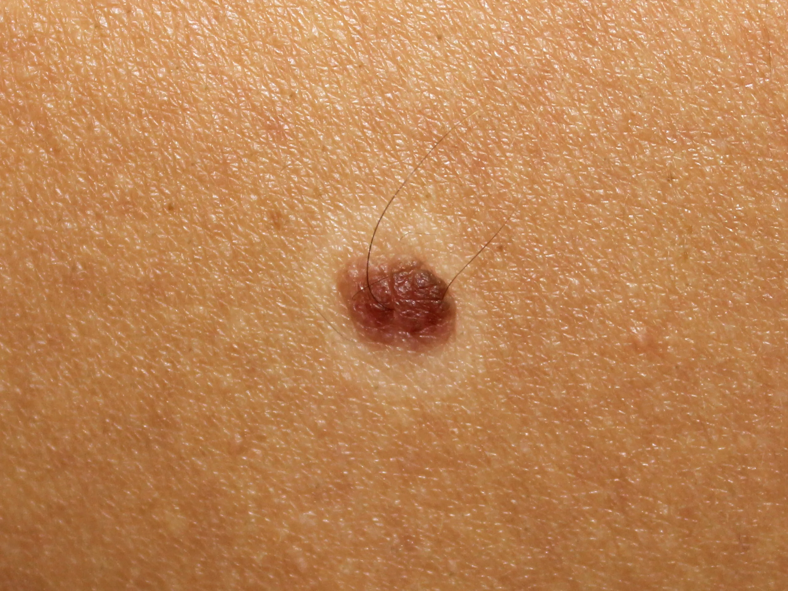
Halo Nevus, a coin-shaped pigmented mole with a surrounding non-pigmented area, is a common skin condition that occurs more often in teenagers and young people. Usually, it is harmless; however, it can become concerning if associated with conditions like melanoma. Understanding its stages, association with vitiligo, and cancer risk is crucial.
What is a Halo Nevus?
A halo nevus is a pigmented mole encircled by a an area that is not pigmented. This non-pigmented area, called a Halo, is uniform in appearance and lacks the cells (melanocytes) that form the pigment melanin. The borders of the halo are well-defined.
In 1916, Sutton described this condition as “leukoderma acquisitum centrifugum,” referring to the acquired loss of pigmentation in the surrounding skin. Later, Happle introduced the term “Grünewald nevus” to describe this phenomenon. It is now more appropriately referred to as Halo Nevus.1Kaminska-Winciorek G, Szymszal J. Dermoscopy of halo nevus in own observation. Postepy Dermatol Alergol. 2014;31:152–8.
Appearance of Halo Nevus
Halo nevi (plural of nevus) are regular brown, pinkish, or tan moles in the center of a white skin patch with regular borders. Nevus is usually of only one color. The halo is usually 1.0-1.5 cm in diameter.
They can appear anywhere on the body but are more common on the back, chest, and abdomen. Sometimes, palms and soles are also involved. A person can have a single halo nevus or multiple nevi simultaneously. They are common in children and young adults.
Development of Halo Nevus
Halo nevi has a progressive nature. The nevus may look different depending upon the duration of the development. People with multiple nevi might notice different stages of development for different nevus. In other words, not all the halo nevi on one’s body have to be in the same stage of development.
Stages of Halo Nevus
There are four stages of development of a halo nevus2De Schrijver, S., Theate, I., & Vanhooteghem, O. (2021). Halo nevi are not trivial: About two young patients of regressed primary melanoma simulate halo nevi. Case Reports in Dermatological Medicine, 2021, 1–5. https://doi.org/10.1155/2021/6672528
Stage One
A classic brown nevus with a surrounding rim of non-pigmented skin.
Stage Two
Central nevus may lose its pigmentation. After that, it might assume a pink appearance with a surrounding halo.
Stage Three
The central papule may disappear altogether, leaving a non-pigmented area on the skin.
Stage Four
The non-pigmented skin patch may undergo pigmentation again, leaving behind normal skin with no evidence of halo nevus.
Causes of Halo Nevus
Halo nevus develops when the body’s immune system acts against a mole. Sometimes, a triggering factor such as sunburn or local trauma damages the existing mole and might result in the activation of the immune system. The exact cause of why the body attacks the mole is not known. Researchers hypothesize that the body and its protective mechanisms perceive the mole as harmful to the body. The white blood cells, especially the T cells, attack the pigment-forming cells in the mole, causing them to disappear.3Babu, A., Bhat, M., Dandeli, S., & Ali, N. (2016). Throwing light onto the core of a halo nevus: A new finding. Indian Journal of Dermatology, 61(2), 238. https://doi.org/10.4103/0019-5154.177801
As a result, the pigmented area disappears, leaving behind a white patch. The white blood cells also attack the skin cells around the mole, forming the white path called the halo.
Antibodies against melanoma cells have also been found in the body. However, it is not yet known whether they are formed after or before the disruption of mole cells.4Naveh, H. P., Rao, U. N. M., & Butterfield, L. H. (2013). Melanoma‐associated leukoderma – immunology in black and white? Pigment Cell & Melanoma Research, 26(6), 796–804. https://doi.org/10.1111/pcmr.12161
Who Develops Halo Nevi?
The majority of Halo nevi occur in adolescents and young adults. Almost 1% of the population develops halo nevus. It affects both genders equally.5Weyant, G. W., Chung, C. G., & Helm, K. F. (2015). Halo nevus: a review of the literature and clinicopathologic findings. International Journal of Dermatology, 54(10). https://doi.org/10.1111/ijd.12843
Halo nevi can also develop in individuals with autoimmune diseases or vitiligo. It is also common in people with generalized pigment loss. It may also develop in people suffering from melanoma or those treated for melanoma.
Halo Nevus Vs. Melanoma
A halo nevus is a benign mole with a white, depigmented ring, often found in teenagers and young adults. It can sometimes be mistaken for melanoma, which is a type of skin cancer that appears as an irregular, asymmetrical mole with different colors and rapid changes. Research shows that adults with newly diagnosed halo nevi have about a 1% risk of developing primary cutaneous melanoma within a year, which is similar to the risk faced by individuals with atypical nevi or a family history of melanoma.6Association between halo nevi and melanoma in adults: A multicenter retrospective case series Haynes, Dylan et al. Journal of the American Academy of Dermatology, Volume 84, Issue 4, 1164 – 1166
Although halo nevi are generally benign, they can rarely coexist with melanoma, highlighting the need for careful monitoring. A biopsy is necessary to confirm a melanoma diagnosis, which usually requires prompt treatment. In contrast, halo nevi typically do not need treatment unless there are signs of malignancy. It is important to check moles for any changes regularly.
Diagnosis of Halo Nevus
The doctor can diagnose Halo Nevi by taking a detailed history and examining the moles.
Clinical Examination
For patients presenting with halo nevus, doctors recommend a complete clinical skin examination to identify other areas of involvement and check for the presence of melanoma elsewhere in the body. This is because halo nevus in one part of the body may be triggered by melanoma elsewhere.
Dermoscopy:
If the diagnosis is uncertain, dermoscopy can be employed. This technique allows for a detailed examination of the mole’s structure and pigmentation, helping differentiate a halo nevus from other skin lesions, including melanoma.
Biopsy
Suppose there is a family history of cancer or a history of skin cancer in the patient. In that case, the doctor might opt for an excisional biopsy. It involves removing all parts of the nevus and checking it for cancer in the lab.
Histopathology
Histopathology of nevus after excision reveals lymphohistiocytic infiltrates under the skin. These infiltrates show the body’s immune response. Biopsy is the best way to rule out melanoma.
Association of Halo Nevi with Vitiligo
Halo Nevus can occur in association with vitiligo but also without it. In simple words, halo nevi are not always associated with vitiligo. Individuals with a family history of vitiligo, multiple nevi, and keratosis pilaris (KP) are more at risk of developing vitiligo in association with halo nevi.7Zhou, H., Wu, L.-C., Chen, M.-K., Liao, Q.-M., Mao, R.-X., & Han, J.-D. (2017). Factors associated with the development of vitiligo in patients with halo nevus. Chinese Medical Journal, 130(22), 2703–2708. https://doi.org/10.4103/0366-6999.218011
Can Halo Nevi be Cancerous?
Halo nevi are almost always benign. A classic halo nevus has a meager chance of developing cancer. Rarely a halo nevus indicates the development of pigmented skin cancer called melanoma.8Haynes, D., Strunck, J. L., Said, J., Tam, I., Varedi, A., Topham, C. A., Olamiju, B., Wei, B. M., Erickson, M. K., Wang, L. L., Tan, A., Stoner, R., Hartman, R. I., Lilly, E., Grossman, D., Curtis, J. A., Westerdahl, J. S., Leventhal, J. S., Choi, J. N., … Greiling, T. M. (2021).
Association between halo nevi and melanoma in adults: A multicenter retrospective case series. Journal of the American Academy of Dermatology, 84(4), 1164–1166. https://doi.org/10.1016/j.jaad.2020.08.056 It most likely happens in older adults. The nevus, in this case, is irregular in shape or color. Changes in their appearance indicate the development of cancer.
How to Keep Track of Moles?
While tracking the moles, the ABCDE rule must be kept in mind.9Duarte, A. F., Sousa-Pinto, B., Azevedo, L. F., Barros, A. M., Puig, S., Malvehy, J., Haneke, E., & Correia, O. (2021). Clinical ABCDE rule for early melanoma detection. European Journal of Dermatology: EJD, 31(6), 771–778. https://doi.org/10.1684/ejd.2021.4171 It includes the following:
Asymmetry
Normally, benign lesions are symmetrical. If the moles become asymmetrical, i.e., the shape of one area does not match the others, it may be a sign of cancer development.
Border
The borders of benign lesions are usually regular and well-defined. When the moles become irregular, undefined, notched, or blurred, they indicate cancer formation. The color might also bleed out into the surrounding skin.
Color
A benign mole is mainly of the same color. Multiple shades of dark brown, tan, pink, red, blue, or white should raise the suspicion of malignant change.
Diameter
The diameter of a benign lesion rarely changes in size. If it increases, it may be due to a cancerous change.
Evolving
A mole that changes progressively over time is also a sign of malignancy and should not be ignored.
Treatment of Halo Nevus
Typically, halo nevus requires no treatment. It is a self-limiting condition. It might take some time, but eventually, it will disappear, leaving behind skin with normal pigmentation and no signs of a previous lesson. Sometimes, patients prefer treatment for cosmetic reasons.
Laser Treatment
Laser treatment can be used if the nevus is large and cosmetically unattractive. Tacrolimus and tattooing have also shown improvement.10Mulekar, S. V., Al Issa, A., & Al Eisa, A. (2007). Treatment of halo nevus with a 308‐nm excimer laser: A pilot study. Journal of Cosmetic and Laser Therapy: Official Publication of the European Society for Laser Dermatology, 9(4), 245–248. https://doi.org/10.1080/14764170701658229
Surgical Excision
Surgery is not necessary. Excision is only recommended when irregular features are seen in the nevus, or it starts changing according to the ABCDE rule. The excised sample is sent to the lab for histopathology.
Precautions
People who have developed a nevus should ensure they do not expose it to the sun. Applying sunscreen directly on the nevus to prevent sunburn is essential. Research indicates that the lack of pigmentation surrounding the halo nevus makes the skin more susceptible to sunburn, increasing the risk of skin damage.11Carrera, C., Puig-Butillè, J. A., Aguilera, P., Ogbah, Z., Palou, J., Lecha, M., Malvehy, J., & Puig, S. (2013). Impact of sunscreens on preventing UVR-induced effects in nevi: In vivo study comparing protection using a physical barrier vs sunscreen. JAMA Dermatology (Chicago, Ill.), 149(7), 803. https://doi.org/10.1001/jamadermatol.2013.398
Differential Diagnoses for Halo Nevus
Several conditions can resemble a halo nevus, including:
- Melanocytic Nevus: Unlike a halo nevus, a melanocytic nevus does not feature a surrounding halo of depigmentation.
- Recurrent Nevus: A recurrent nevus may develop within a scar and can be mistaken for a halo nevus.
- Melanoma: This malignant skin cancer can sometimes mimic the appearance of a halo nevus, making careful evaluation crucial.
- Solar Keratosis: Also known as actinic keratosis, this condition may present with similar features but typically does not include a halo.
Conclusion
In summary, halo nevus is a harmless skin condition featuring a pigmented mole with a surrounding white ring. While it usually doesn’t pose any risks, it’s important to monitor any changes, as it can occasionally be linked to melanoma. Diagnosis typically involves a skin exam and, if needed, a biopsy. Regular check-ups and sun protection are vital for those with halo nevi to minimize potential risks and ensure overall well-being.
Refrences
- 1Kaminska-Winciorek G, Szymszal J. Dermoscopy of halo nevus in own observation. Postepy Dermatol Alergol. 2014;31:152–8.
- 2De Schrijver, S., Theate, I., & Vanhooteghem, O. (2021). Halo nevi are not trivial: About two young patients of regressed primary melanoma simulate halo nevi. Case Reports in Dermatological Medicine, 2021, 1–5. https://doi.org/10.1155/2021/6672528
- 3Babu, A., Bhat, M., Dandeli, S., & Ali, N. (2016). Throwing light onto the core of a halo nevus: A new finding. Indian Journal of Dermatology, 61(2), 238. https://doi.org/10.4103/0019-5154.177801
- 4Naveh, H. P., Rao, U. N. M., & Butterfield, L. H. (2013). Melanoma‐associated leukoderma – immunology in black and white? Pigment Cell & Melanoma Research, 26(6), 796–804. https://doi.org/10.1111/pcmr.12161
- 5Weyant, G. W., Chung, C. G., & Helm, K. F. (2015). Halo nevus: a review of the literature and clinicopathologic findings. International Journal of Dermatology, 54(10). https://doi.org/10.1111/ijd.12843
- 6Association between halo nevi and melanoma in adults: A multicenter retrospective case series Haynes, Dylan et al. Journal of the American Academy of Dermatology, Volume 84, Issue 4, 1164 – 1166
- 7Zhou, H., Wu, L.-C., Chen, M.-K., Liao, Q.-M., Mao, R.-X., & Han, J.-D. (2017). Factors associated with the development of vitiligo in patients with halo nevus. Chinese Medical Journal, 130(22), 2703–2708. https://doi.org/10.4103/0366-6999.218011
- 8Haynes, D., Strunck, J. L., Said, J., Tam, I., Varedi, A., Topham, C. A., Olamiju, B., Wei, B. M., Erickson, M. K., Wang, L. L., Tan, A., Stoner, R., Hartman, R. I., Lilly, E., Grossman, D., Curtis, J. A., Westerdahl, J. S., Leventhal, J. S., Choi, J. N., … Greiling, T. M. (2021).
Association between halo nevi and melanoma in adults: A multicenter retrospective case series. Journal of the American Academy of Dermatology, 84(4), 1164–1166. https://doi.org/10.1016/j.jaad.2020.08.056 - 9Duarte, A. F., Sousa-Pinto, B., Azevedo, L. F., Barros, A. M., Puig, S., Malvehy, J., Haneke, E., & Correia, O. (2021). Clinical ABCDE rule for early melanoma detection. European Journal of Dermatology: EJD, 31(6), 771–778. https://doi.org/10.1684/ejd.2021.4171
- 10Mulekar, S. V., Al Issa, A., & Al Eisa, A. (2007). Treatment of halo nevus with a 308‐nm excimer laser: A pilot study. Journal of Cosmetic and Laser Therapy: Official Publication of the European Society for Laser Dermatology, 9(4), 245–248. https://doi.org/10.1080/14764170701658229
- 11Carrera, C., Puig-Butillè, J. A., Aguilera, P., Ogbah, Z., Palou, J., Lecha, M., Malvehy, J., & Puig, S. (2013). Impact of sunscreens on preventing UVR-induced effects in nevi: In vivo study comparing protection using a physical barrier vs sunscreen. JAMA Dermatology (Chicago, Ill.), 149(7), 803. https://doi.org/10.1001/jamadermatol.2013.398


