Hepatic fibrosis is a chronic liver condition. It occurs when your healthy liver tissue is gradually replaced by scar tissue as a result of long-term liver injury. Hepatic fibrosis causes liver dysfunction and cirrhosis if not addressed early.
What is Hepatic Fibrosis?
Hepatic fibrosis is the buildup of excess fibrous (scar) tissue in the liver, usually as a result of repeated or chronic liver injury. It occurs when the liver tries to repair itself after ongoing inflammation or damage, commonly due to conditions like chronic hepatitis B or C, alcohol abuse, nonalcoholic fatty liver disease (NAFLD), or autoimmune liver disorders.
Doctors first noticed the effects of hepatic fibrosis in the early 20th century. When liver tissue was carefully examined under a microscope, it showed scarring in people with chronic alcohol use or long-term hepatitis. This was called “hepatic fibrosis”, which means your liver is slowly developing scar tissue due to repeated damage.
You usually won’t feel the symptoms of hepatic fibrosis in its early stages. In fact, around 6 to 7% of the global population is believed to have asymptomatic hepatic fibrosis. Overall, clinically significant liver fibrosis affects anywhere from 0.7% to as much as 25% of people worldwide, which makes it a serious global health concern. 1Kim, H. Y., Yu, J. H., Chon, Y. E., Kim, S. U., Kim, M. N., Han, J. W., Lee, H. A., Jin, Y.-J., An, J., Choi, M., & Jun, D. W. (2024). Prevalence of clinically significant liver fibrosis in the general population: A systematic review and meta-analysis. Clinical and Molecular Hepatology, 30(Suppl), S199–S213. https://doi.org/10.3350/cmh.2024.0351
What causes Hepatic Fibrosis to develop?
When your liver gets injured, at first, it tries to repair itself. Some cells regenerate, but over time, more and more healthy cells are replaced with fibrous tissue. This pathogenesis of hepatic fibrosis involves several key elements.
Activation of Hepatic Stellate Cells:
Hepatic stellate cells are found in small spaces between liver cells and blood vessels. In a healthy liver, they simply store vitamin A. But when the liver gets injured repeatedly, these cells get “activated.” They change their shape and function, and start acting like wound-healing cells called fibroblasts. They produce large amounts of scar tissue, especially a protein called collagen. Over time, this leads to liver stiffness and damage.
Inflammation & Oxidative Stress:
Liver injury also causes liver cells to get damaged and release harmful substances. These include molecules called reactive oxygen species, which are unstable and damage surrounding cells. At the same time, immune cells like Kupffer cells release inflammatory chemicals like TNF-alpha and IL-1. These signals stimulate the stellate cells and make them produce more scar tissue.
Damage to Extracellular Matrix:
Normally, the liver can break down and rebuild its structure as needed. But in chronic injury, this balance is lost. Stellate cells make too much extracellular matrix, the surrounding support of your liver cells. At the same time, the enzymes that break down this matrix are blocked by other proteins. As a result, the scar tissue keeps building up, and your normal liver structure is slowly replaced by fibrosis.
Role of Other Cells:
When liver cells die, they send out signals. These signals bring in immune cells like neutrophils and lymphocytes. These cells make the inflammation worse and further activate stellate cells. These activated stellate cells then send out their own signals that attract even more immune cells.
Other cells, like bone marrow–derived fibrocytes and even liver cells that change their identity, may also add to the fibrosis. As more scar tissue builds up, blood flow through your liver gets blocked, oxygen levels drop, and the fibrosis keeps worsening in a vicious cycle. 2Hernandez-Gea, V., & Friedman, S. L. (2011). Pathogenesis of Liver Fibrosis. Annual Review of Pathology Mechanisms of Disease, 6(1), 425–456. https://doi.org/10.1146/annurev-pathol-011110-130246
Risk Factors of Hepatic Fibrosis
Several factors can up your risk of developing liver fibrosis. These factors often involve long-term liver injury from various sources, such as:
Chronic Viral Infections:
If you have a chronic viral infection like hepatitis B or C, you are at a higher risk of developing liver fibrosis. These viruses cause long-term inflammation in your liver. Over time, your liver cells get damaged, and as your liver tries to repair itself, it becomes fibrosed.
Alcohol Consumption:
If you consistently drink more alcohol than what’s considered safe, which is around 14 drinks a week for women and 21 for men, you can develop alcoholic liver disease. This starts with fatty liver, which can progress to alcoholic hepatitis, and eventually hepatic fibrosis.
Non-Alcoholic Fatty Liver Disease or NAFLD:
NAFLD happens when fat builds up in your liver without alcohol being the cause. It often starts without symptoms. If NAFLD progresses, it can become non-alcoholic steatohepatitis or NASH. NASH is a more severe form of the disease and a major cause of liver fibrosis.
Chronic Bile Duct Blockage:
Conditions like primary biliary cholangitis or primary sclerosing cholangitis can block your bile ducts. These blockages cause bile to back up into your liver, leading to damage and fibrosis.
Genetic & Metabolic Disorders:
Certain inherited and metabolic conditions can raise your chances of developing liver fibrosis.
- Hemochromatosis causes iron to build up in your liver and damage it.
- Wilson’s disease leads to copper accumulation in the liver, which can result in fibrosis.
- Alpha-1 antitrypsin deficiency harms your liver and increases the risk of fibrosis.
Obesity & Insulin Resistance:
If you are obese or overweight, you have a higher chance of developing liver fibrosis, especially if you also have insulin resistance. This is a condition where your body doesn’t respond well to insulin, often leading to type 2 diabetes. These conditions give you a fatty liver, which can progress to hepatic fibrosis.
Age & Gender:
As you get older, your risk of liver fibrosis increases because the liver has been exposed to years of damage. Men are also more likely to develop liver fibrosis earlier and with more severity, especially if the fibrosis is due to alcohol abuse or chronic viral infections. However, women with certain autoimmune conditions can develop hepatic fibrosis as well.3Huber, Y., Schulz, A., Schmidtmann, I., Beutel, M., Pfeiffer, N., Münzel, T., Galle, P. R., Wild, P. S., Lackner, K. J., & Schattenberg, J. M. (2022). Prevalence and Risk Factors of Advanced Liver Fibrosis in a Population‐Based Study in Germany. Hepatology Communications, 6(6), 1457–1466. https://doi.org/10.1002/hep4.1899
Stages of Hepatic Fibrosis
Your liver goes through different stages as hepatic fibrosis progresses. These stages help doctors figure out how much damage your liver has endured. Alongside fibrosis, your liver’s level of inflammation or activity grade also matters. This shows how active the liver disease is right now. The METAVIR scoring system is commonly used to assess hepatic fibrosis and inflammation in chronic liver disease.4Weerakkody, Y., Bell, D., & Saber, M. (2017). METAVIR score. Radiopaedia.org. https://doi.org/10.53347/rid-51855
Activity Grade of Liver:
| Activity Grade | Description |
| A0 | There is no significant inflammation in your liver. Liver cells are not being actively attacked by the immune system. |
| A1 | Only mild inflammation is present, only small areas are affected, and there is minimal damage to your liver cells. |
| A2 | Inflammation is more widespread, with moderate immune system activity and increased liver cell injury. |
| A3 | There is severe inflammation, with widespread immune activity and significant damage to your liver cells. |
Stages of Hepatic Fibrosis:
| Stage of Hepatic Fibrosis | Description |
| Stage 0/ F0 | There is no fibrosis in your liver. |
| Stage 1/ F1 | At this early stage, there’s mild scarring in your liver. Your liver’s structure and blood flow are still largely normal, and you probably won’t experience any symptoms. The fibrosis is mainly found in the portal areas, and liver function is generally unaffected. |
| Stage 2/ F2 | Here, the scarring extends beyond the portal areas, and bridges or “septa” begin to form. It causes more disruption in your liver. While you may still have mild or no symptoms, you may start to feel tired or uncomfortable. Liver function usually remains normal, but the damage is becoming more noticeable. |
| Stage 3/ F3 | In this stage, the fibrosis is more advanced, and there are noticeable bridges of scar tissue connecting the portal and central veins of your liver. You might notice more severe symptoms, such as severe fatigue, loss of appetite, and mild liver dysfunction. This stage is a sign that cirrhosis has started. |
| Stage 4/ F4 | This is cirrhosis, the most severe stage. Your liver is severely damaged and no longer functions well. You may experience symptoms like jaundice, abdominal pain, fluid retention, and easy bruising. At this stage, you are at risk for serious complications like liver failure and bleeding. |
Signs & Symptoms of Hepatic Fibrosis
The typical clinical presentation of hepatic fibrosis often depends on how far the condition has progressed.
Early Symptoms of Hepatic Fibrosis:
In the early stages of hepatic fibrosis, you won’t feel much different. The symptoms will be either absent or mild, such as:
- Tiredness
- Loss of appetite
- Mild, dull abdominal pain and discomfort in your upper right abdomen
- Nausea
- Mild jaundice/ yellowing of your skin and mucous membranes
Advanced Symptoms of Hepatic Fibrosis:

As hepatic fibrosis advances, the symptoms become more pronounced:
- Severe jaundice
- Swelling in your abdomen due to fluid buildup (ascites)
- Enlarged liver (hepatomegaly)
- Varices that may bleed
- High pressure in the liver vasculature (portal hypertension)
- Spider-like blood vessels on your skin (spider angiomas)
- Confusion or behavior changes from liver-related brain effects (hepatic encephalopathy)
- Bleeding problems due to inadequate clotting5Kaumudi Somnay, Priyanka Wadgaonkar, Sridhar, N., Prarath Roshni, Rao, N., & Raj Wadgaonkar. (2024). Liver Fibrosis Leading to Cirrhosis: Basic Mechanisms and Clinical Perspectives. Biomedicines, 12(10), 2229–2229. https://doi.org/10.3390/biomedicines12102229
How is a Hepatic Fibrosis diagnosis established?
The earlier the hepatic fibrosis is detected, the better your chances of managing the disease and preventing progression. Since hepatic fibrosis may not cause clear symptoms in its early stages, doctors rely on various methods to detect liver damage and understand its severity.
History & Physical Examination:
Your doctor will first ask about your medical history to identify risk factors for liver disease. Then, they will perform a physical exam. They will check for signs of liver damage such as jaundice, an enlarged liver, or ascites. The doctor may also check for signs of portal hypertension.
Laboratory Tests:
A number of blood tests can help assess liver function in hepatic fibrosis, such as:
- CBC is often done first. A low platelet count suggests advanced liver disease or portal hypertension.
- Liver function tests measure enzymes released by liver cells during injury. They are often increased in hepatic fibrosis.
- Tests like screening or ELISA can identify chronic infections with hepatitis B or C viruses, which are major causes of fibrosis.
- A coagulation profile is usually ordered. Prolonged Prothrombin time can show that your liver is damaged and is not making enough of the clotting proteins.
- Serum albumin levels are checked. Low levels often suggest liver dysfunction.
- The FIB-4 Index is usually ordered. It is a non-invasive test that uses four markers — age, AST, ALT, and platelet count — to estimate liver fibrosis severity.
Liver Biopsy:
Liver biopsy is the gold standard in confirming the degree of hepatic fibrosis. A sample of your liver tissue is removed with a needle. The tissue is then examined under a microscope for scar tissue and liver damage.
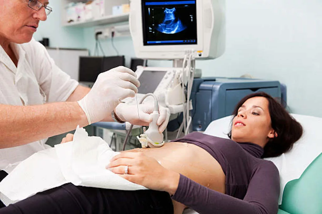
Imaging Studies:
Imaging techniques help your doctor assess liver stiffness and detect complications of hepatic fibrosis:
- Ultrasound is commonly the first imaging test used. It helps detect signs of liver damage, like fatty liver or an enlarged spleen. It can also be used to guide liver biopsies.
- Transient elastography is a non-invasive procedure that uses sound waves and creates an image of your liver’s stiffness. It is used as an alternative to a liver biopsy.
- CT and MRI scans both provide more detailed images of your liver’s structure.6Lee, T. H. (2024, January 4). Hepatic Fibrosis. MSD Manual Professional Edition; MSD Manuals. https://www.msdmanuals.com/professional/hepatic-and-biliary-disorders/fibrosis-and-cirrhosis/hepatic-fibrosis#Diagnosis_v899421
Treatment for Hepatic Fibrosis
There’s no cure for hepatic fibrosis. Treatment can, however, slow down the scarring. Doctors start by targeting whatever’s causing the damage. They do this by:
- Giving you antiviral medication if you have chronic hepatitis B or C
- Asking you to stop drinking alcohol if it’s alcohol-related liver disease
- Prescribing meds to clear out excess iron in hemochromatosis or copper in Wilson’s disease
- Stopping any meds or supplements that might be damaging your liver
- Unblocking your bile ducts if they’re obstructed
- Helping you lose weight and manage blood sugar and cholesterol if the cause is non-alcoholic fatty liver disease
Medications:
- Antifibrinolytic drugs like obeticholic acid and selonsertib are being studied to slow down scarring in hepatic fibrosis.
- In some people with non-alcoholic steatohepatitis, vitamin E has been shown to reduce liver inflammation and fibrosis.
- Pioglitazone is an antidiabetic drug that may help, especially if you also have insulin resistance.7van der Veen JN, Lingrell S, Gao X, Quiroga AD, Takawale A, Armstrong EA, Yager JY, Kassiri Z, Lehner R, Vance DE, Jacobs RL. Pioglitazone attenuates hepatic inflammation and fibrosis in phosphatidylethanolamine N-methyltransferase-deficient mice. Am J Physiol Gastrointest Liver Physiol. 2016 Apr 1;310(7):G526-38. doi: 10.1152/ajpgi.00243.2015. Epub 2016 Jan 21. PMID: 26797396.
Lifestyle Changes:
Regardless of what caused your liver to fibrose, you will need to make healthy changes to your life in order to treat hepatic fibrosis:
- Go for whole foods, more vegetables, less sugar, and healthy fats like olive oil.
- Regular activity, even just walking daily, can help decrease fat in your liver and prevent further hepatic fibrosis.
- Always check medications with your doctor, and avoid unnecessary herbal supplements, as these can burden your liver more.
Monitoring:
Hepatic fibrosis can change over time, and your care plan needs to adjust with it. This is why you’ll need ongoing monitoring and regular follow-ups. Blood tests, FibroScan, and MRI elastography will be done to see if fibrosis is improving or worsening.
Transplant:
If things go too far and your liver stops working properly, a transplant might be your only option. This usually happens when hepatic fibrosis has progressed into cirrhosis or liver failure.8Zhang, C.-Y., Liu, S., & Yang, M. (2023). Treatment of liver fibrosis: Past, current, and future. World Journal of Hepatology, 15(6), 755–774. https://doi.org/10.4254/wjh.v15.i6.755
Hepatic Steatosis Vs Hepatic Fibrosis
Fatty liver, or hepatic steatosis, happens when fat builds up inside your liver cells. If this continues over time, it can trigger inflammation and scarring, which is known as hepatic fibrosis. Without treatment, hepatic fibrosis worsens and turns into liver failure and cirrhosis.
Conclusion
Hepatic fibrosis means scarring of your liver from ongoing damage. It starts with liver injury from viral hepatitis, alcohol, fatty liver, and genetic or metabolic diseases. Without treatment, inflammatory processes speed up the scarring, and fibrosis progresses to advanced stages. There is no cure for hepatic fibrosis, but scarring can be slowed down by treating the specific cause and making lifestyle changes. Advanced hepatic fibrosis requires a liver transplant for treatment.
Refrences
- 1Kim, H. Y., Yu, J. H., Chon, Y. E., Kim, S. U., Kim, M. N., Han, J. W., Lee, H. A., Jin, Y.-J., An, J., Choi, M., & Jun, D. W. (2024). Prevalence of clinically significant liver fibrosis in the general population: A systematic review and meta-analysis. Clinical and Molecular Hepatology, 30(Suppl), S199–S213. https://doi.org/10.3350/cmh.2024.0351
- 2Hernandez-Gea, V., & Friedman, S. L. (2011). Pathogenesis of Liver Fibrosis. Annual Review of Pathology Mechanisms of Disease, 6(1), 425–456. https://doi.org/10.1146/annurev-pathol-011110-130246
- 3Huber, Y., Schulz, A., Schmidtmann, I., Beutel, M., Pfeiffer, N., Münzel, T., Galle, P. R., Wild, P. S., Lackner, K. J., & Schattenberg, J. M. (2022). Prevalence and Risk Factors of Advanced Liver Fibrosis in a Population‐Based Study in Germany. Hepatology Communications, 6(6), 1457–1466. https://doi.org/10.1002/hep4.1899
- 4Weerakkody, Y., Bell, D., & Saber, M. (2017). METAVIR score. Radiopaedia.org. https://doi.org/10.53347/rid-51855
- 5Kaumudi Somnay, Priyanka Wadgaonkar, Sridhar, N., Prarath Roshni, Rao, N., & Raj Wadgaonkar. (2024). Liver Fibrosis Leading to Cirrhosis: Basic Mechanisms and Clinical Perspectives. Biomedicines, 12(10), 2229–2229. https://doi.org/10.3390/biomedicines12102229
- 6Lee, T. H. (2024, January 4). Hepatic Fibrosis. MSD Manual Professional Edition; MSD Manuals. https://www.msdmanuals.com/professional/hepatic-and-biliary-disorders/fibrosis-and-cirrhosis/hepatic-fibrosis#Diagnosis_v899421
- 7van der Veen JN, Lingrell S, Gao X, Quiroga AD, Takawale A, Armstrong EA, Yager JY, Kassiri Z, Lehner R, Vance DE, Jacobs RL. Pioglitazone attenuates hepatic inflammation and fibrosis in phosphatidylethanolamine N-methyltransferase-deficient mice. Am J Physiol Gastrointest Liver Physiol. 2016 Apr 1;310(7):G526-38. doi: 10.1152/ajpgi.00243.2015. Epub 2016 Jan 21. PMID: 26797396.
- 8Zhang, C.-Y., Liu, S., & Yang, M. (2023). Treatment of liver fibrosis: Past, current, and future. World Journal of Hepatology, 15(6), 755–774. https://doi.org/10.4254/wjh.v15.i6.755
