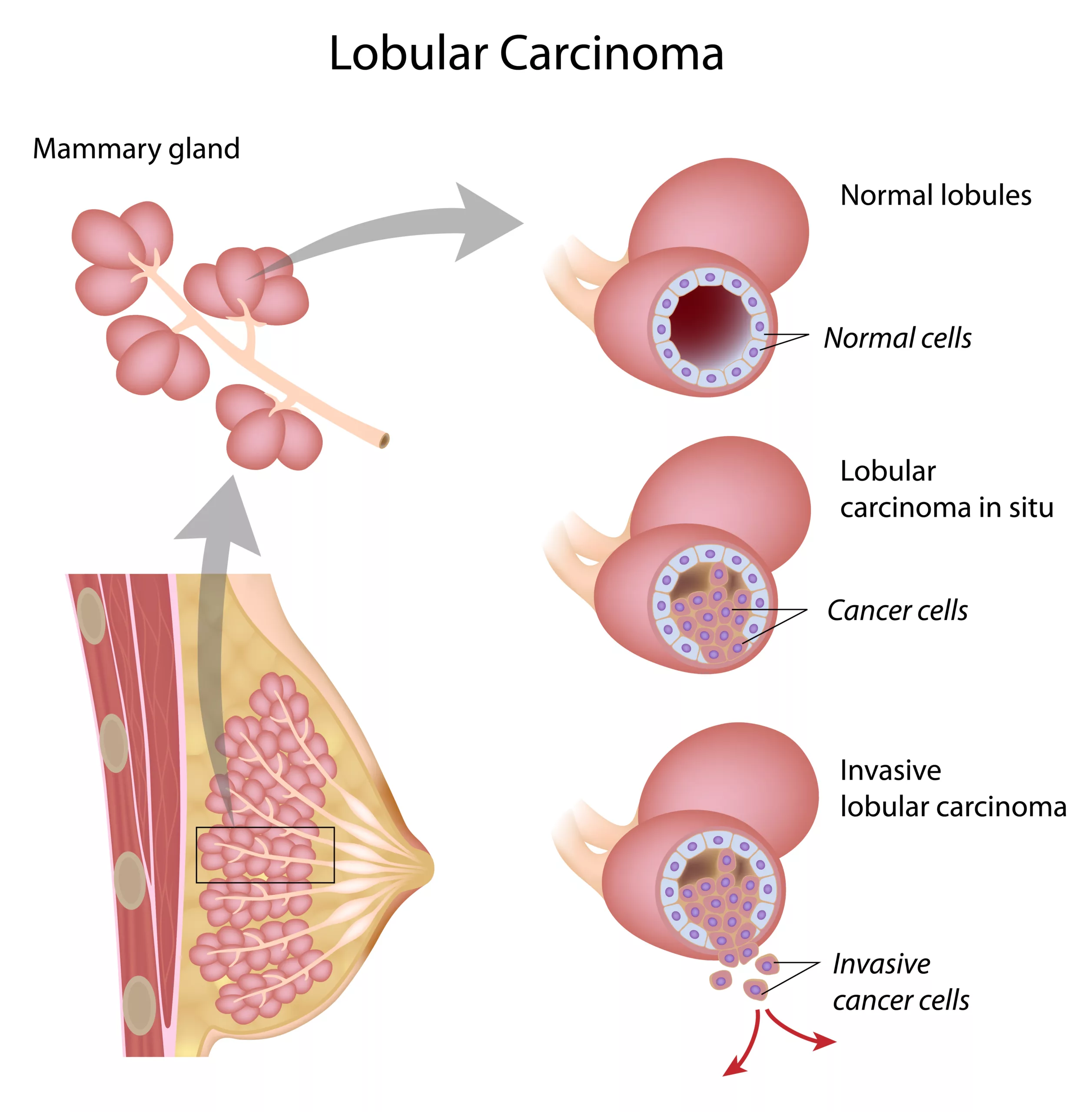
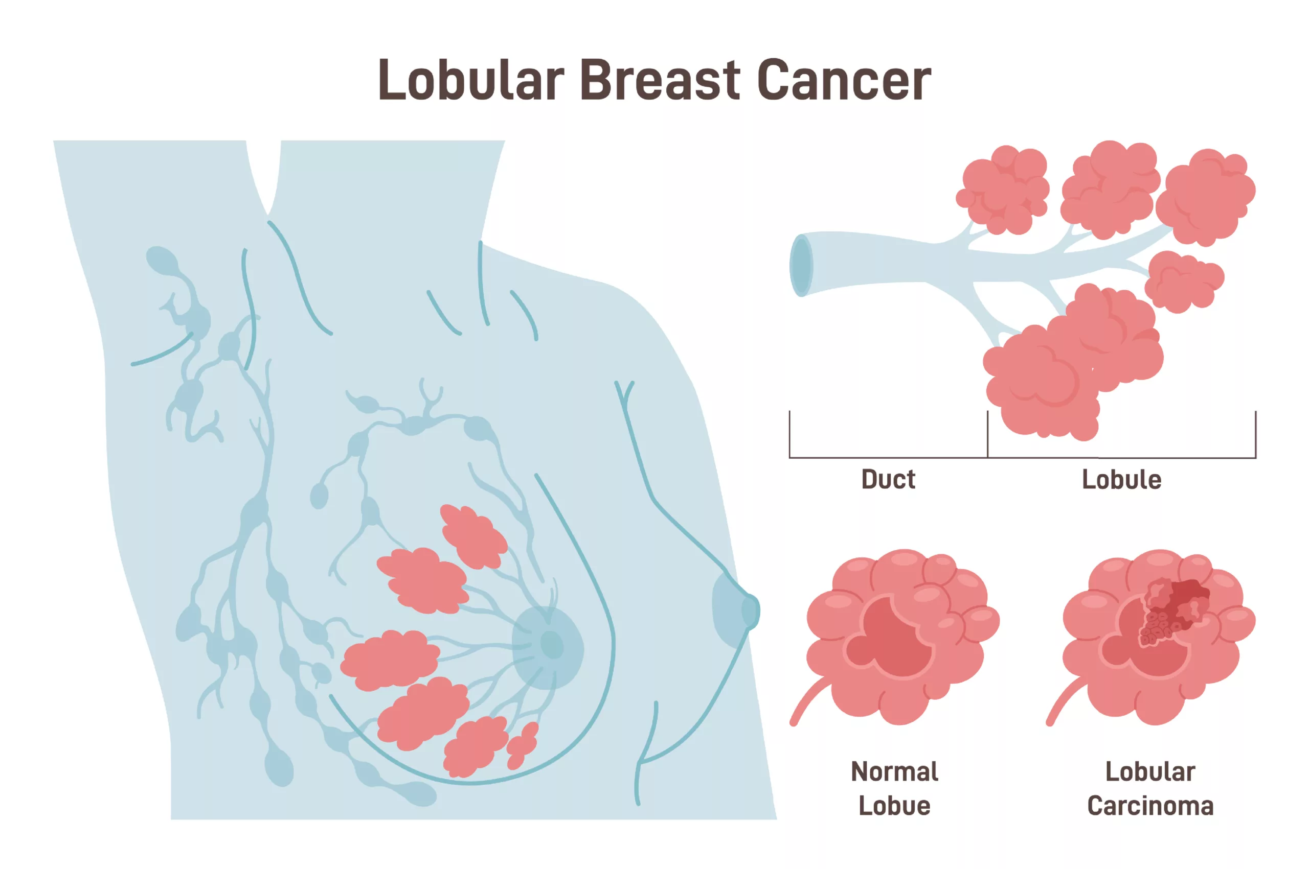
Invasive Lobular Carcinoma (ILC) is a type of breast cancer that starts in the lobules, which are the glands responsible for milk production and spreads to nearby breast tissue. It’s the second most common form of invasive breast cancer after invasive ductal carcinoma, making up 5% to 15% of all cases.1Limaiem F, Khan M, Lotfollahzadeh S. Lobular Breast Carcinoma. [Updated 2023 Jun 3]. In: StatPearls [Internet]. Treasure Island (FL): StatPearls Publishing; 2024 Jan-. Available from: https://www.ncbi.nlm.nih.gov/books/NBK554578/
Breast cancer can develop without obvious signs, often discovered during regular check-ups. Some may notice a lump, changes in breast appearance or feel, nipple fluid, or breast pain. Doctors use physical examinations, mammograms, and tissue samples to diagnose ILC. Detecting it earlier improves survival chances.
What is Invasive Lobular Carcinoma?
Invasive breast cancers are those that have spread from where they started and invaded nearby breast tissue. The most common ones are invasive ductal carcinoma and invasive lobular carcinoma.
Invasive Lobular Carcinoma (ILC) is a kind of breast cancer that specifically begins in the lobules, the milk-producing glands of the breast. Estrogen and progesterone exposure link to ILC. So, its risk increases with large amounts of estrogen and progesterone in the blood, for example, in cases of hormone replacement therapy, menarche, and menopause. Drinking alcohol is another important risk factor for ILC.

Invasive Lobular Carcinoma – Causes
Genetic mutations in the breast’s milk-producing glands (mammary glands) lead to invasive lobular carcinoma. These mutations cause the lobular glands to divide uncontrollably, forming a tumor.
Unlike some other breast cancers, lobular carcinoma doesn’t usually form a solid lump. Instead, the cancer cells spread out in a specific way, making the affected area feel thicker and fuller. It might not feel like a typical lump you can detect easily.
Invasive Lobular Carcinoma – Symptoms
One startling fact about ILC is that it often shows no symptoms, making it challenging to detect.2McCart Reed, A. E., Kutasovic, J. R., Lakhani, S. R., & Simpson, P. T. (2015). Invasive lobular carcinoma of the breast: morphology, biomarkers and ‘omics. Breast cancer research: BCR, 17(1), 12. https://doi.org/10.1186/s13058-015-0519-x Many cases are only discovered when a doctor identifies suspicious areas on a screening mammogram. However, in some instances, there may be noticeable signs. ILC is less likely to form a hard lump, unlike other breast cancers. Instead, its cells grow in straight lines, resembling a sheet rather than a lump.
Additional Signs & Symptoms
Here are some unusual changes in the breast that could be a signal of invasive lobular carcinoma:
- Swelling: This can affect all or a portion of the breast.
- Skin Changes: The skin around your nipples or breasts can be red, scaly, or thick, and it can be irritating and dimpling (like an orange peel).
- Nipple or Breast Tenderness: Inexplicable discomfort in the nipple or breast.
- Nipple Discharge: It may be an indication if you have discharge from your nipples that aren’t from breast milk.
- Lump or Swelling in the Underarm: A lump or swelling in the underarm area may also be a red flag.
Diagnosing Invasive Lobular Carcinoma
Tests and techniques used to diagnose invasive lobular cancer include:
Clinical Examination:
A healthcare provider’s physical examination of the breast may reveal suspicious lumps or changes in breast tissue.
Imaging Studies:
Mammography
A mammogram produces x-ray images of the breast. Invasive lobular cancer is less likely to be detected by a mammogram than other types of breast cancer. However, mammography is a good screening test.3Reeves RA, Kaufman T. Mammography. [Updated 2023 Jul 24]. In: StatPearls [Internet]. Treasure Island (FL): StatPearls Publishing; 2024 Jan-. Available from: https://www.ncbi.nlm.nih.gov/books/NBK559310/
The mammographic features of invasive lobular carcinoma may include:
- The presence of a mass (seen in up to 65% of cases) with irregular margins is often speculated but occasionally circumscribed.4Krecke KN, Gisvold JJ. Invasive lobular carcinoma of the breast: mammographic findings and extent of disease at diagnosis in 184 patients. AJR Am J Roentgenol. 1993 Nov;161(5):957-60
- Architectural distortion and asymmetric focal density.
- Microcalcifications are less common.
- In some instances, mammograms may show normal or benign findings.
Ultrasound
It uses ultrasonic sound waves to create images of the breast. The doctor performs an ultrasound because it is usually one of the first investigations when a lump or mass is suspected in the breast.
Ultrasound findings in invasive lobular carcinoma (ILC) vary, with common presentations including hypoechoic masses with irregular margins and posterior acoustic shadowing and shadowing without an apparent mass. Well-circumscribed mass lesions are observed less frequently, while approximately 10% of cases may be ultrasonographically invisible.5DiCostanzo D, Rosen PP, Gareen I, Franklin S, Lesser M. Prognosis in infiltrating lobular carcinoma. An analysis of “classical” and variant tumors. Am J Surg Pathol. 1990 Jan;14(1):12-23.
Magnetic Resonance Imaging (MRI)
A breast MRI will help evaluate the concern when mammogram and ultrasound results are inconclusive. It can also help find the extent of breast cancer.
Biopsy
It is the removal of tissue for examination. If an abnormality in MRI is detected, your doctor may recommend a biopsy procedure to obtain a sample of breast tissue for testing.
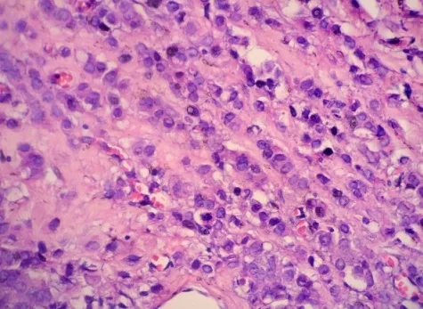
Invasive lobular carcinoma (ILC) on biopsy typically appears as irregular clusters of tumor cells infiltrating the surrounding breast tissue. Under the microscope, these tumor cells often have a signet ring appearance, meaning the cells contain large vacuoles or clear spaces. Additionally, the tumor cells may lack cohesion, forming single-file linear arrangements known as the Indian file pattern. These features, along with the presence of invasive growth into the surrounding tissue, help pathologists diagnose invasive lobular carcinoma on biopsy.6Abdelilah, Belhachmi & Mohamed, Ouazni & Yamoul, Rajae & Elkhiyat, Iman & Bouzidi, A & Alkandry, Sifeddine & Abdelkader, Eheirchou. (2014). Acute cholecystitis as a rare presentation of metastatic breast carcinoma of the Gallbladder: A case report and review of the literature. The Pan African Medical Journal. 17. 216. 10.11604/pamj.2014.17.216.3911.

Histologic Variants of Invasive Lobular Carcinoma
Invasive lobular carcinoma (ILC) is primarily classified into two main types: classic ILC and pleomorphic ILC. Classic ILC typically presents with uniform cells arranged in single-file strands or Indian file patterns, while pleomorphic ILC exhibits more cellular variability and nuclear pleomorphism. Some other less common types include:
- Solid
- Alveolar
- Tubulo-lobular7McCart Reed AE, Kalinowski L, Simpson PT, Lakhani SR. Invasive lobular carcinoma of the breast: the increasing importance of this special subtype. Breast Cancer Res. 2021 Jan 7;23(1):6. doi: 10.1186/s13058-020-01384-6. PMID: 33413533; PMCID: PMC7792208.
Key Factors in Diagnosis of Invasive Lobular Carcinoma
The key factors involved in diagnosing invasive lobular carcinoma are stated below. These play a major role in the process of diagnosis.
- Size of the Cancer: Determining the size of breast cancer.8Danzinger, S., Hielscher, N., Izsó, M., Metzler, J., Trinkl, C., Pfeifer, C., Tendl-Schulz, K., & Singer, C. F. (2021). Invasive lobular carcinoma: clinicopathological features and subtypes. The Journal of International Medical Research, 49(6), 3000605211017039. https://doi.org/10.1177/03000605211017039
- Nottingham Grade: Assessing the grade of the cancer based on cellular appearance and behavior.
- Tumor Characteristics: Examining tumor necrosis, tumor margins, and lymphovascular invasion.
- Lymph Node Status: Identifying the involvement of lymph nodes, which indicates cancer spread.
- Hormone Receptor Status: Determining whether the cancer has hormone receptors for estrogen or progesterone.
- HER2 Status: Checking for HER2 receptors. HER2, or human epidermal growth factor receptor 2, is a protein that, when overproduced, can cause cancer cells to grow more quickly. Knowing if the cancer is HER2-positive is crucial, as specific treatments, such as trastuzumab (Herceptin), can target and inhibit HER2, slowing down or stopping the cancer’s growth.
- Rate of Cell Growth (Ki-67 Levels): Measuring the cell growth rate to assess how quickly the cancer is likely to grow.
Invasive Lobular Carcinoma Staging
The staging of ILC are:
- Stage 0: Cancer is only found in the breast tissue ducts (ductal carcinoma in situ, or DCIS) and has not spread to nearby tissues.
- Stage 1: The tumor is 20mm or smaller and has not spread to lymph nodes, or has only a small spread to nearby lymph nodes (smaller than 2mm).
- Stage 2: The tumor is between 20mm and 50mm. It may or may not have spread to nearby lymph nodes.
- Stage 3: The tumor is larger than 50mm and spreads to nearby areas, such as lymph nodes near the breast.
- Stage 4: Cancer has spread (metastasized) to other body parts like lungs, liver and bones.
Invasive Lobular Carcinoma – Treatment Options
ILC treatment is an interesting topic among physicians. Treatment options depend on the cancer’s stage, and sometimes surgery is done first, sometimes after other treatments.
Surgery, hormonal therapy, radiation, and chemotherapy are all part of the treatment plan, depending on the case.9Mamtani, A., & King, T. A. (2018). Lobular Breast Cancer: Different Disease, Different Algorithms? Surgical oncology clinics of North America, 27(1), 81–94. https://doi.org/10.1016/j.soc.2017.07.005
Surgery – Mastectomy or Lumpectomy
This is often preferred, sometimes with the removal of the opposite breast preventively.10Klumpp, L. C., Shah, R., Syed, N., Fonseca, G., & Jordan, J. (2020). Invasive Lobular Breast Carcinoma Can Be a Challenging Diagnosis Without the Use of Tumor Markers. Cureus, 12(5), e8376. https://doi.org/10.7759/cureus.8376 If tests show the cancer hasn’t spread much, less invasive treatment may be possible.
Other Treatment Options
There are various other options than surgery. Many times, surgery is done in collaboration with the following:
- Surgery & Radiation help control the cancer’s spread in the body.
- Hormone Therapy is often used since many cases of this cancer are hormone-sensitive.
- Chemotherapy isn’t as effective because this cancer tends to grow slowly.
Age, lymph node status, tumor size, and other factors can affect the chances of the cancer coming back or survival, not just the type of cancer it is.11Dessauvagie, B., Thomas, A., Thomas, C., Robinson, C., Combrink, M., Budhavaram, V., Kunjuraman, B., Meehan, K., Sterrett, G., & Harvey, J. (2019). Invasive lobular carcinoma of the breast: assessment of proliferative activity using automated Ki-67 immunostaining. Pathology, 51(7), 681–687. https://doi.org/10.1016/j.pathol.2019.08.004
Invasive Lobular Carcinoma Recurrence
Although invasive lobular carcinoma (ILC) of the breast is associated with a significant risk of late recurrence, some patients recur within 5 years of being diagnosed.12Rothschild, H. T., Clelland, E. N., Mujir, F., Record, H., Wong, J., Esserman, L. J., Alvarado, M., Ewing, C., & Mukhtar, R. A. (2023). Predictors of Early Versus Late Recurrence in Invasive Lobular Carcinoma of the Breast: Impact of Local and Systemic Therapy. Annals of Surgical Oncology, 30(10), 5999–6006. https://doi.org/10.1245/s10434-023-13881-x Identifying characteristics linked with early/late recurrence may aid in therapy and diagnostic procedures.
Prognosis
When invasive lobular carcinoma (ILC) is detected early, it is often highly treatable and may even be curable. The prognosis remains favorable in the later stages, especially if the cancer has not metastasized (spread to other parts of the body). However, prognosis becomes more guarded if the cancer has metastasized, as treatment then focuses on managing symptoms and slowing the spread of the disease.13Ohio State University Comprehensive Cancer Center. (n.d.). Invasive lobular carcinoma prognosis and treatment. Retrieved from https://cancer.osu.edu/for-patients-and-caregivers/learn-about-cancers-and-treatments/cancers-conditions-and-treatment/cancer-types/breast-cancer/types-of-breast-cancer/invasive-lobular-carcinoma
Invasive Ductal Carcinoma vs. Invasive Lobular Carcinoma
IDC is the most frequent form of breast cancer. Invasive (or infiltrating) ductal carcinomas account for approximately eight out of ten invasive breast cancers.
IDC begins in the cells that border the milk ducts in the breast. The malignancy then penetrates the duct’s wall and spreads to the surrounding breast tissues. At this point, it may be able to spread (metastasize) to other regions of the body via the lymphatic and circulation.
On the other hand, Invasive lobular carcinoma (ILC) accounts for approximately one in every ten cases of invasive breast cancer. ILC begins in the breast glands that produce milk (lobules).14Barroso-Sousa R, Metzger-Filho O. Differences between invasive lobular and invasive ductal carcinoma of the breast: results and therapeutic implications. Ther Adv Med Oncol. 2016 Jul;8(4):261-6. doi: 10.1177/1758834016644156. Epub 2016 Apr 25. PMID: 27482285; PMCID: PMC4952020. It has the ability to expand (metastasize) to other regions of the body, as does IDC. Invasive lobular carcinoma may be more difficult to identify by physical examination and imaging, such as mammography, than invasive ductal carcinoma. And, unlike other types of invasive carcinoma, it is more likely to afflict both breasts. Approximately one in every five women with ILC may have cancer in both breasts at the time of diagnosis.
Conclusion
To wrap it up, invasive lobular carcinoma (ILC) is a unique subtype of breast cancer characterized by its distinct growth pattern and presentation. Early detection plays a crucial role in improving prognosis, making awareness of symptoms and risk factors essential for timely diagnosis. While ILC can be more challenging to identify than other breast cancer types, advancements in imaging and treatment options provide hope for effective management.
Refrences
- 1Limaiem F, Khan M, Lotfollahzadeh S. Lobular Breast Carcinoma. [Updated 2023 Jun 3]. In: StatPearls [Internet]. Treasure Island (FL): StatPearls Publishing; 2024 Jan-. Available from: https://www.ncbi.nlm.nih.gov/books/NBK554578/
- 2McCart Reed, A. E., Kutasovic, J. R., Lakhani, S. R., & Simpson, P. T. (2015). Invasive lobular carcinoma of the breast: morphology, biomarkers and ‘omics. Breast cancer research: BCR, 17(1), 12. https://doi.org/10.1186/s13058-015-0519-x
- 3Reeves RA, Kaufman T. Mammography. [Updated 2023 Jul 24]. In: StatPearls [Internet]. Treasure Island (FL): StatPearls Publishing; 2024 Jan-. Available from: https://www.ncbi.nlm.nih.gov/books/NBK559310/
- 4Krecke KN, Gisvold JJ. Invasive lobular carcinoma of the breast: mammographic findings and extent of disease at diagnosis in 184 patients. AJR Am J Roentgenol. 1993 Nov;161(5):957-60
- 5DiCostanzo D, Rosen PP, Gareen I, Franklin S, Lesser M. Prognosis in infiltrating lobular carcinoma. An analysis of “classical” and variant tumors. Am J Surg Pathol. 1990 Jan;14(1):12-23.
- 6Abdelilah, Belhachmi & Mohamed, Ouazni & Yamoul, Rajae & Elkhiyat, Iman & Bouzidi, A & Alkandry, Sifeddine & Abdelkader, Eheirchou. (2014). Acute cholecystitis as a rare presentation of metastatic breast carcinoma of the Gallbladder: A case report and review of the literature. The Pan African Medical Journal. 17. 216. 10.11604/pamj.2014.17.216.3911.
- 7McCart Reed AE, Kalinowski L, Simpson PT, Lakhani SR. Invasive lobular carcinoma of the breast: the increasing importance of this special subtype. Breast Cancer Res. 2021 Jan 7;23(1):6. doi: 10.1186/s13058-020-01384-6. PMID: 33413533; PMCID: PMC7792208.
- 8Danzinger, S., Hielscher, N., Izsó, M., Metzler, J., Trinkl, C., Pfeifer, C., Tendl-Schulz, K., & Singer, C. F. (2021). Invasive lobular carcinoma: clinicopathological features and subtypes. The Journal of International Medical Research, 49(6), 3000605211017039. https://doi.org/10.1177/03000605211017039
- 9Mamtani, A., & King, T. A. (2018). Lobular Breast Cancer: Different Disease, Different Algorithms? Surgical oncology clinics of North America, 27(1), 81–94. https://doi.org/10.1016/j.soc.2017.07.005
- 10Klumpp, L. C., Shah, R., Syed, N., Fonseca, G., & Jordan, J. (2020). Invasive Lobular Breast Carcinoma Can Be a Challenging Diagnosis Without the Use of Tumor Markers. Cureus, 12(5), e8376. https://doi.org/10.7759/cureus.8376
- 11Dessauvagie, B., Thomas, A., Thomas, C., Robinson, C., Combrink, M., Budhavaram, V., Kunjuraman, B., Meehan, K., Sterrett, G., & Harvey, J. (2019). Invasive lobular carcinoma of the breast: assessment of proliferative activity using automated Ki-67 immunostaining. Pathology, 51(7), 681–687. https://doi.org/10.1016/j.pathol.2019.08.004
- 12Rothschild, H. T., Clelland, E. N., Mujir, F., Record, H., Wong, J., Esserman, L. J., Alvarado, M., Ewing, C., & Mukhtar, R. A. (2023). Predictors of Early Versus Late Recurrence in Invasive Lobular Carcinoma of the Breast: Impact of Local and Systemic Therapy. Annals of Surgical Oncology, 30(10), 5999–6006. https://doi.org/10.1245/s10434-023-13881-x
- 13Ohio State University Comprehensive Cancer Center. (n.d.). Invasive lobular carcinoma prognosis and treatment. Retrieved from https://cancer.osu.edu/for-patients-and-caregivers/learn-about-cancers-and-treatments/cancers-conditions-and-treatment/cancer-types/breast-cancer/types-of-breast-cancer/invasive-lobular-carcinoma
- 14Barroso-Sousa R, Metzger-Filho O. Differences between invasive lobular and invasive ductal carcinoma of the breast: results and therapeutic implications. Ther Adv Med Oncol. 2016 Jul;8(4):261-6. doi: 10.1177/1758834016644156. Epub 2016 Apr 25. PMID: 27482285; PMCID: PMC4952020.


