Lichen Planus (LP) is a chronic condition characterized by an inflammatory rash that can appear on the skin or inside the mouth. The lesions can cause itching, discomfort, and, in some cases, significant pain. The exact cause of lichen planus is unknown, but it is believed to be an autoimmune disorder where the body’s immune system mistakenly attacks its own tissues. According to pooled data, the global prevalence of LP is between 0.2% and 1%1Arnold, D. L., & Krishnamurthy, K. (2018). Lichen planus., while that of oral lichen planus was estimated to be 1.01%.2González‐Moles, M. Á., Warnakulasuriya, S., González‐Ruiz, I., González‐Ruiz, L., Ayén, Á., Lenouvel, D., … & Ramos‐García, P. (2021). Worldwide prevalence of oral lichen planus: A systematic review and meta‐analysis. Oral diseases, 27(4), 813-828.
Lichen planus usually develops in the 5th-6th decade of life, and women are more prone to developing this autoimmune disease. The skin of the limbs (arms and legs), genitals, nails, and the inside of the mouth are the most commonly affected sites. It is a self-resolving condition that generally goes away within 1 to 5 years without any treatment.
Lichen Planus Types
Various types of lichen planus have distinct features and require specific modes of treatment.
Cutaneous Lichen Planus:
The most common type of LP is the skin type, i.e., cutaneous lichen planus. The rash can affect different areas of the skin. However, the most commonly involved regions include:
- Wrists
- Lower back
- Ankles
It starts as lesions on the skin that spread to different areas. The rash usually goes away on its own (within a couple of years) and requires no treatment.
Mucosal (Oral) Lichen Planus:
Oral lichen planus (LP) is a common oral pathology known for its recurrence. Patches of LP are generally seen in individuals older than 40. Women are more prone to developing mouth LP than men.
Oral LP is further divided into six types:
- Reticular
- Papular
- Plaque-like
- Atrophic/Erosive
- Ulcerative
- Bullous
Erosive and atrophic types may cause burning pain while consuming spicy foods. The most common type of oral LP is reticular LP.
Lichen Planus Planopilaris:
Scalp LP is called lichen planopilaris (LPP). The auto-immune disease leads to the formation of reddish follicular papules on the scalp. LPP can lay the foundation for hair loss conditions like progressive scarring alopecia and frontal fibrosing alopecia. Therefore, it is important to consult a doctor.
Apart from the cutaneous, planopilaris, and oral types, you may also experience LP lesions on:
- Nails
- Genitals (vagina, vulva and penis)
Lichen Planus Subtypes (Based On Display Patterns)
Experts also classify lichen planus based on the differences in display patterns of the rash. Thus, the following subtypes are identified:
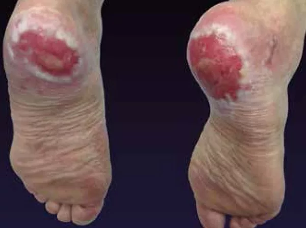
- Hypertrophic LP: Reddish brown to greyish papules form on the shins and ankles.
- Ulcerative LP: Painful erosive lesions form between the toes (or foot soles), making walking difficult.
- Bullous LP: Small-to-large-sized blisters appear on the legs, which can burst and scab.
Other types of LP include LP pemphigoids, LP pigmentosus, and inverse LP.
Lichen Planus Symptoms
The presentation differs based on the type and region involved. In the majority of the cases, lichen planus isn’t painful. When symptomatic, the most commonly encountered symptoms of LP are:
Purple Lesions
A purple-colored bump with a flattened top is a common manifestation of the cutaneous LP. You may notice these lesions on the wrists. However, they may also be present on the genitals. LP bumps start off as tiny, raised dots (measuring 0.4mm). These dots then grow in size (up to 1 cm in width) and may convert into sores. Over the next weeks, the lesions spread to different parts. Hence, maximal spreading is seen within 2-16 weeks of onset.
Itching
Many patients experience itching at the affected skin site. It is seen in cutaneous LP. Skin lesions are rarely painful but can be irritating and itchy.
Nail Cracking
Nail LP patients also report changes in color and splitting of nails. Cracks and permanent changes are seen in nail LP patients.3Minotto, R., Corte, L. D., Millán, T., & Furtado, B. C. (2023). Nail diseases. In Dermatology in Public Health Environments: A Comprehensive Textbook (pp. 1857-1875). Cham: Springer International Publishing.
White Lesions
In most oral LP cases, there is a network of white lines on the buccal mucosa that resembles a lattice. The lacy lesion pattern in the mouth is known as Wickham striae. These white lesions may be painful. In addition to pain, they may also cause burning sensations. Studies have identified multiple oral symptoms in LP patients.4Adamo, D., Calabria, E., Coppola, N., Lo Muzio, L., Giuliani, M., Bizzoca, M. E., … & SIPMO (Italian Society of Oral Pathology, Medicine) a. (2022). Psychological profile and unexpected pain in oral lichen planus: A case-control multicenter SIPMO study. Oral Diseases, 28(2), 398-414. Some people have white dots instead of lines.
Blisters are also seen in a few cases. Over time, these blisters burst and crust, forming scabs.
What Causes Lichen Planus?
Lichen planus is classified as an idiopathic disease whose exact cause is unknown. The pathogenesis is not fully understood; however, studies have revealed that it is an auto-immune disorder. Pathologists believe that various triggers can activate the T-cell-mediated response in the body, including:
- Stress: Psychological/emotional stress is a significant trigger for lichen planus. People living a stressful life are more prone to developing LP.5Gabor, A. I., Guguluș, D. L., Mocanu, M., Văță, D., Pătrașcu, A. I., & Solovăstru, L. G. (2023). Stress-trigger factor in Lichen planus. Bulletin of Integrative Psychiatry, (4).
- Viral infections: Infections like hepatitis C, HPV, EBV, etc. may be linked to LP occurrence.6Lucchese, A., Di Stasio, D., Romano, A., Fiori, F., De Felice, G. P., Lajolo, C., … & Petruzzi, M. (2022). Correlation between oral lichen planus and viral infections other than HCV: a systematic review. Journal of Clinical Medicine, 11(18), 5487.
- Drugs (cause drug-induced lichen planus): Anti-hypertensive drugs are known to cause immune-mediated inflammation of the skin in LP cases.7Boch, K., Langan, E. A., Kridin, K., Zillikens, D., Ludwig, R. J., & Bieber, K. (2021). Lichen planus. Frontiers in medicine, 8, 737813.
- Contact allergens: Metal allergens (such as in dental fillings and crowns) can mediate the development of oral lichen planus.8Yunizar, M. F., Watanabe, M., Liu, L., Minami, N., & Ichikawa, T. (2022). Metal Allergy Mediates the Development of Oral Lichen Planus via TSLP-TSLPR Signaling. Journal of Clinical Medicine, 11(3), 519.
- Genetic Predisposition: New research now indicates a genetic predisposition to the disorder. It is postulated that there is a familial type of LP that constitutes 10% of all LP cases.9Branisteanu, D. E., Pintilie, A., Andres, L. E., DUMITRIU, A., Oanta, A., Stoleriu, G., & Branisteanu, D. C. (2016). Ethiopatogenic hypotheses in lichen planus. The Medical-Surgical Journal, 120(4), 760-767. Women aged 30-60 are more likely to develop lichen planus than men.
Is Lichen Planus Contagious?
No, LP isn’t contagious. The disease does not transmit from infected to healthy individuals by skin or sexual contact.
How To Diagnose LP?
For lichen planus diagnosis, doctors correlate the patient’s history with the physical examination of the lesions. Diagnosticians have identified certain classic features of lichen planus, known as the 6 P’s:10Usatine, R. P., & Tinitigan, M. (2011). Diagnosis and treatment of lichen planus. American Family Physician, 84(1), 53-60.
- Purple: Skin lesions are violet/purple colored.
- Pruritic: Lesions are itchy.
- Polygonal: The lesions of the rash appear polygonal due to their multiple sharp angles.
- Planar: The top of the lesions is flat (planar).
- Papule: The disease appears in the form of bumps or papules.
- Plaques: Raised patches of rash are also seen in LP cases.
In cases of oral lichen planus, dentists can appreciate fine white lines that cover underlying shiny lesions of LP. These white lines are called Wickham striae. However, other manifestations can occur in the buccal mucosa. When present on the tongue, LP lesions can be mistaken for leukoplakia. Therefore, proper history and thorough clinical examination are a must.
Diagnostic Tests
Your healthcare provider may also run some tests based on your presenting condition. Some of the tests for LP include:
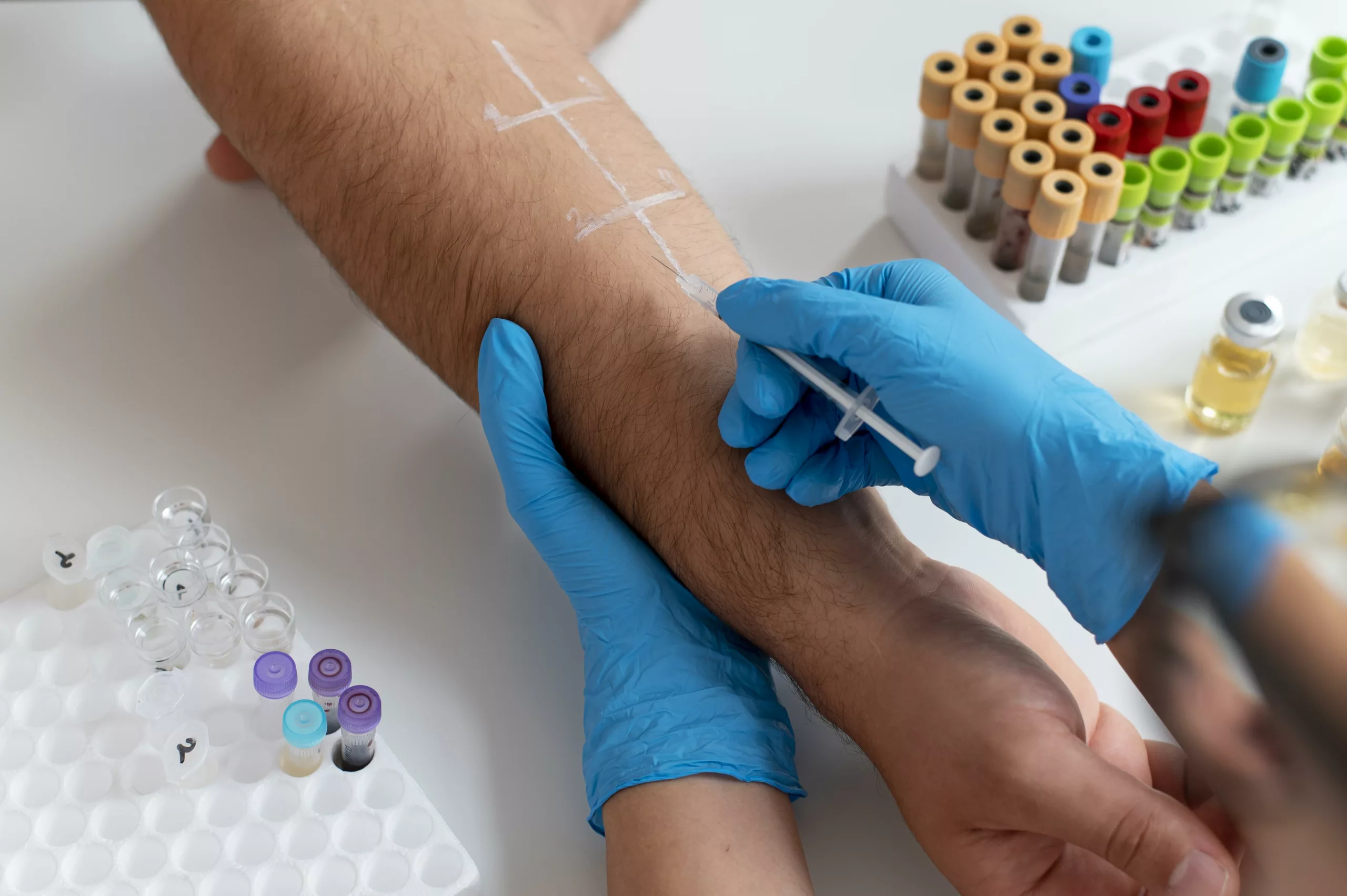
- Allergy Test: As allergens are known to trigger LP, your doctor may order an allergy test to determine the cause of LP flare-ups.
- Dental Patch test: A dental patch test is sometimes done to determine allergy to any metallic restoration in the mouth.
- Biopsy: In a biopsy, the doctor removes a small portion of skin or buccal mucosa (using a scalpel) and sends it for microscopic analysis. Histopathological analysis is done to rule out other pathologies.
Your doctor may also recommend Liver function tests (LFTs) and hepatitis C tests to rule out an underlying infection.
Lichen Planus Vs. Psoriasis
Psoriasis and Lichen planus are auto-immune disorders affecting the skin. The key differentiating feature between both is that psoriasis lesions are scaly while LP lesions are flat. Also, psoriasis does not affect the inside of your mouth.
Lichen Planus Vs. Lichen Sclerosus
Lichen planus and Lichen sclerosis share some similarities, but it is important to tell them apart. Both diseases are idiopathic and affect the mucus membranes of the body. The main target is the skin.
However, Lichen sclerosis is a chronic condition leading to gradual skin thinning. The itchy skin sores soon convert into scars. It predominantly affects the anogenital region (anus and genital) but does not typically involve the oral mucous membranes. It is commonly seen in women going through the menopause.
Lichen Planus Treatment
In the vast majority of cases, lichen planus resorbs without intervention. However, a complete resolution may take a few months to even years. Some patients notice gray-brown hyperpigmentation of skin after resolution.
The following strategies are good for managing symptoms of LP:
First-Line Treatment:
Corticosteroid Cream & Anti-Histamines
The first line of treatment is topical steroid therapy. Studies advocate using oral adhesive gel preparations to alleviate symptoms of oral lichen planus.11Lodi, G., Manfredi, M., Mercadante, V., Murphy, R., & Carrozzo, M. (2020). Interventions for treating oral lichen planus: corticosteroid therapies. Cochrane Database of Systematic Reviews, (2).The different types of topical steroids effective for lichen planus include clobetasol, dexamethasone, betamethasone, and triamcinolone (5-10mg/mL).12Yuan, P., Qiu, X., Ye, L., Hou, F., Liang, Y., Jiang, H., … & Jiang, L. (2022). Efficacy of topical administration for oral lichen planus: a network meta‐analysis. Oral diseases, 28(3), 670-681.
Most doctors also prescribe antihistamines to reduce itching and allergy symptoms.
Second-Line Treatment:
If no changes are seen with topical steroid therapy, doctors opt for second-line treatment. The following therapies are employed in this strategy:
DMARDs
Antibiotics like metronidazole and disease-modifying anti-rheumatic drugs (DMARDs), i.e., sulfasalazine or methotrexate, are advised. DMARDs are immunosuppressants that tweak down the immune system. Thus, there is relief of symptoms.
Retinoids
Vitamin A derivatives, such as retinoids, are widely known for their skin-healing properties. Retinoids are used in acne management and show some positive results in lichen planus treatment.
Calcineurin Inhibitors
Calcineurin is a protein responsible for inflammation in dermal conditions like eczema. Topical calcineurin inhibitor creams such as tacrolimus can halt the inflammatory process initiated by immune responses. Oral application of tacrolimus for oral lichen planus has short-term benefits in symptom management. This very therapy is safe and effective.13Su, Z., Hu, J., Cheng, B., & Tao, X. (2022). Efficacy and safety of topical Administration of Tacrolimus in Oral lichen planus: an updated systematic review and meta‐analysis of randomized controlled trials. Journal of Oral Pathology & Medicine, 51(1), 63-73.
Phototherapy (Light Therapy)
Ultraviolet light (UVA and UVB) has shown some therapeutic benefits in the management of lichen planus. UVB-type light therapy alleviates symptoms and prevents recurrence. Studies conclude that PUVB is ideal for treating female LP patients with lighter skin.14Dawood, M., Sizopoulou, C., Greenberger, S., Barzilai, A., & Pavlotsky, F. (2022). Narrowband ultraviolet B radiation for lichen planus: long-term follow-up of 192 patients. The Journal of Clinical and Aesthetic Dermatology, 15(4), 31. UVA is used to manage nail LP.
Third-Line Treatment:
The third line of treatment is necessary if 2nd line is unfruitful. It involves advising different classes of drugs. You might need to take antibiotics and biologics to halt disease progression. Some drugs belonging to the 3rd line include:
- Anti-malarial drugs
- Tetracyclines
- Terbinafine
- Adalimumab
Home Remedies For LP
At home, you can adopt certain practices to make your LP less worse:
- Maintain cleanliness: You can minimize complications of LP by maintaining good oral hygiene. In addition to good brushing, antibiotic mouthwash prevents bacterial colonization. Thus, both steps are beneficial for oral LP patients.
- Avoid scratching: You should avoid the urge to itch. You can get help from OTC anti-itch creams. Applying cool compresses can potentially reduce the irritation.
- Use aloe vera: Natural products like aloe vera are beneficial for various skin conditions. Thus, oral LP patients may also benefit from it. However, it is essential to consult your dermatologist before applying anything.15Kalaskar, A. R., Bhowate, R. R., Kalaskar, R. R., Walde, S. R., Ramteke, R. D., & Banode, P. P. (2020). Efficacy of herbal interventions in oral lichen planus: A systematic review. Contemporary clinical dentistry, 11(4), 311-319.
- Reduce mental/emotional stress: Stress is a major contributor to LP triggering. Therefore, you should adopt stress-relieving activities like meditation, yoga, etc. This can reduce the frequency of flare-ups.
Complications Of Lichen Planus
Lichen planus is known to increase the risk of squamous cell carcinoma. It was revealed in a study that patients suffering from genital lichen planus had a higher risk of developing valvular squamous cell carcinoma (VSCC).16Leis, M., Singh, A., Li, C., Ahluwalia, R., Fleming, P., & Lynde, C. W. (2022). Risk of vulvar squamous cell carcinoma in lichen sclerosus and lichen planus: a systematic review. Journal of Obstetrics and Gynaecology Canada, 44(2), 182-192. Similarly, there is a possibility of transformation of oral LP into malignant SCC.17Tenore, G., Mohsen, A., Rocchetti, F., Rossi, G., Cassoni, A., Battisti, A., … & Romeo, U. (2023). Risk of oral squamous cell carcinoma in one hundred patients with oral lichen planus: A follow-up study of Umberto I University Hospital of Rome. Cancers, 15(11), 3004.
Final Word
Lichen planus is an auto-immune disorder that causes an inflammatory rash on the skin (cutaneous LP) or the inside of the mouth (oral LP). Mostly, cutaneous LP patients report a purple, itchy rash and papules, while white striated lines are seen in oral LP. The exact causes of LP are unknown, but viral infections, stress, and contact allergens can trigger it. Usually, the disease is asymptomatic and self-resolving. However, complete resolution may take several months to even years. When symptomatic, steroid creams, phototherapy, and immunosuppressant drugs can help reduce symptoms and halt disease activity. Researchers have found an association between LP and cancer (squamous cell carcinoma). Thus, patients should report symptoms to a healthcare provider ASAP.
Refrences
- 1Arnold, D. L., & Krishnamurthy, K. (2018). Lichen planus.
- 2González‐Moles, M. Á., Warnakulasuriya, S., González‐Ruiz, I., González‐Ruiz, L., Ayén, Á., Lenouvel, D., … & Ramos‐García, P. (2021). Worldwide prevalence of oral lichen planus: A systematic review and meta‐analysis. Oral diseases, 27(4), 813-828.
- 3Minotto, R., Corte, L. D., Millán, T., & Furtado, B. C. (2023). Nail diseases. In Dermatology in Public Health Environments: A Comprehensive Textbook (pp. 1857-1875). Cham: Springer International Publishing.
- 4Adamo, D., Calabria, E., Coppola, N., Lo Muzio, L., Giuliani, M., Bizzoca, M. E., … & SIPMO (Italian Society of Oral Pathology, Medicine) a. (2022). Psychological profile and unexpected pain in oral lichen planus: A case-control multicenter SIPMO study. Oral Diseases, 28(2), 398-414.
- 5Gabor, A. I., Guguluș, D. L., Mocanu, M., Văță, D., Pătrașcu, A. I., & Solovăstru, L. G. (2023). Stress-trigger factor in Lichen planus. Bulletin of Integrative Psychiatry, (4).
- 6Lucchese, A., Di Stasio, D., Romano, A., Fiori, F., De Felice, G. P., Lajolo, C., … & Petruzzi, M. (2022). Correlation between oral lichen planus and viral infections other than HCV: a systematic review. Journal of Clinical Medicine, 11(18), 5487.
- 7Boch, K., Langan, E. A., Kridin, K., Zillikens, D., Ludwig, R. J., & Bieber, K. (2021). Lichen planus. Frontiers in medicine, 8, 737813.
- 8Yunizar, M. F., Watanabe, M., Liu, L., Minami, N., & Ichikawa, T. (2022). Metal Allergy Mediates the Development of Oral Lichen Planus via TSLP-TSLPR Signaling. Journal of Clinical Medicine, 11(3), 519.
- 9Branisteanu, D. E., Pintilie, A., Andres, L. E., DUMITRIU, A., Oanta, A., Stoleriu, G., & Branisteanu, D. C. (2016). Ethiopatogenic hypotheses in lichen planus. The Medical-Surgical Journal, 120(4), 760-767.
- 10Usatine, R. P., & Tinitigan, M. (2011). Diagnosis and treatment of lichen planus. American Family Physician, 84(1), 53-60.
- 11Lodi, G., Manfredi, M., Mercadante, V., Murphy, R., & Carrozzo, M. (2020). Interventions for treating oral lichen planus: corticosteroid therapies. Cochrane Database of Systematic Reviews, (2).
- 12Yuan, P., Qiu, X., Ye, L., Hou, F., Liang, Y., Jiang, H., … & Jiang, L. (2022). Efficacy of topical administration for oral lichen planus: a network meta‐analysis. Oral diseases, 28(3), 670-681.
- 13Su, Z., Hu, J., Cheng, B., & Tao, X. (2022). Efficacy and safety of topical Administration of Tacrolimus in Oral lichen planus: an updated systematic review and meta‐analysis of randomized controlled trials. Journal of Oral Pathology & Medicine, 51(1), 63-73.
- 14Dawood, M., Sizopoulou, C., Greenberger, S., Barzilai, A., & Pavlotsky, F. (2022). Narrowband ultraviolet B radiation for lichen planus: long-term follow-up of 192 patients. The Journal of Clinical and Aesthetic Dermatology, 15(4), 31.
- 15Kalaskar, A. R., Bhowate, R. R., Kalaskar, R. R., Walde, S. R., Ramteke, R. D., & Banode, P. P. (2020). Efficacy of herbal interventions in oral lichen planus: A systematic review. Contemporary clinical dentistry, 11(4), 311-319.
- 16Leis, M., Singh, A., Li, C., Ahluwalia, R., Fleming, P., & Lynde, C. W. (2022). Risk of vulvar squamous cell carcinoma in lichen sclerosus and lichen planus: a systematic review. Journal of Obstetrics and Gynaecology Canada, 44(2), 182-192.
- 17Tenore, G., Mohsen, A., Rocchetti, F., Rossi, G., Cassoni, A., Battisti, A., … & Romeo, U. (2023). Risk of oral squamous cell carcinoma in one hundred patients with oral lichen planus: A follow-up study of Umberto I University Hospital of Rome. Cancers, 15(11), 3004.
