Sinus tachycardia occurs when your heart beats more than 100 beats per minute (BPM) in normal adults or above the normal limits in children according to their age.
This rhythm is controlled by impulses coming from the sinoatrial node (SA). An increased number of impulses from the SA node causes sinus tachycardia. A healthy adult’s Normal heart rate is 60-100 beats per minute. This rate is children according to age; in newborn babies, the heart rate is 110-150 bpm. In children two years of age, it is 85-130 bpm; in children four years of age, the heart rate is 75-115 bpm; and in 6-year-old children, the heart rate is about 60-100 bpm.1Buttner, E. B. a. R. (2021). Sinus tachycardia. Life in the Fast Lane • LITFL. https://litfl.com/sinus-tachycardia-ecg-library/
Introduction
Your heart is the most significant organ in your body that drives your other organs by supplying blood and nutrition to them. Like a car engine that uses petrol or diesel to drive your car, your body uses blood to supply oxygen and nutrients to your body. The contraction of the heart pumps blood to different parts of the body. The number of contractions per minute is called the heart rate. Increased heart rate is known as sinus tachycardia. It can be physiological or pathological.
As we have discussed above, the normal heart rate in adults is 60-100 beats per minute, depending upon the fitness of the patient and the presence of comorbidities. However, tachycardia at rest is an alarming feature and should be treated as early as possible according to the cause. This blog will teach you about the causes, risk factors, and clinical approaches to treat the patient. 2Henning A, Krawiec C. Sinus Tachycardia. [Updated 2023 Mar 5]. In: StatPearls [Internet]. Treasure Island (FL): StatPearls Publishing; 2023 Jan-. Available from: https://www.ncbi.nlm.nih.gov/books/NBK553128/
What is the Sinoatrial Node?
The sinoatrial node is a spindle-shaped pacemaker in your heart that is located within the sulcus terminalis at the junction of the upper wall of the right atrium and superior vena cava. In addition, the spontaneous diastolic depolarization is due to the movement of calcium, potassium, and sodium across the cell.
The normal depolarization rate of this node is 60-90 breaths per minute. However, this node adjusts its rate according to the situation, which is modulated by autonomic tone, hypoxia (low level of oxygen), temperature, increase or decrease in blood pH, and hormonal imbalance3Gopinathannair, R. U. H. R. (2013, July 18). “Inappropriate” sinus tachycardia. Medscape. https://www.medscape.com/viewarticle/780588_2
Signs and symptoms of Sinus Tachycardia
Most of the time, patients with sinus tachycardia remain asymptomatic, but complaints of increased heartbeat and palpitations can occur. Therefore, the symptoms of sinus tachycardia depend on the cause and severity. The most common symptoms are:
- Chest pain
- Dyspnea (breathing difficulty)
- Decrease exercise intolerance
- Headache
- Fainting 4Sinus Tachycardia | Cardiology | Mercy Health. (n.d.). https://www.mercy.com/health-care-services/heart-vascular/conditions/sinus-tachycardia
Causes & Risk Factors of Sinus Tachycardia
There are two major causes of sinus tachycardia. It can be pathological or physiological.
Physiological Sinus Tachycardia
It is a normal response to your physical activity that increases your heart rate and returns to normal after taking rest. Physiological sinus tachycardia occurs due to exercise and stress. There is no pathology behind this.
Pathological Sinus Tachycardia
In pathological sinus tachycardia, you have an increased heart even at rest. It can be due to cardiac abnormality or some non-cardiac factors that indirectly affect your heart rate. Pathological sinus tachycardia is usually associated with heart diseases.
Cardiac-Associated Causes:
Cardiac etiologies that mainly affect your cardiac contractility are:
Supraventricular Tachycardia
Supraventricular tachycardia is an abnormal heart rhythm condition produced by ventricles and characterized by a narrow QRS complex on an electrocardiogram. Moreover, if the rhythm is regular, then the possible etiology can be:
- Atrial flutter
- Atrial tachycardia or sinus tachycardia
- Atrioventricular reentrant tachycardia (AVTR)
- AV node reentrant tachycardia (AVNRT)
Additionally, if the rhythm is irregular, then the etiologies are:
- Atrial fibrillation
- Atrial flutter with variable block
- Premature atrial contractions
Ventricular Tachycardia
Like supraventricular tachycardia, ventricular tachycardia originates from the ventricles, but a widened QRS complex on an electrocardiogram characterizes it. This complex is of two types: non-sustained QRS complex, which lasts for less than 30 seconds, and sustained, which lasts for more than 30 seconds. It is associated with hemodynamic instability.
Torsade De Pointes is clinically characterized by prolonged QT interval that can be congenital or acquired.5Sauer AJ, Newton-Cheh C. Clinical and genetic determinants of torsade de pointes risk. Circulation. 2012 Apr 3;125(13):1684-94. doi: 10.1161/CIRCULATIONAHA.111.080887. PMID: 22474311; PMCID: PMC3347483. Risk factors for torsade de Pointes are:
- Female gender
- Hypokalemia (low level of potassium)
- Hypocalcemia (low level of calcium)
- Medications that cause prolongation of QT interval
- Ischemia and structural heart diseases
Myocarditis
According to the word, Myo means muscle, cardia means heart, and itis indicates inflammation. Myocarditis is the inflammation of the heart myocytes that can be due to bacterial or viral infection. In addition, other causes of myocarditis are:
- Hypothermia (decreased body temperature)
- Autoimmune diseases
- Hypersensitive reactions
- Radiations
- Ischemia (decreased blood supply to heart muscles)
Cardiac Tamponade
Cardiac tamponade is an emergency condition in which the fluid is collected inside the pericardial space. This fluid can be transudative exudative or pericardial effusion. Moreover, the effusion is caused by acute chest trauma, aortic dissection, radiation therapy, thoracic surgery, bacterial or viral infection, autoimmune disease, and intrathoracic neoplasm. However, Cardiac tamponade is characterized by multiple symptoms:
- Hypotension (decreased blood pressure)
- Increase jugular venous pressure (engorged neck veins)
- Muffled heart sounds
- Bowing of interventricular septum during inspiration
- Pulsus paradoxus (blood pressure falls by 10 mm Hg during inspiration)
Acute Coronary Syndrome
Acute coronary syndrome is the most commonly presenting disease in a clinical setup every day. It can present in different forms:
- Stable angina (It is a condition in which you feel pain during some activity, but this pain settles down at rest.)
- Unstable angina (In this condition, you feel pain both during activity and at rest.)
- Myocardial infarction (It can be of two types: Non-ST segment elevation MI and ST-segment elevation MI.)
Risk factors6Kumar A, Cannon CP. Acute coronary syndromes: diagnosis and management, part I. Mayo Clin Proc. 2009 Oct;84(10):917-38. doi: 10.1016/S0025-6196(11)60509-0. PMID: 19797781; PMCID: PMC2755812. for acute coronary artery syndrome are :
- Obesity
- Hypertension
- Hypercholesterolemia
- Old age
- Diabetes Mellitus
- Male
- Tobacco
- Smoking
- Alcohol
- Positive family history of heart diseases
Non-Cardiac Causes:
Non-cardiac causes include your other systems and organs. These are:
Respiratory Causes
Pulmonary embolism is one of the life-threatening pathologies of respiratory vasculature. It can be acute, chronic, or both. Acute pulmonary embolism is the most common and is clinically more significant, with a higher rate of mortality and morbidity. Moreover, It is caused by the embolism coming from deep venous thrombosis (DVT). Other causes of embolism are:
- Fat emboli (due to orthopedic injury)
- Amniotic fluid emboli (during pregnancy)
- Air emboli (due to instrumentation)
Hypoxia
It is a condition in which the demand-to-supply ratio of oxygen is reduced, resulting in increased heart workload.7Corrigan D, Prucnal C, Kabrhel C. Pulmonary embolism: the diagnosis, risk-stratification, treatment and disposition of emergency department patients. Clin Exp Emerg Med. 2016 Sep 30;3(3):117-125. doi: 10.15441/ceem.16.146. PMID: 27752629; PMCID: PMC5065342.
Infectious Diseases
Sepsis is the most common inflammatory illness secondary to a bacterial infection that causes organ dysfunction. Additionally, it is the main cause of mortality worldwide. In the early stages of sepsis, systemic vascular resistance is decreased due to a large inflammatory burden, and vessels dilate. However, the patient’s body compensates for this by increasing heart rate and clinically presents with hypertension during this inflammatory period. Sepsis can cause sinus tachycardia.
Electrolyte & Minerals Abnormality
Electrolytes are the most important fuel of our body. Electrolyte abnormality affects your heart rate depending upon the type of electrolyte.
Hypovolemia:
Dehydration is the main cause of volume depletion. Dehydration results when there is an imbalance between fluid input and output. Due to volume depletion, cardiac output and the heart increase as a compensatory response, leading to sinus tachycardia.
Hypoglycemia:
It is a clinical presentation in which your glucose level drops below 70 mg/d. It can be due to the following causes:
- Drug-induced
- Hormonal abnormality
- Excessive alcohol consumption
- Surgery induced (after bariatric surgery)
- Insulinomas
- Tumors of the pituitary and adrenal glands
Hypoglycemia increases the heart rate (sinus tachycardia and QT prolongation)
Hypocalcemia:
It is a clinical state in which the calcium level drops below 8.5mg/dl. Hypocalcemia results due to :
- Malabsorption
- Low exposure to sunlight
- Chronic kidney disease
- Rickets
- Osteomalacia
- Acute pancreatitis
- Liver failure
Hyperkalemia:
It is a condition in which the potassium level is greater than normal. Hemolysis is the most common cause of hyperkalemia.
Shock:
Shock is a lethal condition in which the tissue perfusion ratio is reduced. There are four types of shock which can cause sinus tachycardia:
- Distributive shock (due to decreased systemic vascular resistance, e.g., sepsis.
- Cardiogenic shock (due to ischemia)
- Hypovolemic which (due to intravascular volume distribution)
- Obstructive shock (due to respiratory pathology)
Hematologic Causes:
Anemia is a condition in which your hemoglobin drops below normal. It can be acute or chronic and is divided into microcytic (red blood cells are of smaller size), normocytic (normal size and shape of red blood cells), and megaloblastic ( larger size and abnormal shape of red blood cells). Additionally, anemia increases the heart rate to maintain cardiac output, leading to sinus tachycardia.
Hemorrhage is a clinical condition in which a significant amount of blood loss occurs due to vascular damage. It is classified into four classes:
- Class 1 (when the blood loss is less than 15 % of total blood)
- Class 2 (when the blood loss is 15-30% of total blood )
- Class 3 (when the blood loss is 30-40 % of the total blood)
- Class 4 (when the blood loss is greater than 40%)
Hemorrhage leads to hypovolemia, which in turn causes sinus tachycardia.
Endocrinological Causes
It is a condition with an abnormal level of circulating hormone. It involves the following disorders:
Hyperthyroidism:
It is the most commonly presenting illness in which the thyroid hormone level is significantly increased. However, Hyperthyroidism increases the activation of calcium-activated ATPase activity, resulting in sinus tachycardia.
Pheochromocytoma:
A neuroendocrine tumor originates from chromaffin cells in the adrenal glands. The clinical triad of pheochromocytoma is :
- Headache
- Sweating
- Tachycardia (Increased heart rate)
Pregnancy:
Pregnancy8Klein HH, Pich S. Physiologische Anderungen des Herz-Kreislauf-Systems in der Schwangerschaft [Cardiovascular changes during pregnancy]. Herz. 2003 May;28(3):173-4. German. doi: 10.1007/s00059-003-2455-2. PMID: 12756474. induces many physiological changes like:
- Increase in heart rate
- Increase cardiac output
- Increase vascular volume
Toxicology:
Multiple medications cause tachycardia if taken at higher doses. Some of these medications are the following:
- Albuterol
- Antihistamine
- Atropine
- Cocaine
- Nicotine
- Dopamine
- Dobutamine
- Potassium
- Theophylline
Tachycardia9Henning A, Krawiec C. Sinus Tachycardia. [Updated 2023 Mar 5]. In: StatPearls [Internet]. Treasure Island (FL): StatPearls Publishing; 2023 Jan-. Available from: https://www.ncbi.nlm.nih.gov/books/NBK553128/ can also result in withdrawal from a substance or medication. Withdrawal-induced tachycardia include:
- Alcohol withdrawal
- Calcium channel blocker
- Digoxin
- Organophosphate
- Opioids
- Sedatives
- Digitoxin
- Magnesium
Diagnosis of Sinus Tachycardia
A careful examination is required to diagnose the sinus tachycardia, whether it is due to a physiological response or due to underlying pathology. The diagnosis of sinus tachycardia is made by the following:
- History
- Physical examination
- Laboratory investigation
- Imaging
History:
Understanding a patient’s medical history is vital for the diagnostic process, providing essential insights into the condition’s nature and its underlying causes. Various factors must be considered and systematically evaluated when gathering a patient’s history. These factors include patient demographics, such as age and gender, and weight, which can often play a significant role in certain conditions. The patient’s history should also explore symptoms, including chest pain, palpitations, difficulty breathing, and headaches, which can offer crucial clues to the diagnosis. Additionally, assessing any history of drug abuse is essential, as substance use can contribute to various medical issues.
Beyond immediate symptoms, a comprehensive history encompasses past medical history, which may reveal relevant pre-existing conditions or chronic illnesses. Family medical history can unveil hereditary factors, increasing the risk of certain diseases. Occupational history sheds light on potential exposure to occupational hazards, while personal history, including smoking habits, alcohol consumption, and relationship issues, can all impact a patient’s overall health. By meticulously examining these historical elements and utilizing them as building blocks in the diagnostic process, healthcare providers can arrive at a more accurate diagnosis.
Physical Examination:
A detailed physical examination follows history. In physical examination, the following steps are involved:
- Inspection of the chest to check for abnormality
- Palpation of the chest to check for any swelling and tenderness
- Auscultation of the chest to check the heart sounds
- Pulse examination
- Blood pressure examination
- Signs of anemia and jaundice
- Chest auscultation
- Check for edema
- Pulse oximetry (A quick way to identify hypoxia)
Laboratory Investigations:
When there’s suspicion of illness, essential steps involve conducting laboratory investigations, which encompass:
- Complete blood count (CBC) with ESR to check for anemia and infection
- Liver function tests (LFTs)
- Renal parameters
- Cardiac markers
- Arterial blood gasses (ABGs)
- Lactic acid level (To determine tissue hypoperfusion)
- D-dimers
- Random blood sugar level
- HBA1C
- Toxicology screen
- Electrolyte level
Imaging:
Radiological investigations are required to confirm the disease or rule out the other causes. The radiological investigation is:
- Chest X-ray to identify heart and lung problems like heart failure and pneumothorax
- CT chest to confirm the presence of embolus
- Echocardiography to assess the function of the heart.
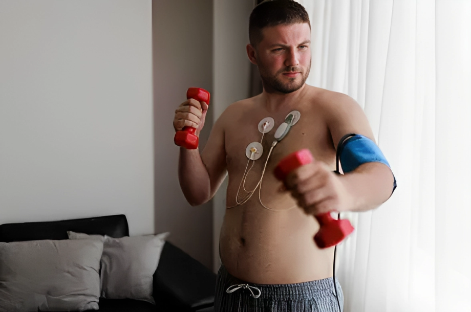
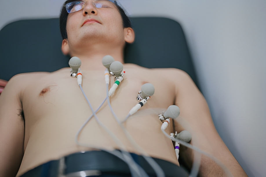
Electrocardiogram
ECG is one of the important baseline investigations to rule out or diagnose cardiac abnormalities. Sinus tachycardia on an ECG shows a regular heartbeat (sinus rhythm) with an elevated heart rate above 100 beats per minute.
For episodic or intermittent tachycardia, a Holter monitor might be used. It tracks the heart’s rhythm over a more extended period to capture irregularities that might not be evident during a short-term ECG.
Ventilation-Perfusion Scan
It is a type of imaging that is used to evaluate the lungs of the patients to check the circulation of air and blood.10Henning A, Krawiec C. Sinus Tachycardia. [Updated 2023 Mar 5]. In: StatPearls [Internet]. Treasure Island (FL): StatPearls Publishing; 2023 Jan-. Available from: https://www.ncbi.nlm.nih.gov/books/NBK553128/
When to see your Doctors?
Immediately consult with your doctor and book your appointment if you feel persistent chest pain and breathing difficulty. Immediate diagnosis and treatment reduce the chance of severity of the disease.
How to Treat Sinus Tachycardia?
If the sinus tachycardia is due to a physiological response, it returns to normal without taking medical interventions. Reassurance and patient education are enough to manage sinus tachycardia. In comparison, If the sinus tachycardia is due to some pathology, then treatment of the underlying cause is necessary.
Lifestyle Modifications:
Some lifestyle modifications help treat heart-related problems. These modifications are:
- Weight reduction
- A healthy exercise daily
- Stop smoking
- Stop alcohol
- Good sleep pattern
- Reduce stress and anxiety
- Use a healthy diet plan
Difference between Sinus and Ventricular Tachycardia
As we have discussed above, sinus tachycardia is most commonly due to a physiological response like exercise, emotional stress, and pregnancy. However, It rarely needs treatment unless no underlying pathology is present.
Ventricular tachycardia most commonly occurs due to Myocardial infarction, cardiomyopathy, and coronary artery diseases. It is characterized by impaired functionality of the left ventricle and ventricular aneurysm. Moreover, the ECG pattern shows an independent activity of atria and ventricles works and a broad QRS complex.
Conclusion
To conclude, sinus tachycardia is characterized by an increased heart rate. Causes of sinus tachycardia can be physiological or pathological. Physiological tachycardia does not necessitate the use of medications; reassurance and patient education are enough. However, pathological sinus tachycardia indicates an underlying pathology and needs to be treated promptly to avoid adverse consequences. If you have a higher chance of sinus tachycardia, immediately consult with a doctor for prompt diagnosis and management.
Refrences
- 1Buttner, E. B. a. R. (2021). Sinus tachycardia. Life in the Fast Lane • LITFL. https://litfl.com/sinus-tachycardia-ecg-library/
- 2Henning A, Krawiec C. Sinus Tachycardia. [Updated 2023 Mar 5]. In: StatPearls [Internet]. Treasure Island (FL): StatPearls Publishing; 2023 Jan-. Available from: https://www.ncbi.nlm.nih.gov/books/NBK553128/
- 3Gopinathannair, R. U. H. R. (2013, July 18). “Inappropriate” sinus tachycardia. Medscape. https://www.medscape.com/viewarticle/780588_2
- 4Sinus Tachycardia | Cardiology | Mercy Health. (n.d.). https://www.mercy.com/health-care-services/heart-vascular/conditions/sinus-tachycardia
- 5Sauer AJ, Newton-Cheh C. Clinical and genetic determinants of torsade de pointes risk. Circulation. 2012 Apr 3;125(13):1684-94. doi: 10.1161/CIRCULATIONAHA.111.080887. PMID: 22474311; PMCID: PMC3347483.
- 6Kumar A, Cannon CP. Acute coronary syndromes: diagnosis and management, part I. Mayo Clin Proc. 2009 Oct;84(10):917-38. doi: 10.1016/S0025-6196(11)60509-0. PMID: 19797781; PMCID: PMC2755812.
- 7Corrigan D, Prucnal C, Kabrhel C. Pulmonary embolism: the diagnosis, risk-stratification, treatment and disposition of emergency department patients. Clin Exp Emerg Med. 2016 Sep 30;3(3):117-125. doi: 10.15441/ceem.16.146. PMID: 27752629; PMCID: PMC5065342.
- 8Klein HH, Pich S. Physiologische Anderungen des Herz-Kreislauf-Systems in der Schwangerschaft [Cardiovascular changes during pregnancy]. Herz. 2003 May;28(3):173-4. German. doi: 10.1007/s00059-003-2455-2. PMID: 12756474.
- 9Henning A, Krawiec C. Sinus Tachycardia. [Updated 2023 Mar 5]. In: StatPearls [Internet]. Treasure Island (FL): StatPearls Publishing; 2023 Jan-. Available from: https://www.ncbi.nlm.nih.gov/books/NBK553128/
- 10Henning A, Krawiec C. Sinus Tachycardia. [Updated 2023 Mar 5]. In: StatPearls [Internet]. Treasure Island (FL): StatPearls Publishing; 2023 Jan-. Available from: https://www.ncbi.nlm.nih.gov/books/NBK553128/
