
Otosclerosis is a condition that can lead to progressive hearing loss due to abnormal bone growth occurring in the middle ear. Other symptoms include ringing in the ears, monotonous speech, and dizziness in some cases. The treatment is mainly surgical. Without treatment, hearing deteriorates over time, and the individual becomes socially handicapped. The prognosis of otosclerosis surgery is fairly good.
What is Otosclerosis?
“Oto” means “ear,” and “sclerosis” means “hardening.” Otosclerosis is a condition that affects the bony structures of the middle ear. It is characterized by the abnormal growth of bone tissue, which interferes with the movement of the tiny bones in the ear, leading to hearing loss. While otosclerosis can affect both ears, it often starts in one ear and is usually more severe in that ear.
What Causes Otosclerosis?
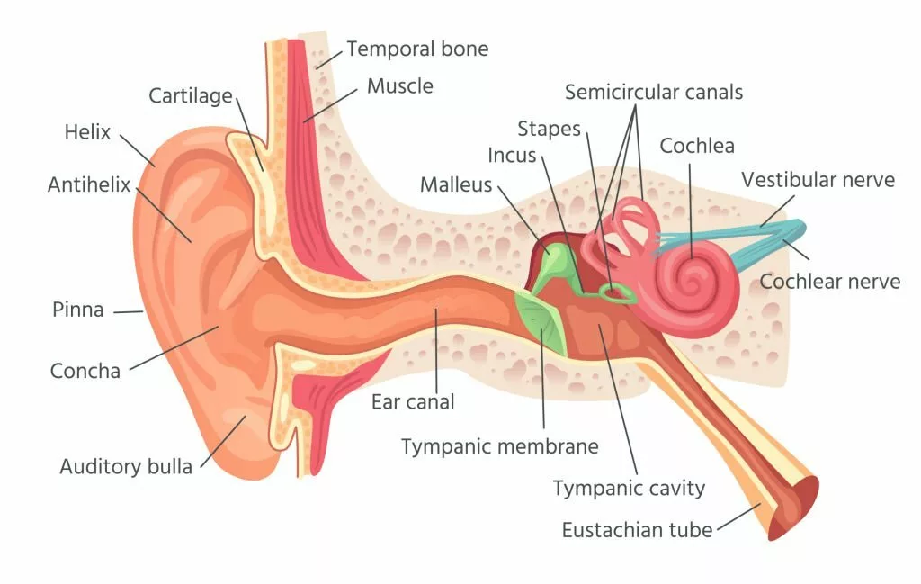
Otosclerosis involves abnormal bone growth in the otic capsule, which is the bony structure of the inner ear. The otic capsule has three layers: the innermost endosteal layer, the middle endochondral layer (which develops from cartilage into bone), and the outer periosteal layer. In otosclerosis, some areas of the otic capsule may develop abnormal spongy bone instead of the normal dense bone.
This abnormal bone growth most commonly affects the stapes, one of the three tiny bones in the middle ear that helps transmit sound from the middle ear to the inner ear. In otosclerosis, the stapes can become fixed or less mobile at their connection to the oval window, the membrane against which they normally move. Although the stapes is the most frequent site of otosclerosis, the abnormal bone growth can also occur in other parts of the otic capsule. This condition may cause hearing loss, which can vary in severity and may not always produce noticeable symptoms.
In front of the oval window, where the stapes footplate rests, lies an area called fistula ante fenestram. This area is prone to developing otosclerosis.
What are the Risk Factors of Otosclerosis?
The exact causes of this disease are not known; however, three small bones in the middle ear vibrate when sound reaches them. These bones amplify the sound, which then travels to the inner ear. The inner ear converts the sound into signals and sends them to the brain. Otosclerosis occurs when the surrounding bony tissue fuses with the stapes, preventing proper vibration. As a result, sound does not travel effectively to the inner ear, resulting in reduced hearing.
The following are the risk factors that increase the chances of this disease:
Family History
Genetic studies have revealed that otosclerosis is an Autosomal Dominant trait in which 50% to 60% of people have a positive family history, and the rest are sporadic.1Rudic, M., Keogh, I., Wagner, R., Wilkinson, E., Kiros, N., Ferrary, E., Sterkers, O., Bozorg Grayeli, A., Zarkovic, K., & Zarkovic, N. (2015). The pathophysiology of otosclerosis: Review of current research. Hearing Research, 330(Pt A), 51–56. https://doi.org/10.1016/j.heares.2015.07.014 It means that people with otosclerosis have family members suffering from the same disease 50 percent of the time.
Race
According to trials and studies, it is more common in white people than black and in Indians than in Japanese and Chinese. So, race plays a part in the development of this condition.
Age and Sex
Otosclerosis is most common in the second and third decades of life. Females are more susceptible to developing otosclerosis.2Clayton, A. E., Mikulec, A. A., Mikulec, K. H., Merchant, S. N., & McKenna, M. J. (2004). Association between osteoporosis and otosclerosis in women. The Journal of laryngology and otology, 118(8), 617–621. https://doi.org/10.1258/0022215041917790
Hormonal Condition
Hormonal conditions, including puberty, menopause, and pregnancy, also contribute to exacerbating this condition.3Rudic, M., Keogh, I., Wagner, R., Wilkinson, E., Kiros, N., Ferrary, E., Sterkers, O., Bozorg Grayeli, A., Zarkovic, K., & Zarkovic, N. (2015). The pathophysiology of otosclerosis: Review of current research. Hearing Research, 330(Pt A), 51–56. https://doi.org/10.1016/j.heares.2015.07.014
Viral Infections
It might be caused by a viral infection, especially measles exposure, which is considered a risk factor for developing otosclerosis. Research has also found viral nucleic acid(a genetic material) in the stapes(ear bone) footplate. Vaccination against measles has also acted as a protective factor against otosclerosis.4Sagar, P. R., Shah, P., Bollampally, V. C., Alhumaidi, N., & Malik, B. H. (2020). Otosclerosis and Measles: Do Measles Have a Role in Otosclerosis? A Review Article. Cureus, 12(8), e9908. https://doi.org/10.7759/cureus.9908
Inflammation and Autoimmunity
Inflammatory mediators and cytokines are released from the disease area in otosclerosis, implying that inflammation plays a role in its development.5Rudic, M., Keogh, I., Wagner, R., Wilkinson, E., Kiros, N., Ferrary, E., Sterkers, O., Bozorg Grayeli, A., Zarkovic, K., & Zarkovic, N. (2015). The pathophysiology of otosclerosis: Review of current research. Hearing Research, 330(Pt A), 51–56. https://doi.org/10.1016/j.heares.2015.07.014
Van Der Hoeve Syndrome
When otosclerosis occurs with multiple bone fractures and blue sclera (a bluish tint to the whites of the eyes), the triad of symptoms is called Van Der Hoeve Syndrome. People with brittle bone disease (osteogenesis imperfecta) are more likely to get this condition. Osteogenesis imperfecta and otosclerosis both occur due to defects in type I collagen-encoding genes.
Types of Otosclerosis
The following are the major types of otosclerosis
-
Stapedial Otosclerosis
It is the most common type of otosclerosis characterized by fixation of the stapes footplate on the oval window, which results in deafness. The disease process can involve a small part or the whole footplate.
-
Cochlear Otosclerosis
It involves other areas of the otic capsule except the stapes footplate and oval window. It causes hearing loss by releasing toxic materials in the inner ear fluid.
-
Histologic Otosclerosis
The disease process in this type is microscopic, and it does not produce any symptoms.
Symptoms of Otosclerosis
It may be present without any symptoms in some patients. In the case of symptomatic disease, patients present with the following complaints.
Hearing loss
The most common presentation of otosclerosis is hearing loss. Hearing loss is progressive and worse for lower-frequency sounds. For example, the patient complains of difficulty in hearing male voices. Some patients present with hearing loss in both ears, which developed over many years. Some people have hearing loss in only one ear.
Tinnitus – Ringing in Ear
Tinnitus is the ringing sound in the ear, which is psychologically very disturbing. Otosclerosis patients complain of tinnitus. It is more common in it’s cochlear type.
Paracusis Willisii
A patient with otosclerosis reports that he can hear better in noisy surroundings. This phenomenon is called paracusis willisii. Although it is not specific to otosclerosis, it shows deafness due to a disturbance in the sound wave traveling mechanism.
Speech
The patient himself can talk at low frequencies, but his speech becomes very monotonous and soft.
Vertigo
Vertigo is the sensation that the environment around you is spinning. It is not a common symptom of otosclerosis, but it becomes more pronounced as the disease progresses.
How will the Physicians Diagnose Otosclerosis?
The first step in diagnosing otosclerosis is taking adequate history and doing proper clinical examination. In history, the patient will complain of the symptoms described above.
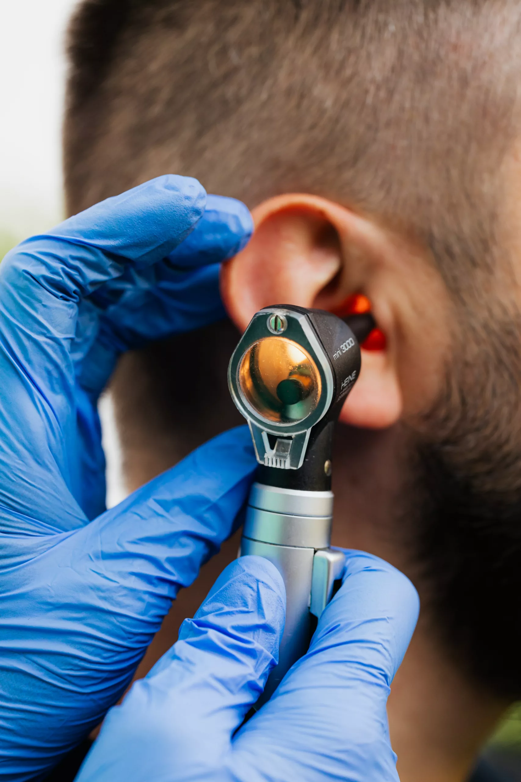
Otoscopy
Otoscopic examination shows that the tympanic membrane is normal in appearance and mobile. An important sign on otoscopy is the “Schwartze Sign” – a reddish hue is seen on the promontory through the tympanic membrane. It is characteristic of otosclerosis.6Koenen, L., & Gupta, G. (2022). Schwartze Sign. In StatPearls. StatPearls Publishing. The function of the eustachian tube is also normal.
Tuning Fork Tests
In cases of conductive deafness, the Weber Test indicates that hearing trouble is mainly in one ear, and the Rinne Test shows that hearing through bone vibrations is better than through the air in that ear. This suggests the issue lies in the ear’s ability to conduct sound, but the inner ear is fine.
Audiometry and Tympanometry
Pure tone audiometry will measure hearing loss at different frequencies. There is a dip in the graph obtained, which is the most pronounced at 2000 Hz. It is called Carhart’s notch, and it was used as the diagnostic point for otosclerosis, but recent studies have proved otherwise.7Cheon, D., Kim, D., Kim, S. H., Choi, J. Y., & Bae, S. H. (2023). Diagnostic Value of the Air-Bone Threshold Gap in Stapes Fixation. Audiology & neuro-otology, 28(4), 255–261. https://doi.org/10.1159/000528826 Tympanometry tells the physician about the working of the eardrum. It is normal initially but shows stiffness as the disease progresses.
CT Scan
The standard choice for diagnosis of otosclerosis is high-resolution CT of temporal bones.
Treatment Options for Otosclerosis
Following are the various acknowledged treatments of otosclerosis:
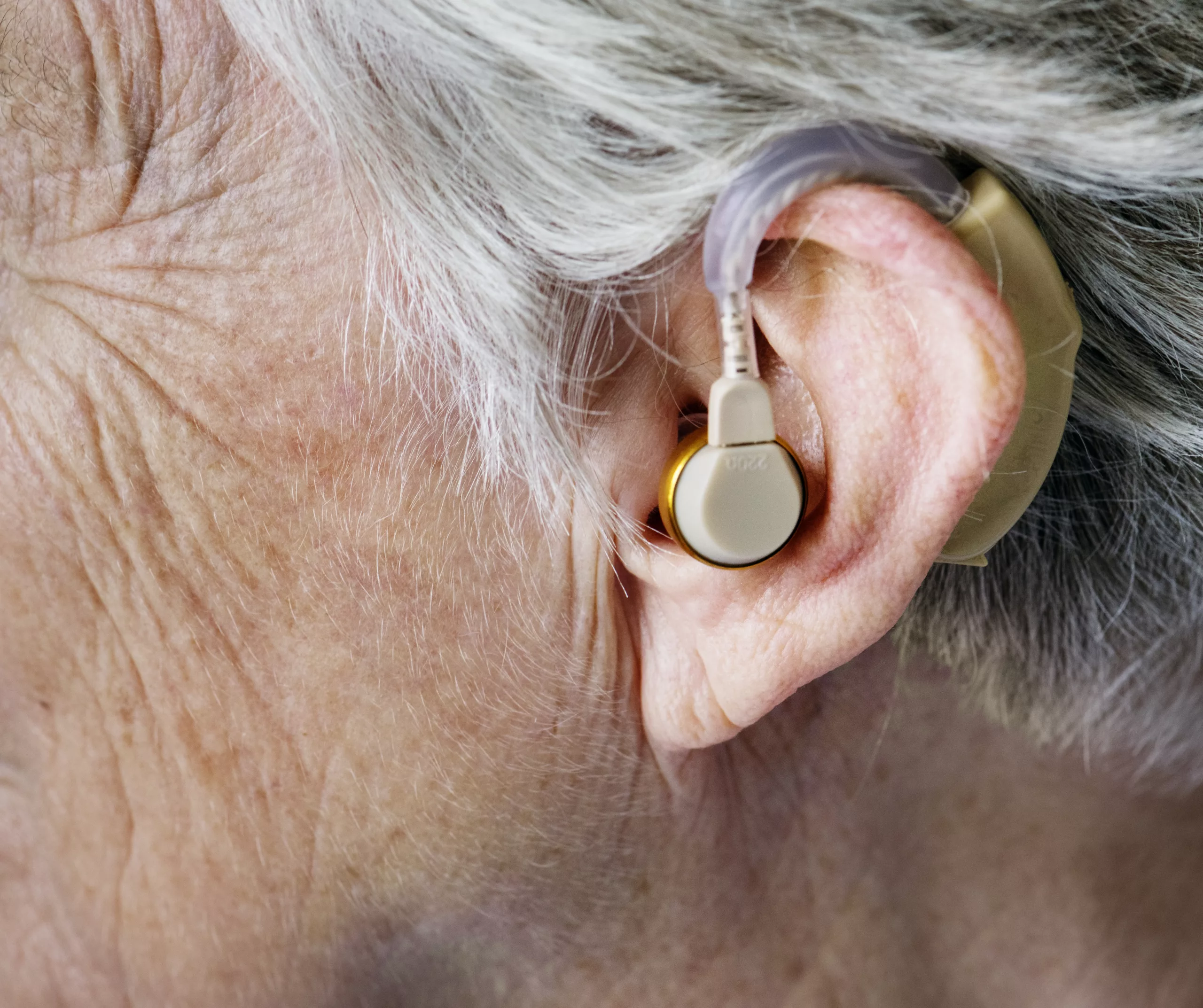
Hearing Aids
Hearing aids are devices placed in the ear to amplify sound. They have different setting options that the physician can optimize according to patient requirements. Hearing aids provide relief because the patient can hear again, but they do not stop the disease from getting worse.
Medical Treatment
There is no curative medical treatment for otosclerosis. The use of sodium fluoride and bisphosphonates is the mainstay of medical treatment. Sodium fluoride combined with calcium carbonate and vitamin D reduces the deterioration of conductive and sensorineural deafness in otosclerosis. Bisphosphonates are used if the patient is intolerant of sodium fluoride or as an adjuvant to sodium fluoride.8Gogoulos, P. P., Sideris, G., Nikolopoulos, T., Sevastatou, E. K., Korres, G., & Delides, A. (2023). Conservative Otosclerosis Treatment With Sodium Fluoride and Other Modern Formulations: A Systematic Review. Cureus, 15(2), e34850. https://doi.org/10.7759/cureus.34850
Surgical Managment
The treatment of choice for otosclerosis is stapedectomy/stapedotomy with the placement of the prosthesis. The fixed footplate of the stapes is removed in stapedectomy, and a prosthesis is inserted between the incus and the oval window.9Toscano, M. L., & Shermetaro, C. (2023). Stapedectomy. In StatPearls. StatPearls Publishing. It allows the sound to travel effectively to the inner ear, resulting in better hearing.
In stapedotomy, the surgeon makes a hole in the footplate of the stapes(bone in the ear). The surgeon uses prosthetic materials such as Teflon, stainless steel, or platinum-Teflon to replace or stabilize the stapes. If both ears are affected, the surgeon will perform the operation on one ear at a time, allowing each ear sufficient time to heal. In the case of cochlear otosclerosis, the surgeon conducts cochlear implant surgery, creating a pathway for sound to travel to the brain.
Contradictions of Otosclerosis Surgery
Following are the various contraindications of surgery:
- Surgery is typically not recommended for individuals who have hearing loss in only one ear
- Athletes
- Associated Meniere’s disease
- Young children
- People working in noisy surroundings
- Any middle ear or external ear infection
- Patients not suitable for surgery can use hearing aids
Prognosis of Otosclerosis
In the majority of cases, the surgical treatment is effective and improves hearing loss.10Souza, J. C., Bento, R. F., Pereira, L. V., Ikari, L., Souza, S. R., Della Torre, A. A., & Fonseca, A. C. (2016). Evaluation of Functional Outcomes after Stapes Surgery in Patients with Clinical Otosclerosis in a Teaching Institution. International archives of otorhinolaryngology, 20(1), 39–42. https://doi.org/10.1055/s-0035-1563540 In some cases, surgery does not provide any beneficial results. The recurrence of the disease can occur in a few cases due to postoperative complications, such as displacement of the prosthesis.
Conclusion
In summary, Otosclerosis is a hearing loss-inducing condition with not completely understood causes, but multiple factors contribute to its development and progression. Timely surgery is crucial for a symptom-free life. Consulting a doctor as soon as any hearing changes occur is essential for early diagnosis and improved prognosis.
Refrences
- 1Rudic, M., Keogh, I., Wagner, R., Wilkinson, E., Kiros, N., Ferrary, E., Sterkers, O., Bozorg Grayeli, A., Zarkovic, K., & Zarkovic, N. (2015). The pathophysiology of otosclerosis: Review of current research. Hearing Research, 330(Pt A), 51–56. https://doi.org/10.1016/j.heares.2015.07.014
- 2Clayton, A. E., Mikulec, A. A., Mikulec, K. H., Merchant, S. N., & McKenna, M. J. (2004). Association between osteoporosis and otosclerosis in women. The Journal of laryngology and otology, 118(8), 617–621. https://doi.org/10.1258/0022215041917790
- 3Rudic, M., Keogh, I., Wagner, R., Wilkinson, E., Kiros, N., Ferrary, E., Sterkers, O., Bozorg Grayeli, A., Zarkovic, K., & Zarkovic, N. (2015). The pathophysiology of otosclerosis: Review of current research. Hearing Research, 330(Pt A), 51–56. https://doi.org/10.1016/j.heares.2015.07.014
- 4Sagar, P. R., Shah, P., Bollampally, V. C., Alhumaidi, N., & Malik, B. H. (2020). Otosclerosis and Measles: Do Measles Have a Role in Otosclerosis? A Review Article. Cureus, 12(8), e9908. https://doi.org/10.7759/cureus.9908
- 5Rudic, M., Keogh, I., Wagner, R., Wilkinson, E., Kiros, N., Ferrary, E., Sterkers, O., Bozorg Grayeli, A., Zarkovic, K., & Zarkovic, N. (2015). The pathophysiology of otosclerosis: Review of current research. Hearing Research, 330(Pt A), 51–56. https://doi.org/10.1016/j.heares.2015.07.014
- 6Koenen, L., & Gupta, G. (2022). Schwartze Sign. In StatPearls. StatPearls Publishing.
- 7Cheon, D., Kim, D., Kim, S. H., Choi, J. Y., & Bae, S. H. (2023). Diagnostic Value of the Air-Bone Threshold Gap in Stapes Fixation. Audiology & neuro-otology, 28(4), 255–261. https://doi.org/10.1159/000528826
- 8Gogoulos, P. P., Sideris, G., Nikolopoulos, T., Sevastatou, E. K., Korres, G., & Delides, A. (2023). Conservative Otosclerosis Treatment With Sodium Fluoride and Other Modern Formulations: A Systematic Review. Cureus, 15(2), e34850. https://doi.org/10.7759/cureus.34850
- 9Toscano, M. L., & Shermetaro, C. (2023). Stapedectomy. In StatPearls. StatPearls Publishing.
- 10Souza, J. C., Bento, R. F., Pereira, L. V., Ikari, L., Souza, S. R., Della Torre, A. A., & Fonseca, A. C. (2016). Evaluation of Functional Outcomes after Stapes Surgery in Patients with Clinical Otosclerosis in a Teaching Institution. International archives of otorhinolaryngology, 20(1), 39–42. https://doi.org/10.1055/s-0035-1563540
: Causes, Symptoms & Care")


