
Blue baby syndrome, also referred to as infant methemoglobinemia (when caused by nitrate exposure), is a rare condition characterized by a bluish or purplish discoloration of a baby’s skin, known as cyanosis, due to insufficient oxygen in the blood. Several pathologies like heart issues (tetralogy of Fallot), lung diseases, nitrate poisoning, and birth defects can lead to cyanosis. As per estimates, 2-5/1000 infants suffer from blue baby syndrome, and the disease is more common in rural areas (especially in developing countries). Most patients require early diagnosis and treatment to prevent serious complications.
Blue Baby Syndrome Symptoms
In addition to the blue color of the skin and the mucosa, infants experience the following symptoms:
Breathing Difficulties:
Babies presenting with cyanosis have poor oxygenation. Improper perfusion of oxygen leads to breathing difficulties. To compensate for the lack of oxygen, infants breathe rapidly and more deeply. Prolonged breathing trouble can be life-threatening. Therefore, the restoration of optimal breathing is the main aim of healthcare providers.
Feeding Issues & Inability to Gain Weight:
Such infants also encounter feeding problems. This eventually leads to the child’s inability to gain weight. Often, methemoglobinemia patients present with poor weight gain, feeding difficulty, and hypotonia.1Johnson, S. F. (2019). Methemoglobinemia: Infants at risk. Current problems in pediatric and adolescent health care, 49(3), 57-67.
Vomiting & Diarrhea:
Affected babies may also present with vomiting and diarrhea, which further contributes to difficulty in weight gain.
Rapid Heartbeat:
A rapid heartbeat accompanies breathing abnormalities. Pediatricians have noted multiple cases with a faster-than-usual beating of the heart, i.e., tachycardia.
Irritability & Lethargy:
Unlike infants of the same age, blue babies are lethargic and sluggish. In most cases, this is attributed to a lack of oxygen and energy for the muscles. Due to their health issues, such infants are also highly irritable.
Developmental Issues:
If defects (that cause cyanosis) persist for a longer period, infants tend to encounter developmental issues. Poor oxygenation hinders proper nourishment and can lay the foundation for developmental delays.
Other presentations of blue baby syndrome include seizures and clubbing of fingers (and toes).
What Causes Blue Baby Syndrome?
As already mentioned, the bluish/purple coloration of the skin (especially present around the lips and the fingernails) is the result of low oxygen levels in the blood. Normally, the heart receives oxygenated blood from the lungs and distributes it to the body (including the skin). However, when the lungs, the heart, or even the blood, become erratic, there is suboptimal oxygen delivered to the body. This lack of oxygen displays through the skin as cyanosis. There are multiple potential causes for this, including:
Heart Defects:
Tetralogy Of Fallot (TOF)
The most prevalent cause of blue baby syndrome is a rare congenital heart condition, i.e., tetralogy of Fallot. This anomaly is a combination of four defects (thus the name tetralogy!) which include a ventricular septal defect (a hole in the wall separating the lower heart chambers), pulmonary artery stenosis (narrowing of the pulmonary artery), ventricular hypertrophy (oversized right ventricle), and an overriding aorta.
A communication between the two ventricles causes mixing of oxygenated and deoxygenated blood. Therefore, mixed blood and lesser oxygen reach the body, leading to cyanosis. Together, these defects also reduce the blood flow from the heart to the rest of the body. The intensity of cyanosis varies between individuals depending on the extent of blood flow obstruction.2Bailliard, F., & Anderson, R. H. (2009). Tetralogy of fallot. Orphanet journal of rare diseases, 4, 1-10.
Other conditions, like Down syndrome, can cause congenital malformations in the heart. These malformations contribute to poor oxygenation, and neonates present with cyanosis.3Azzahra, S., Utari, A., & Soetadji, A. (2022). Clinical Characteristics of Down Syndrome with Congenital Heart Disease. eJournal Kedokteran Indonesia, 33-8.
Diabetic mothers are at a high risk of giving birth to children with heart defects. In a study, more than 10% of diabetic mothers’ children suffered from a ventricular septal defect, which can present as cyanosis.4Abhirami, P. (2018). Clinical Study of Infants of Diabetic Mother (Doctoral dissertation, Rajiv Gandhi University of Health Sciences (India)). Thus, newborns of mothers with uncontrolled diabetes can have poor oxygenation due to congenital heart defects.
Nitrate Poisoning:
Nitrate ingestion causes methemoglobinemia, another cause of blue baby syndrome. The abnormality arises when infants are fed formula milk mixed with nitrate-rich water. It most often occurs in babies under 6 months of age. Developing countries face this issue more frequently than developed countries because of excess nitrates in their water supplies. Your child may also develop methemoglobinemia if you give homemade foods teeming with nitrates, like spinach or beets.5Yoon JC, Kim SE. Suicide attempt using sodium nitrite ordered on the internet: Two case reports. Medicine (Baltimore). 2022 Jul 15;101(28):e29355. doi: 10.1097/MD.0000000000029355. PMID: 35839015; PMCID: PMC11132383.
The immature GIT tracts of infants convert the ingested nitrate into nitrite, which reacts with the hemoglobin in the blood, leading to the formation of methemoglobin. This very structure renders hemoglobin incapable of binding with oxygen (to form oxyhemoglobin, which imparts the red color to oxygenated blood), and a lack of oxygenation shows as blue skin color.6Johnson, S. F. (2019). Methemoglobinemia: Infants at risk. Current problems in pediatric and adolescent health care, 49(3), 57-67. Some antimicrobial drugs, like sulfonamides, chloroquine, and dapsone, can also induce methemoglobinemia in pediatric patients.7Alotaibi, S., Mously, A., Alnujaimy, Z., Alotaibi, A., & Alharthi, A. (2023). Trimethoprim/sulfamethoxazole‐induced methemoglobinemia in pediatric patient: A case report. Clinical Case Reports, 11(3), e7124.
Blue Baby Syndrome Diagnosis
Pediatricians take a full history of the symptoms from the parents. They will ask you about the breathing and feeding patterns of the newborn, and a physical examination follows. The doctor will check the skin discoloration and hear lung/heart sounds through a stethoscope. Your healthcare provider might order some additional tests to identify the underlying problem causing the cyanosis. Commonly performed tests include:
Blood Test:
It helps reveal methemoglobin levels in the blood. When methemoglobinemia is suspected, doctors may also order a CYB5R enzyme activity test. Low activity of the enzyme may indicate underlying methemoglobinemia because it is responsible for converting methemoglobin back to hemoglobin. Further blood tests include hemoglobin electrophoresis.
Oxygen Saturation:
A pulse oximeter test is done to analyze the oxygen saturation of the infant.
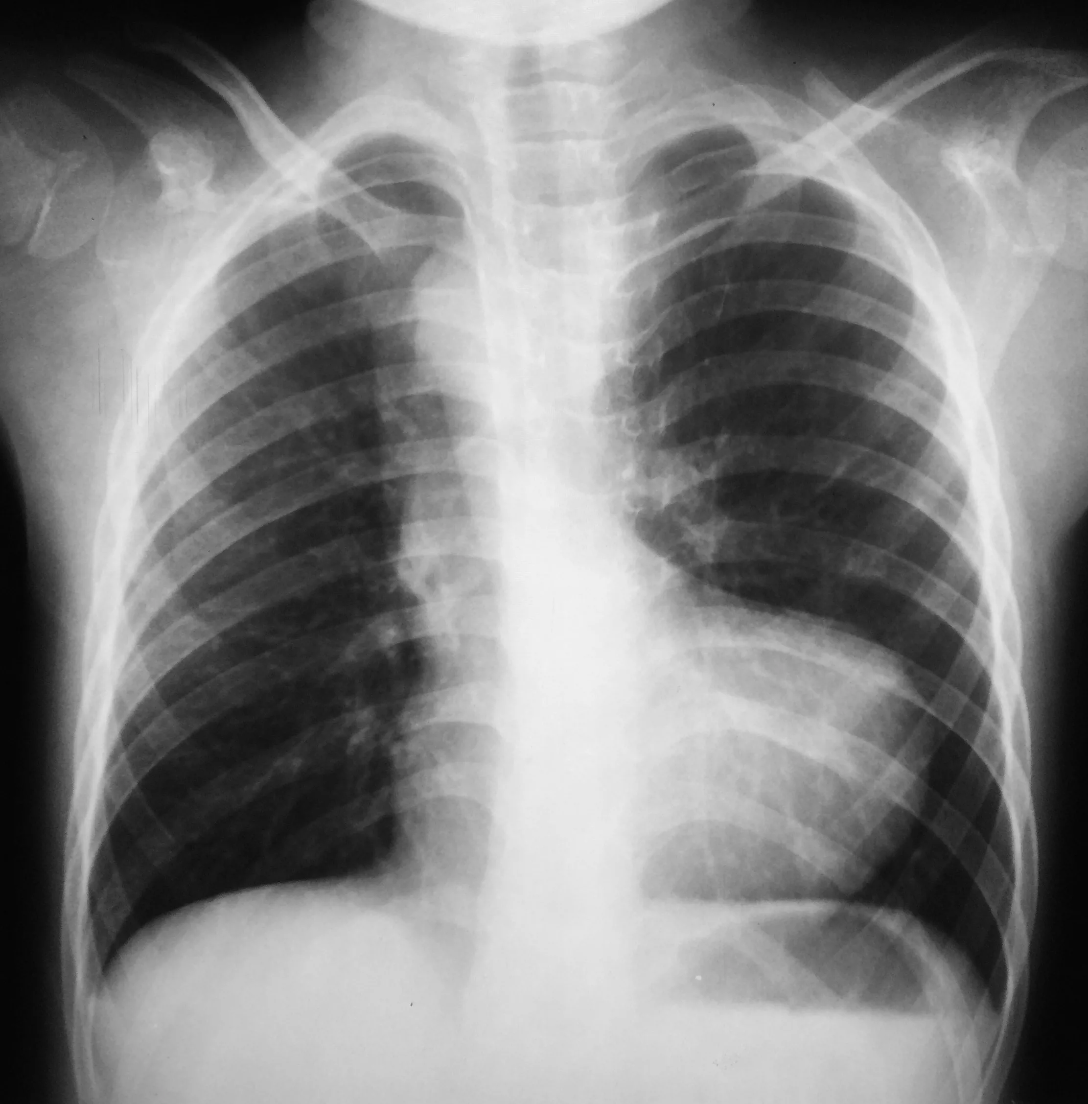
From the case rID: 43059.
Chest X-rays:
Radiographs of the chest are done to check the size of the heart and lungs in infants. This aids in the diagnosis of congenital malformations.
Electrocardiogram (ECG/EKG) & Echocardiogram:
ECG and echo tests are done to analyze the electrical activity and anatomical anomalies of the heart, respectively.
Cardiac Catheterization:
For a better understanding of the heart issues, doctors may also perform a catheterization (a tube is inserted via the arm/groin and reaches the heart through a vessel). Most of the time, this is done as an emergency procedure to improve the heart’s performance in the newborn.8Romer, A. J., Johng, S., Hsia, J., Scott, S., Reddy, A., & Gardner, M. M. (2022). Cyanosis in a Newborn Immediately after Birth. NEJM evidence, 1(2), EVIDmr2100060.
Blue Baby Syndrome Treatment
Treatment strategies depend on the underlying condition causing the cyanosis and the severity of the condition. Congenital heart abnormalities like ventricular septal defect (VSD) and tetralogy of Fallot require surgical interventions. Most pediatric cardiac surgeries are performed before the patient’s first birthday so that the oxygenation (and life quality) can improve.
In cases of nitrate poisoning, pediatricians team up with toxicologists to flush out the nitrates from the body. Doctors aim to quickly diagnose the condition and remove the source of exposure. In severe cases, health professionals may use methylene blue to reverse methemoglobinemia. The drug is delivered via an intravenous injection.
How To Prevent Blue Baby Syndrome?
Congenital deformities can not be prevented. However, you can avoid acquired cases of methemoglobinemia by adopting the following steps:
Avoid well/supply Water:
Residents of developing countries should avoid preparing baby formula milk with local supply/well water. Try using bottled water even for drinking. Be cautious about water intake until your kid is over 12 months old. An important point to note here is that boiling doesn’t remove nitrates from the water!
Limit intake of Nitrate-rich Foods:
You should limit the amount of nitrate foods like carrots, spinach, and beets in your child’s diet. Follow a cautious routine till your baby is 7 months old.
Be cautious during Pregnancy:
Pregnant ladies should avoid alcohol, smoking, drugs, and unnecessary medications. Uncontrolled diabetes is linked to heart malformations, so keep your sugar levels in check.
Final Word
Blue baby syndrome is a condition in which an infant’s skin appears blue-purple due to poor oxygenation. It arises due to congenital heart defects (tetralogy of Fallot, maternal diabetes, etc.) or due to nitrate poisoning. Mixing of blood in the heart (due to a ventricular septal defect) leads to the delivery of deoxygenated blood to the body. Excessive nitrate in the diet/water leads to the formation of methemoglobin, which hinders the blood’s capacity to carry oxygen. Patients present with irritability, breathing difficulties, feeding issues, fatigue, and developmental delays. Doctors diagnose it by physical examination and diagnostic tests like blood tests (checks methemoglobin levels), chest X-rays, ECG, and echocardiograms etc. Treatment of congenital cardiac malformations requires surgery, while nitrate poisoning can be corrected with methylene blue injection. Parents can avoid methemoglobinemia by avoiding mixing supply/well water with formula milk and limiting the infant’s intake of nitrate-rich foods like beets, spinach, etc.
Refrences
- 1Johnson, S. F. (2019). Methemoglobinemia: Infants at risk. Current problems in pediatric and adolescent health care, 49(3), 57-67.
- 2Bailliard, F., & Anderson, R. H. (2009). Tetralogy of fallot. Orphanet journal of rare diseases, 4, 1-10.
- 3Azzahra, S., Utari, A., & Soetadji, A. (2022). Clinical Characteristics of Down Syndrome with Congenital Heart Disease. eJournal Kedokteran Indonesia, 33-8.
- 4Abhirami, P. (2018). Clinical Study of Infants of Diabetic Mother (Doctoral dissertation, Rajiv Gandhi University of Health Sciences (India)).
- 5Yoon JC, Kim SE. Suicide attempt using sodium nitrite ordered on the internet: Two case reports. Medicine (Baltimore). 2022 Jul 15;101(28):e29355. doi: 10.1097/MD.0000000000029355. PMID: 35839015; PMCID: PMC11132383.
- 6Johnson, S. F. (2019). Methemoglobinemia: Infants at risk. Current problems in pediatric and adolescent health care, 49(3), 57-67.
- 7Alotaibi, S., Mously, A., Alnujaimy, Z., Alotaibi, A., & Alharthi, A. (2023). Trimethoprim/sulfamethoxazole‐induced methemoglobinemia in pediatric patient: A case report. Clinical Case Reports, 11(3), e7124.
- 8Romer, A. J., Johng, S., Hsia, J., Scott, S., Reddy, A., & Gardner, M. M. (2022). Cyanosis in a Newborn Immediately after Birth. NEJM evidence, 1(2), EVIDmr2100060.


