Brain Contusions are hemorrhagic brain tissue lesions caused by a direct blow to the head. It commonly occurs along the poles or under the temporal and frontal lobes, presenting with bruises at the injury site. A person may experience confusion, loss of consciousness, headache, nausea, vomiting, ear crackling, and other related symptoms.
Brain Contusion Causes
A contusion usually occurs when the head hits a hard object. The injury may be due to the following causes1Pellot, J.E. (2023) Cerebral Contusion. Retrieved 29 May 2023 from: https://www.ncbi.nlm.nih.gov/books/NBK562147/:
- Vigorous physical activity
- Road accidents
- Domestic violence
- Child abuse
- Falls
- Fights
- Sports Injury
- Cycling
- Blast injury
Types of Brain Contusion
Following are the three common types of brain contusion based on the severity of the injury.
Mild Contusion
Head injuries or common blows often result in surface bruising at the impact site. Mild contusions usually present with only swelling without bleeding. However, a person may feel some pain but with only short-term effects.
Moderate Contusion
Moderate contusions often occur due to severe injury, resulting in internal bleeding. The patient experiences severe symptoms, including short-term memory loss, unconsciousness, and difficulty focusing. Also, some symptoms may not go away even after recovery.
Severe Contusion
It can be a fatal condition that requires immediate medical intervention. The victim may experience seizures, repeated vomiting, worsening headache, loss of consciousness, and impaired vision. Severe contusion should be managed immediately to avoid permanent damage. Also, proper care can help reduce the severity of symptoms.
Contusion may be classified into two types based on the movement of intracranial content and impingement on the interior surface of the skull.
- Coup Contusion: It presents with a contusion directly at the impact site.
- Contrecoup Contusion: It presents with a contusion directly opposite to the impact site of the brain.
Brain Contusion Risk Factors
Anyone can be vulnerable to a brain injury. However, you must know certain risk factors for a brain contusion.
- Gender: Men are twice as risky of contusion as women due to their risk-taking behavior.
- Age Group: Young children and elderly individuals are more vulnerable to injury.
- Alcohol Use: It slows the thought process and increases the risk of accidents.
- Certain Sports: Contact sports like soccer and basketball increase the risk of brain trauma.
Brain Contusion Symptoms
Clinical signs may vary depending on the severity and site of the head injury2Mao, G. (2023) Overview of head injuries – injuries and poisoning, MSD Manual Consumer Version. Retrieved 04 June 2023 from: https://www.msdmanuals.com/home/injuries-and-poisoning/head-injuries/overview-of-head-injuries#v739890. Here are some common symptoms of a brain contusion.
- Reduced cognitive abilities
- Impaired coordination
- Localized tingling or numbness
- Personality disorders
- Memory changes
- Cerebral swelling
- Loss of consciousness
Diagnosis of Brain Contusion
Understanding the problem after a head injury requires a comprehensive medical evaluation. The doctor performs a physical examination and asks for a medical history to determine the cause. Besides the physical examination, your healthcare provider may order certain imaging tests to check the extent of the injury.
Neurological Assessment:
A comprehensive neurological evaluation is conducted to assess the patient’s mental status, cognitive function, coordination, and sensory responses. The healthcare provider may use standardized scales, such as the Glasgow Coma Scale, to quantify the patient’s level of consciousness and neurological deficits.
Glasgow Coma Scale (GCS)3Jain, S. (2022) Glasgow coma scale – statpearls – NCBI bookshelf. Retrieved 26 June 2023 from https://www.ncbi.nlm.nih.gov/books/NBK513298/ is a widely used tool to assess patients’ responsiveness for the early management of head injuries. It has three parameters: best eye response (E), best verbal response (V), and best motor reaction (M). GCS levels are scored from 1, representing no response, to normal values of 4, 5, and 6 for an eye-opening, verbal, and motor response. The overall GCS score ranges from 3 to 15, with 3 denoting the worst and 15 denoting the best responsiveness level.
Imaging Studies
Imaging tests are a primary component in diagnosing brain contusions. Neuroimaging is a crucial component in diagnosing brain contusions. The most commonly used imaging modalities are:
Computed Tomography (CT) Scan:
CT scans are often the initial imaging modality of choice for acute head injuries. CT can quickly identify and visualize acute intracranial hemorrhages, such as contusions, and can help determine the location and extent of the injury.
Magnetic Resonance Imaging (MRI):
MRI provides detailed images of the brain’s soft tissues and is particularly useful for detecting subtle brain injuries and assessing contusions that might not be as apparent on a CT scan. Magnetic resonance imaging (MRI) is more sensitive and accurate than CT for detecting contusions because of its multiplanar capability and greater sensitivity for edema.4Paterakis K, Karantanas AH, Komnos A, Volikas Z. Outcome of patients with diffuse axonal injury: the significance and prognostic value of MRI in the acute phase. J Trauma 2000;49(6):1071–1075.
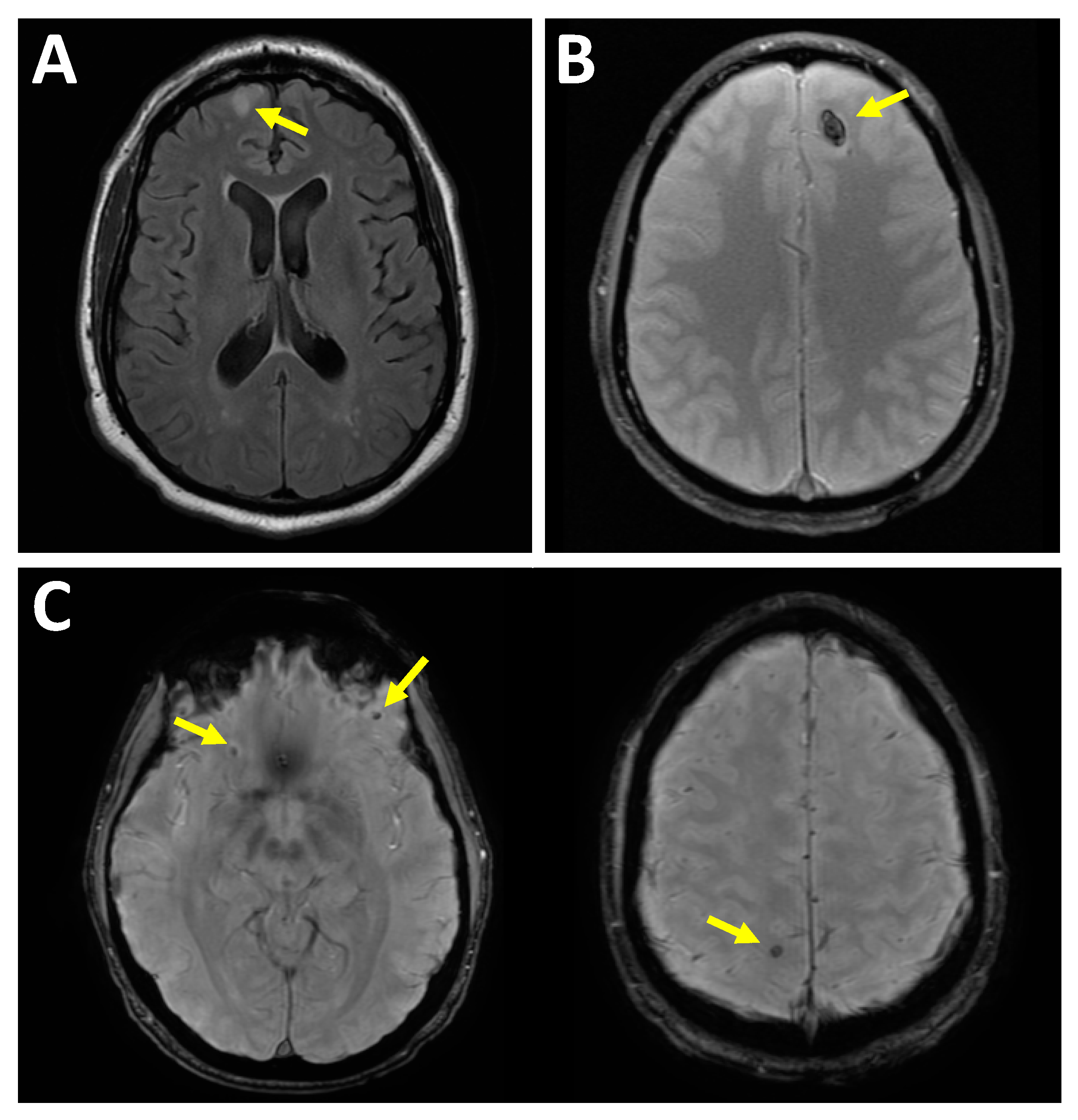
However, In the acute stage, CT is more sensitive than MRI, as the clot signal can be indistinguishable from brain parenchyma on MRI. On non-contrast CT scans, contusions are visualized as areas of altered attenuation within the brain tissue. If there is no hemorrhage present, these contusions appear as regions of low attenuation, indicating a decrease in tissue density compared to the surrounding normal brain tissue. However, if hemorrhage is present within the contusion, it can manifest as mixed or high attenuation areas, signifying a higher density due to the presence of blood.
Brain Contusion Treatment
The severity and site of injury determine the treatment. It may include ice, rest, and painkillers. Patients usually stay in the hospital and are observed for up to a week. Some common treatment approaches for brain contusions are:
Controlling & Monitoring Intracranial Pressure (ICP):
Managing intracranial pressure is a critical aspect of brain contusion treatment. Elevated pressure within the skull can exacerbate the injury and lead to complications. Medical professionals closely monitor and take measures to control ICP to prevent additional damage to brain tissue.
Ensuring Sufficient Blood Flow to the Brain:
Adequate blood flow to the brain is essential for delivering oxygen and nutrients necessary for healing. Measures are taken to optimize blood circulation and maintain cerebral perfusion.
Pain Management and Swelling Reduction:
Pain medications, including nonsteroidal anti-inflammatory drugs (NSAIDs), may be administered to alleviate discomfort and reduce swelling associated with the contusion.
Craniotomy for Surgical Intervention:
In severe cases, neurosurgeons may perform a craniotomy, surgically opening a section of the skull. This procedure enables direct access to the cerebral contusion, allowing them to remove it and thereby reduce pressure on the brain.
Addressing Coexisting Infections:
In situations involving concurrent infections, healthcare providers administer suitable treatments to combat these infections and avert potential complications. A personalized treatment strategy is tailored to each patient, taking into account the unique characteristics of their brain contusion. Additionally, the recovery journey is bolstered by the implementation of rehabilitation and essential supportive care. Throughout their hospitalization period, which typically extends up to a week, patients undergo close monitoring.
It is crucial to emphasize that the management of brain contusions adopts a multidisciplinary approach, fostering collaboration among neurologists, neurosurgeons, and other healthcare specialists, with the overarching goal of optimizing patient outcomes.
Brain Contusion Recovery Time
Recovery time usually depends on the severity of symptoms and the treatment plan. Mild contusions can heal quickly with simple bed rest and mild medications. Alternatively, severe contusions can take weeks to months to heal completely. Post-operative recovery can be prolonged for severe bruises as patients slowly restore their stamina and coordination power.
Brain Contusion Vs. Concussion
Both contusion and concussion are types of traumatic brain injury (TBI) that affect normal brain function. However, there lies a difference in their symptoms and treatment. A contusion is localized damage, whereas a concussion affects a more significant part of the brain.
Brain contusions can cause altered thinking ability, poor concentration, local numbness, dilated pupils, and difficulty speaking and moving. In contrast, people with concussions may experience behavioral changes, intense headaches, nausea, vomiting, and crackling in the ear.
Brain Contusion Vs. Hematoma
A cerebral hematoma is bleeding under the skull in or around the brain that can form a clot. However, a contusion is a type of hematoma characterized by bruising on the brain, resulting in swelling and bleeding5Saling, J. (2022) Head injuries (contusion, hematoma, skull fracture): Causes, treatments, headaches after, WebMD. Retrieved 26 May 2023 from: https://www.webmd.com/fitness-exercise/guide/head-injuries-causes-and-treatments.
Prevention of Brain Contusion
Here are some common suggestions for preventing brain contusions.
- Using helmets for cycling and other such sports.
- Use seatbelts while traveling to prevent severe head injury
- Use child control measures to avoid falls
- Monitor kids at all times, especially while playing.
- Avoid cycling or skating on uneven surfaces.
Final Words
A brain contusion is any traumatic head injury that causes blood to collect under the skin. It may result in impaired cognition, numbness, and inability to concentrate. Mild contusions usually resolve after some time without treatment. However, you must seek medical help to avoid permanent damage in case of severe head injury.
Refrences
- 1Pellot, J.E. (2023) Cerebral Contusion. Retrieved 29 May 2023 from: https://www.ncbi.nlm.nih.gov/books/NBK562147/
- 2Mao, G. (2023) Overview of head injuries – injuries and poisoning, MSD Manual Consumer Version. Retrieved 04 June 2023 from: https://www.msdmanuals.com/home/injuries-and-poisoning/head-injuries/overview-of-head-injuries#v739890
- 3Jain, S. (2022) Glasgow coma scale – statpearls – NCBI bookshelf. Retrieved 26 June 2023 from https://www.ncbi.nlm.nih.gov/books/NBK513298/
- 4Paterakis K, Karantanas AH, Komnos A, Volikas Z. Outcome of patients with diffuse axonal injury: the significance and prognostic value of MRI in the acute phase. J Trauma 2000;49(6):1071–1075.
- 5Saling, J. (2022) Head injuries (contusion, hematoma, skull fracture): Causes, treatments, headaches after, WebMD. Retrieved 26 May 2023 from: https://www.webmd.com/fitness-exercise/guide/head-injuries-causes-and-treatments
