
Cholangiocarcinoma is a rare neoplasm that affects your bile duct. It is characterized by abdominal pain, a yellow discoloration of the skin, and weight loss.
What is Cholangiocarcinoma?
Cholangiocarcinoma (CC) is a disease of the bile duct in which the normal epithelial cells (cholangiocytes) of the bile duct transform into cancerous or malignant cells. This transformation can occur anywhere in the bile duct (extrahepatic, intrahepatic, and distal extrahepatic). Cholangiocarcinoma usually presents with abdominal pain, yellowish color of the skin, fever, and weight loss. However, it is a rare pathology of the hepatobiliary system, constituting only 10-15 % of hepatobiliary carcinoma.
It is an uncommon disease that constitutes about 3% of the total gastrointestinal pathologies. The overall incidence and frequency of intrahepatic cholangiocarcinoma are rising day by day, while the frequency of extrahepatic cholangiocarcinoma is falling. In the United States of America (USA), out of 15000 cases of hepatocellular carcinoma, only 2500 are diagnosed with cholangiocarcinoma. Moreover, the average incidence of this cancer is about 1/100000 individuals per year.
The incidence of cholangiocarcinoma is directly proportional to age. Additionally, it most commonly targets an older population between the ages of 60 and 70. It targets men more than women.1Garikipati, S. C. (2023, February 6). Biliary tract cholangiocarcinoma. StatPearls – NCBI Bookshelf. https://www.ncbi.nlm.nih.gov/books/NBK560708/
Anatomy of the Bile Duct
Ducts are small tubules that connect the liver, gallbladder, and small intestine (2nd part of the duodenum). The ducts that are present inside the liver are called intrahepatic and that leave the liver are called extrahepatic bile ducts.
These ducts form right and left hepatic ducts after leaving the liver. Right and left hepatic ducts join to form a common hepatic duct. Moreover, the gallbladder is drained by a cystic duct. The cystic duct and common hepatic duct combine to give a common bile duct that opens into the 2nd part of the duodenum (ampulla of Vater).
Bile travels through the Bile duct to aid digestion. Bile is a green fluid that is produced in the gallbladder. It mainly helps in the digestion of fatty foods.
Types of Bile Duct Cancer
The bile duct cancer is divided into two types. These are intrahepatic and extrahepatic bile duct cancer.
Intrahepatic Bile Duct Cancer:
The cancer that affects the ducts inside the liver is called intrahepatic cholangiocarcinoma. However, the diagnosis of intrahepatic carcinoma is sometimes confused with hepatocellular carcinoma (HCC).
Extrahepatic Bile Duct Cancer:
This type affects the ducts that are outside the liver. Extrahepatic cholangiocarcinoma has subdivisions, perihilar and distal bile duct cancer.
Perihilar (Klatskin Tumor)
This type of extrahepatic bile duct cancer affects the area where right and left hepatic ducts leave the liver and join to form a common hepatic duct.
Distal Bile Duct Cancer
This type of cancer affects the distal portion where the common hepatic duct and cystic duct combine to give a common bile duct.2National Cancer Institute (US). (2023, June 2). Bile duct cancer (Cholangiocarcinoma) treatment. PDQ Cancer Information Summaries – NCBI Bookshelf. https://www.ncbi.nlm.nih.gov/books/NBK65851/3What is bile duct cancer? | What is cholangiocarcinoma? (n.d.). American Cancer Society. https://amp.cancer.org/cancer/types/bile-duct-cancer/about/what-is-bile-duct-cancer.html
Who is at risk of having Cholangiocarcinoma?
Some individuals have a greater chance of having bile duct cancer due to certain risk factors. However, this cancer can also occur in people who don’t have any risk factors. People who are at greater risk are those who have a positive history of:
- Primary sclerosing cholangitis
- Cholelithiasis/acute cholecystitis
- Chronic liver disease
- Hepatocellular carcinoma
- Inflammatory bowel disease (IBD)
- Hepatocellular infection (Liver fluke)
- Exposure to toxins
- Formation of the cyst bile duct (choledochal cyst)
- Genetic mutation
- Obesity
- Alcohol abuse
- Smoking
- History of diabetes
Out of these risk factors, primary sclerosing cholangitis is the most common. Moreover, almost 30% of the cases of cholangiocarcinoma have a positive history of primary sclerosing cholangitis.4Song J, Li Y, Bowlus CL, Yang G, Leung PSC, Gershwin ME. Cholangiocarcinoma in Patients with Primary Sclerosing Cholangitis (PSC): a Comprehensive Review. Clin Rev Allergy Immunol. 2020 Feb;58(1):134-149. doi: 10.1007/s12016-019-08764-7. PMID: 31463807.
Signs & Symptoms of Cholangiocarcinoma
The signs and symptoms of cholangiocarcinoma are the same as other gastrointestinal pathologies. The clinical examination of the patient with cholangiocarcinoma is vague. The presentation signs and symptoms depend upon the location of the tumor. Additionally, The symptoms of extrahepatic bile duct cancer manifest earlier (jaundice) than intrahepatic because of bile outflow obstruction.
However, the signs and symptoms of intrahepatic cholangiocarcinoma appear later when the size becomes large enough to obstruct bile outflow. However, jaundice is usually not present in intrahepatic carcinoma. The signs and symptoms include:
Jaundice:
Jaundice (yellow coloration of the eyes) is the most common feature of bile duct cancer. It is prominent under the sunlight. However, jaundice is a marker of advanced disease because it occurs at later stages of extrahepatic or intrahepatic carcinoma.
Dark Color Urine:
In cholangiocarcinoma, the level of bilirubin increases in your blood and is eliminated from the body through urine. Moreover, it makes urine darker than normal.
Clay Color Stool:
People with bile duct cancer can not produce bile. The extra fat gets excreted from the stool. Therefore, due to excessive fat, the stools are foamy and clay-colored.
Pruritus (Skin Itching):
Itching is the initial feature of bile duct cancer. It can be severe or mild.
Abdominal Pain:
It is also the initial clinical feature but can be mixed with other gastrointestinal pathologies.
Nausea & Vomiting:
It is also common in this disease because of digestion problems.
Weight Loss:
The appetite of the patient who is diagnosed with cholangiocarcinoma is decreased. Therefore Weight loss is also a feature of bile duct cancer that occurs later.
How to diagnose Cholangiocarcinoma?
Diagnosis of cholangiocarcinoma is based on history, physical examination, laboratory investigation, and imaging.
History:
A detailed history will let your doctor know about the disease and its severity. In history, he will ask you about the signs and symptoms, including (pain in the abdomen, fever, weight loss, nausea, and vomiting) of the disease and risk factors. Moreover, he will ask you about any previous health issues or positive family history.
Physical Examination:
During the physical examination, your doctor will closely examine your eyes to look for signs of jaundice. He will examine your abdomen to see any abdominal mass or any tenderness. He will also examine your lymph nodes. However, lymphadenopathy is uncommon in bile duct cancer. The physical examination shows hepatomegaly (enlargement of the liver) in 25% of the cases and positive courvoisier sign.
Courvoisier Sign: Courvoisier sign is positive when a patient has a swollen gallbladder due to a collection of bile and mild jaundice. It is positive in patients with cholangiocarcinoma. However, people with bile duff cancer distal to the cystic duct have jaundice and palpable gallbladder.
Laboratory Investigations:
Your doctor will advise laboratory investigations to make a diagnosis and initiate treatment accordingly. However, these investigations include:
Complete Blood Count
A complete blood picture is advised to look for bleeding disorders to rule out other pathological conditions.
White Blood Cell Count
Your doctor will advise a white blood cell count to see any ongoing infection. In the case of liver infection, the white blood cell count is higher than normal.
Liver Function Test
Liver function tests (LFTs) assess the level of bilirubin and alkaline phosphatase (ALP). The levels of bilirubin and ALP are higher in patients with bile duct cancer. Levels of ALT and AST can also be elevated.
Prothrombin Level
Prothrombin levels also increase due to abnormality in Vitamin K absorption.
Tumor Markers
Your doctor will advise carcinoembryonic antigen CEA and CA 19-9 tests to confirm his diagnosis. People with bile duct cancer have increased levels of CEA and CA19-9 levels.
Imaging Studies:
Imaging studies are advised to confirm the diagnosis. These investigations include:
Ultrasound
It is the first-line imaging study that a doctor advises in patients with bile duct cancer. The ultrasound of a patient with bile duct cancer shows obstruction and biliary duct dilation.
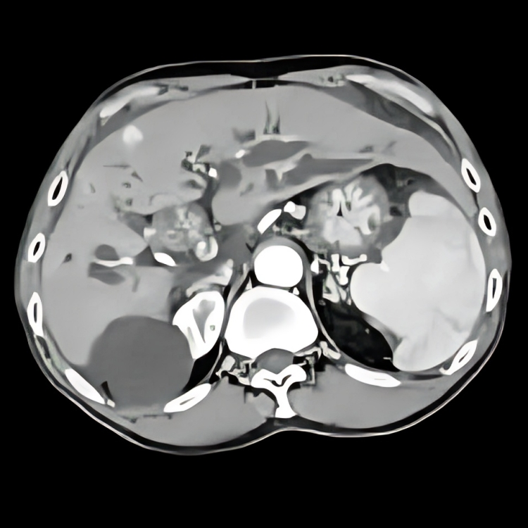
CT Scan
CT abdomen is the diagnostic test to confirm the diagnosis of bile duct cancer. It gives the three-dimensional imaging of the abdominal viscera. Moreover, the findings of the CT scan depend upon the type of bile duct cancer.
MRI
MRI of the abdomen gives a dimension structure of the liver parenchyma. Additionally, it also helps in the staging of bile duct cancer.
Percutaneous Transhepatic Cholangiography
It is advised for patients who are not fit for ERCP. In this test, a radiologist injects dye through a needle directly into your bile ducts and liver and takes an image.
Moreover, other imaging tests include endoscopic ultrasonography, ERCP, and MRCP.5
Darwin, P. E., MD. (n.d.). Cholangiocarcinoma treatment & management: approach considerations, stent placement, photodynamic therapy. https://emedicine.medscape.com/article/277393-treatment#d1
Staging of the Cholangiocarcinoma
The TNM staging of the bile duct cancer depends upon the location of the tumor. Moreover, it shows the spread of the tumor. The staging of bile duct cancer is as follows.
Intrahepatic Bile Duct Cancer:
- Stage 0: carcinoma in situ
- Stage 1A: Intrahepatic bile duct tumor less than 5mm
- Stage 1B: Intrahepatic bile duct tumor size greater than 5mm
- Stage 2: Tumor spreads into blood vessels
- Stage 3A: The tumor involves the capsule of the liver
- Stage 3B: The tumor involves nearby tissue and organs of the liver
- Stage 4: The distant spread of the tumor including bone, lungs, and lymph nodes
Perihilar Bile Duct Cancer:
- Stage 0: Carcinoma in situ
- Stage 1: Tumor in the inner layer of the perihilar bile duct involving the muscular wall of the perihilar bile duct wall
- Stage 2: The tumor spreads to the nearby fatty tissue
- Stage 3A: The tumor involves one side of the hepatic artery
- Stage 3B: The tumor involving the portal vein, common hepatic artery, and right hepatic duct
- Stage 3C: The tumor involves 1-3 lymph nodes
- Stage 4: The tumor involves more than 3 lymph nodes
Distal Bile Duct Cancer:
- Stage 0: Carcinoma in situ
- Stage 1: Tumor in the distal bile duct and involve less than 5 mm of the wall
- Stage 2A: Tumor involving less than 5mm of the wall and 1-3 lymph node
- Stage 2B: The tumor spread more than 5 mm of the wall
- Stage 3: The tumor involves more than 5 mm of the wall along with 3-4 lymph nodes and large vessels
- Stage 4: The tumor spreads to other parts and organs of the body
Histopathology:
Diagnosis of cholangiocarcinoma through tissue samples is difficult. However, the sample is taken through fine needle aspiration, brush cytology, and CT-guided biopsy. The tissue diagnosis is advised in patients having primary sclerosing cholangitis. About 95% of the cholangiocarcinoma are adenocarcinoma and 5 % are squamous.6Chung T, Park YN. Up-to-Date Pathologic Classification and Molecular Characteristics of Intrahepatic Cholangiocarcinoma. Front Med (Lausanne). 2022;9:857140.
How to treat Cholangiocarcinoma?
The treatment of cholangiocarcinoma depends upon the location and extent of the tumor. If the tumor is localized, it can be removed surgically. If it is unlocalized, surgery can not be done. Therefore, palliative care is given to patients with unresectable cholangiocarcinoma.
Surgical Treatment:
If the tumor is small and localized and is in the place of surgical safety, then surgical resection of the tumor is the only option. In surgical resection, the whole tumor, along with normal surrounding tissue, is removed to reduce recurrence, followed by chemotherapy or radiotherapy. The surgical procedures involving removing the tumor are:
Resection of the Bile Duct
If the tumor is small and is localized in the bile duct then surgery is done to remove the affected part of the bile duct followed by lymph node clearance. Finally, the resected part of the bile duct is sent for histopathology.
Partial Hepatectomy
In this surgical procedure, your doctor removes some parts of the liver where the tumor is present. The part of the liver that is removed depends upon the size of the tumor.
Whipple Procedure
In this procedure, your doctor will remove multiple organs. These organs are:
- Head of the pancreas
- Some parts of the stomach
- Some parts of the small intestine
- Gallbladder
- Bile duct
In the Whipple procedure, the surgeons do not remove the whole pancreas to maintain digestion and insulin release.7D’Cruz JR, Misra S, Menon G, et al. Pancreaticoduodenectomy (Whipple Procedure) [Updated 2024 Oct 6]. In: StatPearls [Internet]. Treasure Island (FL): StatPearls Publishing; 2025 Jan-. Available from: https://www.ncbi.nlm.nih.gov/books/NBK560747/
Liver Transplant
If the tumor has involved the maximum area of the liver, then the only option is to transplant the diseased liver with a healthy one. Moreover, a liver transplant is suitable for patients who have perihilar bile duct cancer.
Palliative Surgery
The purpose of this surgery is to relieve the symptoms of cholangiocarcinoma and to improve the quality of life. It is also suitable for patients who are not fit for surgery. It includes the following palliative procedures.
Biliary Bypass Procedure
Bypass surgery is done to change the normal route. In some patients, the bile duct is blocked, and bile accumulates in the bile duct and gallbladder. In the bypass procedure, surgeons cut off the bile duct above the blockage and fit it with the post-blockage duct or with the small intestine.
Stenting
In this procedure, the surgeon places a stent with the help of a camera in the bile duct where blockage is present. This stent helps drain the bile duct.
Photodynamic Therapy
It is a two-step procedure in which the blocked bile duct is restored. In this therapy, during the first step, a photosensitizer is administered to the body through an IV route, and in the second step, it is activated by light illumination at a specific wavelength.
Additionally, this therapy is indicated in patients in which a tumor is non-resectable. It improves the quality of life of the patients.
Radiation Therapy
Radiation therapy aims to kill the remaining cancerous particles of the tumor that are not suitable for removal with the help of surgery or in non-resectable tumors. Therefore, radiation therapy includes:
- External beam radiation therapy involves the bombardment of radiation from outside of the body on the affected area to destroy the cancerous cells.
- Radiosensitizer drugs are given to the patients which makes cancer cells more sensitive to radiation.
- Internal radiation therapy in which a radioactive material in the form of seed is placed at the site of the tumor.
Chemotherapy:
Some chemotherapeutic drugs can kill cancerous cells or to stop their proliferation. The chemotherapy includes:
Systemic Chemotherapy
In this therapy, the doctors give chemotherapeutic agents orally or inject through veins into the body to kill the cancerous cells. Therefore, systemic chemotherapeutic agents include:
- Cisplatin
- Capecitabine and oxaliplatin
- Gemcitabine and oxaliplatin
Regional Chemotherapy:
In this therapy, the chemotherapeutic agents are placed in a region where targeted cells are present.8Bile duct Cancer (Cholangiocarcinoma) Treatment (PDQ®). (2023, December 22). National Cancer Institute. https://www.cancer.gov/types/liver/hp/bile-duct-treatment-pdq
How does targeted therapy treat Cholangiocarcinoma?
In targeted therapy, specific drugs are used that target specific cells. Moreover, in this therapy, the normal cells are not damaged like in chemo and radiotherapy. These drugs are:
- Ivosidenib
- Pemigatinib
- Infigratinib
How does immunotherapy treat cholangiocarcinoma?
In immunotherapy, specific substances are used that use your body’s immune system to kill the cancerous cells. However, these substances are designed to boost the body’s natural immune system. These are:
- Pembrolizumab
- Durvalumab
How can I prevent having Cholangiocarcinoma?
You can not prevent having cholangiocarcinoma but you can reduce your risk of getting cholangiocarcinoma by:
- Vaccination against viruses such as Hep B, Hep C
- Avoid drinking alcohol
- Stop smoking
- Healthy exercise
- Maintain your body mass index
What is the Outcome?
The prognosis of the cholangiocarcinoma is not good. The prognosis of this disease depends upon the type and extent of the tumor. The 5-year survival rate of the patients in which the distant spread does not occur is about 10-15 %. However, it drops to 2% with the involvement of the vasculature and lymph nodes.
Conclusion
To conclude, cholangiocarcinoma is a cancer of the bile duct that manifests as abdominal pain, jaundice, yellowish discoloration of the skin, and weight loss. Moreover, the Treatment and prognosis of cholangiocarcinoma depend on the type of tumor and the staging and grading of the disease. If a tumor involves vessels and lymph nodes, the survival rate is very low.
Refrences
- 1Garikipati, S. C. (2023, February 6). Biliary tract cholangiocarcinoma. StatPearls – NCBI Bookshelf. https://www.ncbi.nlm.nih.gov/books/NBK560708/
- 2National Cancer Institute (US). (2023, June 2). Bile duct cancer (Cholangiocarcinoma) treatment. PDQ Cancer Information Summaries – NCBI Bookshelf. https://www.ncbi.nlm.nih.gov/books/NBK65851/
- 3What is bile duct cancer? | What is cholangiocarcinoma? (n.d.). American Cancer Society. https://amp.cancer.org/cancer/types/bile-duct-cancer/about/what-is-bile-duct-cancer.html
- 4Song J, Li Y, Bowlus CL, Yang G, Leung PSC, Gershwin ME. Cholangiocarcinoma in Patients with Primary Sclerosing Cholangitis (PSC): a Comprehensive Review. Clin Rev Allergy Immunol. 2020 Feb;58(1):134-149. doi: 10.1007/s12016-019-08764-7. PMID: 31463807.
- 5
Darwin, P. E., MD. (n.d.). Cholangiocarcinoma treatment & management: approach considerations, stent placement, photodynamic therapy. https://emedicine.medscape.com/article/277393-treatment#d1
- 6Chung T, Park YN. Up-to-Date Pathologic Classification and Molecular Characteristics of Intrahepatic Cholangiocarcinoma. Front Med (Lausanne). 2022;9:857140.
- 7D’Cruz JR, Misra S, Menon G, et al. Pancreaticoduodenectomy (Whipple Procedure) [Updated 2024 Oct 6]. In: StatPearls [Internet]. Treasure Island (FL): StatPearls Publishing; 2025 Jan-. Available from: https://www.ncbi.nlm.nih.gov/books/NBK560747/
- 8Bile duct Cancer (Cholangiocarcinoma) Treatment (PDQ®). (2023, December 22). National Cancer Institute. https://www.cancer.gov/types/liver/hp/bile-duct-treatment-pdq


