What is Demodex Blepharitis?
Blepharitis is a chronic inflammatory disease of the eyelid margins. It most commonly affects the ocular surface of your eye. Common symptoms include eye discharge, itching, redness, and abnormalities in eyelashes. In severe cases, it can involve your cornea, resulting in keratitis, and can also exacerbate the coexisting eye disease.1Rhee, M. K., Yeu, E., Barnett, M., Rapuano, C. J., Dhaliwal, D. K., Nichols, K. K., Karpecki, P., Mah, F. S., Chan, A. W., Mun, J., & Gaddie, I. B. (2023). Demodex Blepharitis: A Comprehensive review of the disease, current management, and emerging therapies. Eye & Contact Lens-science and Clinical Practice, 49(8), 311–318. https://doi.org/10.1097/icl.0000000000001003
There can be multiple causes of blepharitis, such as bacterial, fungal, and allergic, but one of the most common causes is Demodex infestation. Firstly, this case (Demodex infestation) was reported in 1876, and its association with blepharitis was established in 1976.2Fpao, M. R. R. M. M. (n.d.). Ocular Demodicosis (Demodex Infestation): background, pathophysiology, epidemiology. https://emedicine.medscape.com/article/1203895-overview#a4
Demodex is a microscopic ectoparasite (a parasite that lives externally) inhabiting the human body. There are two types of Demodex (Demodex folliculorum and Demodex brevis). Demodex folliculorum lives in small hair follicles, while Demodex brevis lives in sebaceous and meibomian glands. Both of these Demodex species are implicated in causing blepharitis. However, Demodex mites not only cause blepharitis but also induce other skin manifestations like perioral dermatitis, pityriasis folliculorum, demodicosis gravis, scabies, and basal cell carcinoma.3Liu, J., Sheha, H., & Tseng, S. C. (2010). Pathogenic role of Demodex mites in blepharitis. Current Opinion in Allergy and Clinical Immunology, 10(5), 505–510. https://doi.org/10.1097/aci.0b013e32833df9f4
Epidemiology
The prevalence of Demodex blepharitis increases with age. Notably, it affects more than 80 percent of individuals older than 60 and about 100 percent of individuals older than 70.4Rhee, M. K., Yeu, E., Barnett, M., Rapuano, C. J., Dhaliwal, D. K., Nichols, K. K., Karpecki, P., Mah, F. S., Chan, A. W., Mun, J., & Gaddie, I. B. (2023). Demodex Blepharitis: A Comprehensive review of the disease, current management, and emerging therapies. Eye & Contact Lens-science and Clinical Practice, 49(8), 311–318. https://doi.org/10.1097/icl.0000000000001003
Recently, in the US, two studies reported similar results of Demodex blepharitis in people of all ages. According to a study performed by Titan, it was found that about 58% of 1032 patients (out of which 69% are diagnosed cases of blepharitis) presented with discharge and debris at the base of lashes, which is the pathological sign of the Demodex blepharitis.5Cheng, A. M. S., Hwang, J., Dermer, H., & Galor, A. (2020). Prevalence of ocular demodicosis in an older population and its association with symptoms and signs of dry eye. Cornea, 40(8), 995–1001. https://doi.org/10.1097/ico.0000000000002542
In addition, despite frequent associations between blepharitis and demodex infestation, it remains undetectable. 6Liu, J., Sheha, H., & Tseng, S. C. (2010). Pathogenic role of Demodex mites in blepharitis. Current Opinion in Allergy and Clinical Immunology, 10(5), 505–510. https://doi.org/10.1097/aci.0b013e32833df9f47Fpao, M. R. R. M. M. (n.d.). Ocular Demodicosis (Demodex Infestation): background, pathophysiology, epidemiology. https://emedicine.medscape.com/article/1203895-overview#a4
Pathogenesis of Demodex Blepharitis
As we have discussed above, two species of Demodex, Demodex folliculorum and Demodex brevis, inhabit human skin. Both species have a glassy appearance and four short claw legs on microscopic examination.
Demodex folliculorum is about 0.3-0.4 mm and is found in cluster form on small hair follicles and eyelashes. It feeds on sebaceous fluid and lash epithelium, resulting in follicular distention and hyperplasia. Meanwhile, Demodex brevis is shorter and is found in sebaceous and meibomian glands. Moreover, the adult and immature forms of Demodex brevis consume the glands.
Demodex folliculorum is more prevalent than demodex brevis and is found in type 1 allergic reactions. According to research, it was noted that male and female demodex mites mate outside the hair follicles and glands, and female Demodex lay eggs inside the follicles and glands. Besides this, Demodex mites secrete digestive enzymes on adjacent sites that break the epithelial cells for feeding purposes. This digestive material remains in the gut of the mites and is spilled into the surroundings at the end of the life cycle.
The life cycle of Demodex mites is 14-23 days, and the progression is as follows:
Several possible mechanisms of Demodex mite contribute to blepharitis. These are:
Direct Damage:
As Demodex mites feed on epithelial cells, their short and sharp-edged claws cause microabrasions on the epithelial cells, leading to epithelial hyperplasia. These mites lay eggs at the base of the lashes, contributing to reactive conjunctivitis and persistent damage to the cornea, ultimately resulting in keratitis. However, in case of heavy infestation, the structure of hair follicles is distorted, and lashes become misdirected.
Furthermore, mites digest epithelial cells and excrete debris on nearby structures. This debris contains digestive enzymes (proteases and lipases) that cause direct damage to ocular structures. As a result, this damage may cause inflammation, irritation, and type 1 allergic reaction.
Follicular inflammation results in edema formation that leads to epilation of the eyelashes. Madarosis is the severe form in which the lashes are lost. It occurs when a large number of mites inhabit the eyelashes. Additionally, these mites cause edema of the hair shaft, resulting in hair loss.
Demodex brevis live in and feed on meibomian glands, resulting in gland loss (meibomian gland disease). These mites change the glands’ overall environment and architecture, making them favorable for infestation.
Bacterial Dysbiosis:
The mechanism of microbial association with Demodex mites is somewhat complex. According to the study by Fu et al., it was noted that Demodex mites interfere with the normal microbial environment of the conjunctiva. Demodex mites contain some bacteria on the surface (staphylococcus and streptococcus) and inside the mites’ abdomen (Bacillus oleronius). 8Fu, Y., Wu, J., Wang, D., Li, T., Shi, X., Li, L., Zhu, M., Zhang, Z., Yu, X., & Dai, Q. (2022). Metagenomic profiling of ocular surface microbiome changes in Demodex blepharitis patients. Frontiers in Cellular and Infection Microbiology, 12. https://doi.org/10.3389/fcimb.2022.922753
Hypersensitivity Reaction (Delayed):
In Demodex blepharitis, digestive enzymes (chitin) and other waste products boost inflammatory response, increasing the number of Macrophages, CD4 cells, and Langerhans cells. However, Demodex mites also trigger the formation of pro-inflammatory cytokines (Interleukin-1b and interleukin 17).9Fpao, M. R. R. M. M. (n.d.). Ocular Demodicosis (Demodex Infestation): background, pathophysiology, epidemiology. https://emedicine.medscape.com/article/1203895-overview#a4
According to Tarkowski et al., the debris of Demodex mites disrupts the epithelial safety barriers that cause chronic inflammation, resulting in the development of pterygium. These pro-inflammatory and inflammatory cells contribute to causing blepharitis.10Georgala, S., Katoulis, A. C., Kylafis, G., Koumantaki-Mathioudaki, E., Georgala, C., & Aroni, K. (2001). Increased density of Demodex folliculorum and evidence of delayed hypersensitivity reaction in subjects with papulopustular rosacea. Journal of the European Academy of Dermatology and Venereology, 15(5), 441–444. https://doi.org/10.1046/j.1468-3083.2001.00331.x
Risk Factors of Demodex Blepharitis
There can be multiple risk factors that contribute to Demodex blepharitis. These are rosacea, diabetes type 1 and type 2, blepharitis, immunosuppressant drugs, prolonged exposure to sun, alcoholism, and smoking. The following are the most common risk factors of Demodex blepharitis.
Dry Eyes:
Dry Eye is a disease in which the tear film decreases, making the environment favorable for Demodex mites. According to research studies, about 60-70 % of people with dry eye disease also present with Demodex Blepharitis.
Dysfunction of Meibomian Glands:
Demodex mites live inside the meibomian glands and damage the structure of the meibomian glands. Damage to meibomian glands depends upon the intensity of mites; the greater the number of mites, the more severe the damage is. According to studies, several authors have reported the association between Demodex blepharitis and meibomian gland dysfunction.
Additionally, patients with Demodex blepharitis have low meibum levels in the gland and high hydroxy fatty acids, which exacerbate meibomian gland dysfunction and result in tear film instability.
Rosacea:
Demodex mites and rosacea are closely related. Once Demodex mites increase in number, they begin to multiply and inhabit themselves around the eyes, causing ocular rosacea. Ocular rosacea is a clinical condition characterized by irregular lid margins, meibomian gland dysfunction, and itching. If ocular rosacea remains neglected, it can cause serious complications like chronic conjunctivitis and corneal perforation.
Moreover, patients suffering from rosacea are more prone to Demodex blepharitis than others. Demodex mites also cause other dermatological manifestations, such as acne vulgaris and seborrheic dermatitis.
Chalazion & Pterygium:
Demodex mites also have associations with chalazion and pterygium. Moreover, it is also noteworthy that inflammatory reactions induced by mite debris (chitin) may cause chalazion formation.11Rhee, M. K., Yeu, E., Barnett, M., Rapuano, C. J., Dhaliwal, D. K., Nichols, K. K., Karpecki, P., Mah, F. S., Chan, A. W., Mun, J., & Gaddie, I. B. (2023). Demodex Blepharitis: A Comprehensive review of the disease, current management, and emerging therapies. Eye & Contact Lens-science and Clinical Practice, 49(8), 311–318. https://doi.org/10.1097/icl.0000000000001003
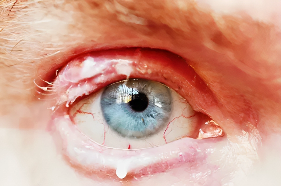
Signs & Symptoms of Demodex Blepharitis
Clinically, Demodex blepharitis is characterized by itching, conjunctival erythema, instability of tear films, irregular lid margin, vision problems, and foreign body sensation. Moreover, the signs of Demodex blepharitis are eyelash abnormality, meibomian gland dysfunction, and corneal and conjunctival inflammation. Symptoms of Demodex Blepharitis are:
Itching:
Itching is considered one of the most common symptoms of Demodex blepharitis. However, it occurs at night or early morning, while allergy-associated itching occurs during the daytime.
Collarette formation:
Collarettes are solidified exudative secretions that form a cylindrical collar around the base of the eyelash follicle. They are a key sign of Demodex blepharitis. In the literature, collarettes are often called cylindrical dandruff (CD), but they are also referred to as sleeves, cuffs, crusting, or lash debris.12Rhee, M. K., Yeu, E., Barnett, M., Rapuano, C. J., Dhaliwal, D. K., Nichols, K. K., Karpecki, P., Mah, F. S., Chan, A., Mun, J., & Gaddie, I. B. (2023). Demodex Blepharitis: A Comprehensive Review of the Disease, Current Management, and Emerging Therapies. Eye & Contact Lens, 49(8), 311-318. https://doi.org/10.1097/ICL.0000000000001003

Disorder of the Eyelashes:
Prolonged infestation of Demodex mites results in malignant transformation (trichiasis and madarosis). Moreover, the characteristic feature of trichiasis is foreign body sensation, which results in corneal ulceration and perforation if it remains untreated.
Meibomian Glands Abnormality:
Demodex mites block the meibomian gland duct. It results in swelling, inflammation, and infection of the gland that interferes with the rear film formation. However, the granulomatous reaction in sebaceous glands leads to the formation of hordeolum or Chalazion.
Inflammation of Lid Margin:
As discussed above, Demodex mites induce cascades of inflammatory reactions resulting in inflammation and lid margin edema.13Rhee, M. K., Yeu, E., Barnett, M., Rapuano, C. J., Dhaliwal, D. K., Nichols, K. K., Karpecki, P., Mah, F. S., Chan, A. W., Mun, J., & Gaddie, I. B. (2023). Demodex Blepharitis: A Comprehensive review of the disease, current management, and emerging therapies. Eye & Contact Lens-science and Clinical Practice, 49(8), 311–318. https://doi.org/10.1097/icl.0000000000001003
Conjunctival Manifestations:
Prolonged lid inflammation and an unhygienic environment cause the spread of inflammation in the conjunctiva, resulting in blepharoconjunctivitis.
Corneal Manifestations:
Long-standing inflammation damages your cornea. Demodex-induced corneal manifestations are :
- Superficial corneal vascularization
- Corneal opacity
- Corneal ulceration
- Nodule formation14Fpao, M. R. R. M. M. (n.d.). Ocular Demodicosis (Demodex Infestation): background, pathophysiology, epidemiology. https://emedicine.medscape.com/article/1203895-overview#a4
How to Diagnose Demodex Blepharitis?
Diagnosis of Demodex blepharitis is mainly based on history, physical examination, and laboratory investigation.
History:
When diagnosing demodex blepharitis, doctors inquire about various aspects of the patient’s history. They usually ask about the biodata, onset, and timing of eye itching, presence of eye discharge, history of previous eye infections, sensation of foreign bodies in the eyes, and occurrence of blurry vision.
Additionally, they might also gather information on current medications, existing medical conditions (such as diabetes or asthma), alcohol consumption, smoking habits (including the number of packs per day), family medical history, socioeconomic status, and environmental factors. This comprehensive approach helps gather relevant information for a thorough diagnosis.
Physical Examination:
Gross physical observations include:
- Inspection of the eyes to check for any discharge, irregularity of lid margin, loss of eyelashes, and other dermatological manifestations.
- Check for vision abnormality.
Slit Lamp Examination:
Slit lamp examination shows cylindrical dandruff at the base of eyelashes, which is a characteristic feature of Demodex blepharitis.
Microscopic Examination:
During the microscopic examination, your doctor may be able to see the intensity of infestations, such as the number of eggs, larvae, and adult mites.

Laboratory Investigations:
Laboratory investigations are usually not required but can be advised in case of infections. These are :
- Complete blood count
- C reactive protein (CRP)
- HBA1C
- White blood cell count
- Nasal skin scraping
What is the Collarette Grading Scale?
The collarette grading scale is used to analyze the efficacy of the treatment. There are about five grades. These are:
|
when there is involvement of 0-2 lashes with collar formation
3-10 lashes with collar formation indicate grade 1 More than ten but less than ⅓ of lashes with collar formation ⅓ of lashes with collar formation indicate grade 3 ⅔ or more lashes with collar formation indicates grade 4 |
How to Treat Demodex Blepharitis?
It is critical to diagnose and treat Demodex blepharitis at early stages to avoid complications. The management of Demodex blepharitis is divided into in-office management and at-home management.
In-office Management:
In-office Demodex blepharitis management includes using volatile fluids (ether) followed by brushing and 0.5 % proparacaine instillation, followed by washing with 70 percent alcohol after 5 minutes. This treatment successfully reduced the symptoms and number of eggs after three weeks of follow-up. However, it is necessary to use this volatile substance with caution to avoid corneal involvement.
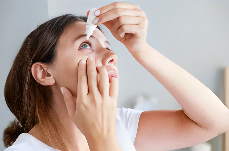
At-Home Management:
Your doctor may also suggest at-home management of Demodex blepharitis, such as scrubbing the eyes with baby shampoo (to maintain a hygienic environment) and applying topical antibiotics until symptoms resolve.
1% Mercury oxide is used to control the spread of mites. This ointment is applied over the base of eyelashes, trapping mites as they emerge from the follicles. Pilocarpine gel also helps reduce the number of mites because it halts the respiratory movement and motility of mites, resulting in their death.
Ivermectin 1% (Antihelmintic):
It is an FDA-approved drug for treating rosacea. It is applied to eyelashes and eyelids and dramatically reduces the symptoms of Demodex mites.
Tea Tree Oil:
Tea tree oil is the best treatment regime for Demodex blepharitis. After debridement, tea tree oil is applied over the root of the lashes and eyelid to kill the eggs. Moreover, it should be applied thrice after a 10-minute gap to get maximum results. Some studies also report that daily massage with 5% tea tree oil can significantly reduce symptoms.
Pharmacological Management of Demodex Blepharitis:
The pharmacological management includes the following medications to treat blepharitis. These are the following:
Antibiotics:
To control the spread of the disease, comprehensive antibiotic therapy is advised. Erythromycin is a potent antibiotic that can treat ocular infections.
Lotilaner Ophthalmic:
It is a gamma amino butyric acid chloride channel selective inhibitor for mites. This drug causes paralysis of the target organism, resulting in the death of mites. Besides this, it is suitable for adult patients to treat Demodex blepharitis.15 Gaddie I, Donnenfeld E, Karpecki P, et al. “Review of: Lotilaner ophthalmic solution 0.25% for Demodex blepharitis: Randomized, vehicle-controlled, multicenter, phase 3 trial (Saturn-2).” Ophthalmology, in press, 2023.
Patient Education:
After giving a prescription for the treatment of Demodex blepharitis, patient education is necessary to educate about the use of medications and adapt precautionary measures:
- Scrub your eyelashes twice a day with the help of baby shampoo by using a cotton swab
- Do not rub your eyes with your hands to further inflammation
- Consult a dermatologist if there are any dermatological manifestation
If the patient is not responding to treatment, consider non-compliance; if he is compliant, rule out other possible causes.
What is the Prognosis of Demodex Blepharitis?
The symptomatic relief of Demodex blepharitis is excellent. While the total eradication of the Demodex mites is unlikely, proper hygiene can help reduce the number of mites to an acceptable level.
Some reports show 100 percent eradication of the mites with the help of tea tree oil and ivermectin 1%. Recurrence can occur due to poor hygiene.16Liu, J., Sheha, H., & Tseng, S. C. (2010). Pathogenic role of Demodex mites in blepharitis. Current Opinion in Allergy and Clinical Immunology, 10(5), 505–510. https://doi.org/10.1097/aci.0b013e32833df9f4
Conclusion
To conclude, Demodex Blepharitis is a chronic inflammation of the eyelids that involves a reduction in tear film, discharges from eyes, itching, redness, and lash abnormalities. Bacteria, fungi, and allergens can cause it. Moreover, in-office management, at-home treatment, and other pharmacological medications can treat it. Demodex Blepharitis has an excellent prognosis if the patient practices hygienic measures.
Refrences
- 1Rhee, M. K., Yeu, E., Barnett, M., Rapuano, C. J., Dhaliwal, D. K., Nichols, K. K., Karpecki, P., Mah, F. S., Chan, A. W., Mun, J., & Gaddie, I. B. (2023). Demodex Blepharitis: A Comprehensive review of the disease, current management, and emerging therapies. Eye & Contact Lens-science and Clinical Practice, 49(8), 311–318. https://doi.org/10.1097/icl.0000000000001003
- 2Fpao, M. R. R. M. M. (n.d.). Ocular Demodicosis (Demodex Infestation): background, pathophysiology, epidemiology. https://emedicine.medscape.com/article/1203895-overview#a4
- 3Liu, J., Sheha, H., & Tseng, S. C. (2010). Pathogenic role of Demodex mites in blepharitis. Current Opinion in Allergy and Clinical Immunology, 10(5), 505–510. https://doi.org/10.1097/aci.0b013e32833df9f4
- 4Rhee, M. K., Yeu, E., Barnett, M., Rapuano, C. J., Dhaliwal, D. K., Nichols, K. K., Karpecki, P., Mah, F. S., Chan, A. W., Mun, J., & Gaddie, I. B. (2023). Demodex Blepharitis: A Comprehensive review of the disease, current management, and emerging therapies. Eye & Contact Lens-science and Clinical Practice, 49(8), 311–318. https://doi.org/10.1097/icl.0000000000001003
- 5Cheng, A. M. S., Hwang, J., Dermer, H., & Galor, A. (2020). Prevalence of ocular demodicosis in an older population and its association with symptoms and signs of dry eye. Cornea, 40(8), 995–1001. https://doi.org/10.1097/ico.0000000000002542
- 6Liu, J., Sheha, H., & Tseng, S. C. (2010). Pathogenic role of Demodex mites in blepharitis. Current Opinion in Allergy and Clinical Immunology, 10(5), 505–510. https://doi.org/10.1097/aci.0b013e32833df9f4
- 7Fpao, M. R. R. M. M. (n.d.). Ocular Demodicosis (Demodex Infestation): background, pathophysiology, epidemiology. https://emedicine.medscape.com/article/1203895-overview#a4
- 8Fu, Y., Wu, J., Wang, D., Li, T., Shi, X., Li, L., Zhu, M., Zhang, Z., Yu, X., & Dai, Q. (2022). Metagenomic profiling of ocular surface microbiome changes in Demodex blepharitis patients. Frontiers in Cellular and Infection Microbiology, 12. https://doi.org/10.3389/fcimb.2022.922753
- 9Fpao, M. R. R. M. M. (n.d.). Ocular Demodicosis (Demodex Infestation): background, pathophysiology, epidemiology. https://emedicine.medscape.com/article/1203895-overview#a4
- 10Georgala, S., Katoulis, A. C., Kylafis, G., Koumantaki-Mathioudaki, E., Georgala, C., & Aroni, K. (2001). Increased density of Demodex folliculorum and evidence of delayed hypersensitivity reaction in subjects with papulopustular rosacea. Journal of the European Academy of Dermatology and Venereology, 15(5), 441–444. https://doi.org/10.1046/j.1468-3083.2001.00331.x
- 11Rhee, M. K., Yeu, E., Barnett, M., Rapuano, C. J., Dhaliwal, D. K., Nichols, K. K., Karpecki, P., Mah, F. S., Chan, A. W., Mun, J., & Gaddie, I. B. (2023). Demodex Blepharitis: A Comprehensive review of the disease, current management, and emerging therapies. Eye & Contact Lens-science and Clinical Practice, 49(8), 311–318. https://doi.org/10.1097/icl.0000000000001003
- 12Rhee, M. K., Yeu, E., Barnett, M., Rapuano, C. J., Dhaliwal, D. K., Nichols, K. K., Karpecki, P., Mah, F. S., Chan, A., Mun, J., & Gaddie, I. B. (2023). Demodex Blepharitis: A Comprehensive Review of the Disease, Current Management, and Emerging Therapies. Eye & Contact Lens, 49(8), 311-318. https://doi.org/10.1097/ICL.0000000000001003
- 13Rhee, M. K., Yeu, E., Barnett, M., Rapuano, C. J., Dhaliwal, D. K., Nichols, K. K., Karpecki, P., Mah, F. S., Chan, A. W., Mun, J., & Gaddie, I. B. (2023). Demodex Blepharitis: A Comprehensive review of the disease, current management, and emerging therapies. Eye & Contact Lens-science and Clinical Practice, 49(8), 311–318. https://doi.org/10.1097/icl.0000000000001003
- 14Fpao, M. R. R. M. M. (n.d.). Ocular Demodicosis (Demodex Infestation): background, pathophysiology, epidemiology. https://emedicine.medscape.com/article/1203895-overview#a4
- 15Gaddie I, Donnenfeld E, Karpecki P, et al. “Review of: Lotilaner ophthalmic solution 0.25% for Demodex blepharitis: Randomized, vehicle-controlled, multicenter, phase 3 trial (Saturn-2).” Ophthalmology, in press, 2023.
- 16Liu, J., Sheha, H., & Tseng, S. C. (2010). Pathogenic role of Demodex mites in blepharitis. Current Opinion in Allergy and Clinical Immunology, 10(5), 505–510. https://doi.org/10.1097/aci.0b013e32833df9f4
