Dystrophic Epidermolysis Bullosa is a rare hereditary skin disorder that causes the skin, mucous membranes, and nails to become highly fragile. It presents with marked blistering and scarring all over the body. It is also known as the “butterfly skin disease” or the “zebra skin” condition.
What is Dystrophic Epidermolysis Bullosa?
It is a severe condition. It is one of the many forms of epidermolysis bullosa. Epidermolysis bullosa is a group of skin conditions lacking proteins that hold the skin together. As a result, your body’s epithelial surfaces undergo significant damage.
The term has roots in Greek and Latin. “Dystrophic” means “abnormal growth” in Greek, referring to the abnormal growth pattern of a tissue in your body. “Epidermolysis” is a Greek term that refers to “the breaking down of skin layers.” “Bulla” means a bubble or blister in Latin.
It is generally a lifelong condition, and the prognosis depends on the severity of your disease.1NCI Dictionary of Cancer Terms. (2024). Cancer.gov; Cancer.gov. https://www.cancer.gov/publications/dictionaries/cancer-terms/def/dystrophic-epidermolysis-bullosa
Prevalence of Dystrophic Epidermolysis Bullosa
Currently, over 25,000 to 50,000 people in the United States are estimated to have this condition.2Epidermolysis Bullosa Clinic Frequently Asked Questions. (2019). Dermatology. https://med.stanford.edu/dermatology/resources/gsdc/eb_clinic/eb-faqs.html
However, new estimates show that the incidence of dystrophic epidermolysis recessive is 95 per a million live births. It is a rare disorder. 3Eichstadt, S., Tang, J. Y., Solis, D. C., Zurab Siprashvili, M Peter Marinkovich, Whitehead, N., Schu, M., Fang, F., Erickson, S. W., Ritchey, M. E., Colao, M., Spratt, K., Amir Shaygan, Ahn, M. J., & Sarin, K. Y. (2019). From Clinical Phenotype to Genotypic Modelling: Incidence and Prevalence of Recessive Dystrophic Epidermolysis Bullosa (RDEB). Clinical, Cosmetic and Investigational Dermatology, Volume 12, 933–942. https://doi.org/10.2147/ccid.s232547
Causes of Dystrophic Epidermolysis Bullosa
In this condition, there is a mutation in the COL7A1 gene. The COL7A1 gene is a gene inside your cells. It is vital for the production of collagen, especially type 7 collagen. Type 7 collagen is important for the integrity of your skin, as it binds together the different skin layers.
As a result of abnormal genetic mutations, antibodies against collagen type 7 are formed. The various skin layers do not bind well together. The autoimmune response also leads to inflammatory changes in the body.
Therefore, there is the formation of blisters and skin changes all over the body. In dystrophic epidermolysis bullosa, blisters specifically grow in the middle of the “dermis” layer of your skin. Even the slightest mechanical stress can cause skin layers to separate.4Satoru Shinkuma. (2015). Dystrophic epidermolysis bullosa: a review. Clinical, Cosmetic and Investigational Dermatology, 275–275. https://doi.org/10.2147/ccid.s54681
Types of Dystrophic Epidermolysis Bullosa
There are two main types which are determined by its inheritance pattern.
Recessive Dystrophic Epidermolysis Bullosa
In recessive DEB, you receive two abnormal copies of the COL7A1 gene. It is known for being more severe than its dominant counterpart. There are four subtypes.
| Subtype | Manifestation |
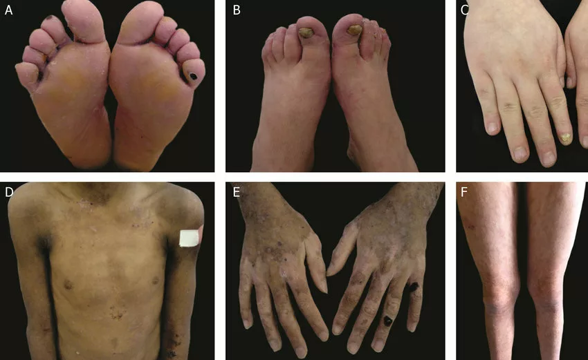
| Recessive Dystrophic Epidermolysis Bullosa Hallopeau-Siemens | It is the most serious form of dystrophic epidermolysis bullosa. Usually diagnosed in infancy, it causes severe blistering of the skin, nails, and mucosal surfaces, e.g., the mouth and esophagus. |
| Recessive Dystrophic Epidermolysis Bullosa non-Hallopeau-Siemens | This, too, causes severe blistering but does not follow the pattern of the Hallopeau-Siemens variant. It is slightly milder. |
| Recessive Dystrophic Epidermolysis Bullosa Inversa | The blistering and scarring occur at the flexures of your skin. Regions include skinfolds like the armpit, groin, and back of the knees. |
| Recessive Dystrophic Epidermolysis Bullosa Centripetalis | Symptoms begin at your extremities, like your hands and feet, and then spread towards your trunk. |
Dominant Dystrophic Epidermolysis Bullosa
In dominant DEB, just receiving one copy of the mutated COL7A1 gene results in clinical manifestations of the disease. It has the following subtypes: 5Klausegger, A., Niklas Jeschko, Grammer, M., Cemper-Kiesslich, J., Franz Neuhuber, Diem, A., Breitenbach-Koller, H., Sander, G., Dieter Kotzot, Johann Wolfgang Bauer, & Laimer, M. (2022). Recessive Dystrophic Epidermolysis bullosa due to Hemizygous 40 kb Deletion of COL7A1 and the Proximate PFKFB4 Gene Focusing on the Mutation c.425A>G Mimicking Homozygous Status. Diagnostics, 12(10), 2460–2460. https://doi.org/10.3390/diagnostics12102460
| Subtype | Manifestation |
| Dominant Dystrophic Epidermolysis Bullosa Pasini | It causes blisters on different parts of your body that leave scars upon resolution. It is milder than the recessive form of the disorder. |
| Dominant Dystrophic Epidermolysis Bullosa Cockayne-Touraine | It causes blistering as well as other signs such as brittle nails, neurological abnormalities and damage to mucosal membranes. |
| Dominant Dystrophic Epidermolysis Bullosa Pretibial | It causes blistering and skin damage to the shin area. |
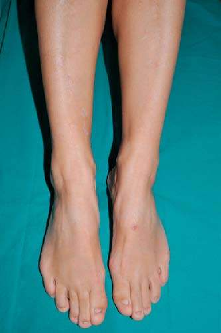
Symptoms of Dystrophic Epidermolysis Bullosa
The symptoms are mostly related to the skin, mucosal membranes, and surrounding organs. They include:
- Marked blistering on the skin
- Even gentle pressure and minor trauma can lead to blisters and open wounds
- Scars that form on the skin after the blisters heal
- Small-sized white bumps on the skin
- Itching or pruritus
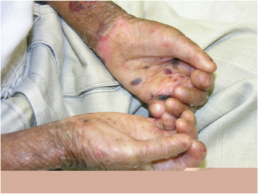
- Contractures or shortening of muscles after repeated damage to the skin and nearby soft tissue
- Brittle nails
- Lesions and ulcers of lips, gums, mouth, and esophagus that cause trouble eating and swallowing6Oliveira, T. M., Vivien Thiemy Sakai, Candido, L. A., Silva, & Aparecida, M. (2008). Clinical management for epidermolysis bullosa dystrophica. Journal of Applied Oral Science, 16(1), 81–85. https://doi.org/10.1590/s1678-77572008000100016
Complications of Dystrophic Epidermolysis Bullosa
If left unmanaged, the symptoms of epidermolysis can cause a number of complications:
- Gastrointestinal problems can cause malnutrition. In children, they can also lead to failure to thrive and developmental delays.
- Open sores cause infections, especially in immunocompromised individuals
- Bleeding from wounds and mucosal surfaces of internal organs can cause anemia, and symptoms of anemia include fatigue, dizziness, and breathlessness.
- Eye problems are caused by damage to mucosal surfaces of the eyes. E.g., corneal abrasion, blindness.
- Recessive dystrophic epidermolysis bullosa has been linked with squamous carcinoma of the skin.
Diagnosis of Dystrophic Epidermolysis Bullosa
Your doctor will suspect you have DEB, if you have a history of recurrent blistering on minor frictional trauma, especially if it started when you were an infant or child.
History
Your doctor will take a detailed history of your complaints. They will ask you questions such as “When did you first notice you were blistering?” and “How much pressure triggers your blisters?”
In addition, you will be asked about any symptoms similar to those in your family. As with any other hereditary disorder, dystrophic epidermolysis runs in families.
Physical Exam
You will undergo a thorough skin assessment. Your doctor will check your skin for any blisters, wounds, thinness, and fragility. They may induce the formation of blisters near the affected areas, too. They will check the sites, size, and severity of your symptoms, as well as the associated pain and loss of function.
To differentiate between dystrophic epidermolysis bullosa and other skin conditions, your doctor will specifically look for:
- Scarring
- White spots/ milia
- Brittleness and breakage of nails
- Involvement of the mouth and oral cavity
- Involvement of other organs
They also check for nutritional status and delayed developmental milestones in children. In the case of infants, they specifically check for diaper rashes because they are areas of constant friction and can lead to the symptoms of this condition.
Laboratory Investigations
Lab investigations are the mainstay diagnosis. A small tissue sample is taken from affected sites and sent for analysis.
Skin Biopsy
First, a skin biopsy is performed to identify the extent of involvement of various skin layers. The doctor induces the formation of a blister by rubbing the eraser end of a pencil against your skin. They then study a small sample from the area under a microscope.
Immunofluorescence Mapping
Immunofluorescence mapping employs the use of fluorescent antibodies. These antibodies attach to various biomolecules involved in the pathogenesis of dystrophic epidermolysis bullosa. It gives information about the skin layers involved, the extent of skin damage as well as the underlying defects.
Transmission Electron Microscopy
Skin samples are viewed under a high-resolution electron microscope in transmission electron microscopy. This way, subcellular changes caused by dystrophic epidermolysis bullosa can be visualized more easily. It helps determine the exact subtype of epidermolysis and monitor the effectiveness of treatment plans.
Genetic Testing
Genetic testing often involves taking a blood or saliva sample. Then, DNA is extracted from this sample and analyzed using different techniques. It identified any mutations present in the COL7A1 gene, even if you are only an asymptomatic carrier. 7Has, C., Liu, L., Bolling, M. C., Charlesworth, A. V., M. El Hachem, M.J. Escámez, Fuentes, I., S. Büchel, R. Hiremagalore, G. Pohla‐Gubo, van, K. Wertheim‐Tysarowska, & G. Zambruno. (2019). Clinical practice guidelines for laboratory diagnosis of epidermolysis bullosa. British Journal of Dermatology/British Journal of Dermatology, Supplement, 182(3), 574–592. https://doi.org/10.1111/bjd.18128
How is Dystrophic Epidermolysis Bullosa Treated?
There is no cure for dystrophic epidermolysis bullosa. However, it can be managed well with different treatments for symptom relief.
Topical Therapy
A number of moisturizers, creams, wound dressings, and ointments can be applied to the blisters to soothe and heal the affected areas of your skin. They often include emollients, soothing gels, and silver compounds. Wounds need to be cleaned and dressed regularly.
Analgesics
For mild cases, acetaminophen and NSAIDs are used. However, severe dystrophic epidermolysis bullosa may require opioids such as fentanyl. In addition, for nerve damage and its resultant pain, your doctor may prescribe antidepressants and antiepileptic drugs.
Corticosteroids
Corticosteroids help reduce inflammation and swelling. They can be applied topically to improve healing as well as taken orally in case of severe disease.
Antihistamines
Antihistamines or antiallergics reduce the usual itching and discomfort caused by epidermolysis bullosa.
Antibiotics
Open wounds are a gateway for bacteria and other microorganisms. Therefore, it is necessary to use topical antibiotics. Sometimes, oral therapy may also be required. Commonly used antibiotics include:
- Polymyxin
- Bacitracin
- Gentamicin
- Minocycline
Laser Therapy
Laser therapy is used to improve scar reduction and wound healing. They help the skin regenerate. They target specific regions of your skin and can reduce symptoms, e.g., itching caused by dystrophic epidermolysis bullosa.
Lifestyle Changes
This condition can become manageable with changes in your lifestyle and habits.
- Avoid contact sports
- Wear loose, soft clothes
- Avoid abrasive fabrics
- Use gentle, antibacterial bath salts, and always moisturize your skin
- Avoid any activities that can cause a break in your mucous membranes e.g., checking your temperature rectally
- Take care of your bandages and dressings
Surgery
Surgery is helpful in treating and preventing the complications of dystrophic epidermolysis bullosa, such as:
- Releasing contractures
- Separating fused fingers
- Dilating narrowed esophagus
- Skin grafting
Gene Therapy
Gene therapy is the most studied new treatment for dystrophic epidermolysis bullosa. It has recently been FDA-approved as a topical treatment for the disorder. It involves the introduction of new genetic material into the body as a replacement for defective genes.
Cell-Based Therapies
Cell-based therapies are being tested as a treatment for this condition. For instance, injections of fibroblasts can improve the anchoring of different skin layers to each other.
Protein Therapy
Recessive DEB causes a deficiency in C7 proteins. So, in protein therapy, doctors intravenously administer these to the affected areas of your skin. 8UpToDate. (2024). Uptodate.com. https://www.uptodate.com/contents/overview-of-the-management-of-epidermolysis-bullosa
Prevention of Dystrophic Epidermolysis Bullosa
The only way to prevent this condition is through genetic counseling and early termination of pregnancy after prenatal testing. Because there is no cure, genetic counseling is vital for all families who are affected by it.
It has both dominant and autosomal presentation. If you have the dominant variant, you are 50% likely to pass it down to your children, even if your partner doesn’t have it.
Similarly, for recessive DEB, two faulty copies of each gene are required for the disease to manifest. If you are a carrier whose partner is another carrier, there is a 25% chance you will pass the recessive trait onto your children.
It can also be diagnosed in utero. Fetal samples are obtained through sampling methods such as chorionic villi sampling. Then, a gene analysis is conducted to identify possible mutations in the COL7A1 gene. 9Dystrophic epidermolysis bullosa – About the Disease – Genetic and Rare Diseases Information Center. (2024). Nih.gov. https://rarediseases.info.nih.gov/diseases/2150/dystrophic-epidermolysis-bullosa
Life Expectancy
If you have mild DEB, your life expectancy will remain unaffected. However, in cases of severe disease, life expectancy usually hovers between infancy and mid-thirties. Severe recessive disease is associated with a much poorer prognosis. 10Fine, J.-D. (2010). Inherited epidermolysis bullosa. Orphanet Journal of Rare Diseases, 5(1). https://doi.org/10.1186/1750-1172-5-12
Conclusion
This is a rare skin condition that runs in families. It is also known as the butterfly skin disease because of how fragile it makes a person’s skin. DEB causes widespread blistering and scarring all over the body. It is manageable with treatment, and different medical advancements can provide relief from symptoms. There is no cure for it, but mild cases have an excellent prognosis.
Refrences
- 1NCI Dictionary of Cancer Terms. (2024). Cancer.gov; Cancer.gov. https://www.cancer.gov/publications/dictionaries/cancer-terms/def/dystrophic-epidermolysis-bullosa
- 2Epidermolysis Bullosa Clinic Frequently Asked Questions. (2019). Dermatology. https://med.stanford.edu/dermatology/resources/gsdc/eb_clinic/eb-faqs.html
- 3Eichstadt, S., Tang, J. Y., Solis, D. C., Zurab Siprashvili, M Peter Marinkovich, Whitehead, N., Schu, M., Fang, F., Erickson, S. W., Ritchey, M. E., Colao, M., Spratt, K., Amir Shaygan, Ahn, M. J., & Sarin, K. Y. (2019). From Clinical Phenotype to Genotypic Modelling: Incidence and Prevalence of Recessive Dystrophic Epidermolysis Bullosa (RDEB). Clinical, Cosmetic and Investigational Dermatology, Volume 12, 933–942. https://doi.org/10.2147/ccid.s232547
- 4Satoru Shinkuma. (2015). Dystrophic epidermolysis bullosa: a review. Clinical, Cosmetic and Investigational Dermatology, 275–275. https://doi.org/10.2147/ccid.s54681
- 5Klausegger, A., Niklas Jeschko, Grammer, M., Cemper-Kiesslich, J., Franz Neuhuber, Diem, A., Breitenbach-Koller, H., Sander, G., Dieter Kotzot, Johann Wolfgang Bauer, & Laimer, M. (2022). Recessive Dystrophic Epidermolysis bullosa due to Hemizygous 40 kb Deletion of COL7A1 and the Proximate PFKFB4 Gene Focusing on the Mutation c.425A>G Mimicking Homozygous Status. Diagnostics, 12(10), 2460–2460. https://doi.org/10.3390/diagnostics12102460
- 6Oliveira, T. M., Vivien Thiemy Sakai, Candido, L. A., Silva, & Aparecida, M. (2008). Clinical management for epidermolysis bullosa dystrophica. Journal of Applied Oral Science, 16(1), 81–85. https://doi.org/10.1590/s1678-77572008000100016
- 7Has, C., Liu, L., Bolling, M. C., Charlesworth, A. V., M. El Hachem, M.J. Escámez, Fuentes, I., S. Büchel, R. Hiremagalore, G. Pohla‐Gubo, van, K. Wertheim‐Tysarowska, & G. Zambruno. (2019). Clinical practice guidelines for laboratory diagnosis of epidermolysis bullosa. British Journal of Dermatology/British Journal of Dermatology, Supplement, 182(3), 574–592. https://doi.org/10.1111/bjd.18128
- 8UpToDate. (2024). Uptodate.com. https://www.uptodate.com/contents/overview-of-the-management-of-epidermolysis-bullosa
- 9Dystrophic epidermolysis bullosa – About the Disease – Genetic and Rare Diseases Information Center. (2024). Nih.gov. https://rarediseases.info.nih.gov/diseases/2150/dystrophic-epidermolysis-bullosa
- 10Fine, J.-D. (2010). Inherited epidermolysis bullosa. Orphanet Journal of Rare Diseases, 5(1). https://doi.org/10.1186/1750-1172-5-12
