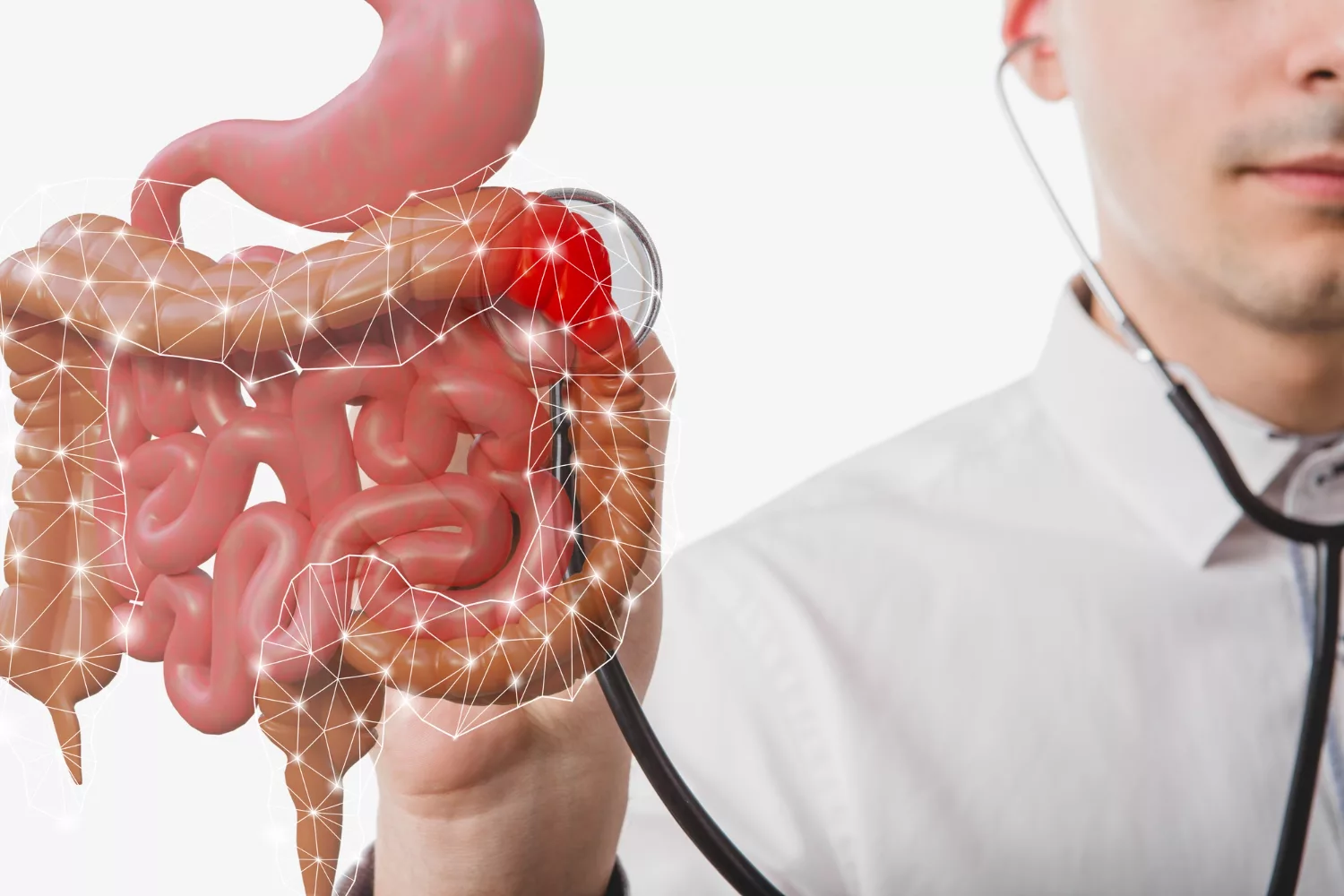
Esophageal adenocarcinoma is a type of esophageal cancer that arises from the glandular cells in the lower part of the esophagus, near the junction with the stomach. The esophagus, also known as the food pipe, is a hollow muscular tube responsible for moving food down from the throat to the stomach. Esophageal cancers usually begin in the inner lining (mucosa) and can affect any part of the esophagus. The types of esophageal cancer are:1 Types of Esophageal Cancer. Memorial Sloan Kettering Cancer Center. Published 2020. https://www.mskcc.org/cancer-care/types/esophageal/types-esophageal
- Squamous cell carcinoma
- Adenocarcinoma
- Small cell carcinoma
Generally, tumors start as mucosal lesions (the mucosa is the inner lining of the esophagus), but disease progression leads to the involvement of deeper layers such as the submucosa, muscular layer, etc. Unlike squamous cell carcinoma, which develops from the normal squamous lining of the esophagus, adenocarcinoma originates from columnar cells that have undergone abnormal changes due to chronic acid reflux (GERD) or Barrett’s esophagus.
How Does Esophageal Adenocarcinoma Develop?
The esophagus, or food pipe, is normally lined with stratified squamous cells, which are not designed to withstand repeated exposure to stomach acid.
However, chronic acid reflux (GERD) can cause long-term irritation, triggering a process called intestinal metaplasia, in which squamous cells are replaced by columnar glandular cells that resemble those in the intestine. This condition, known as Barrett’s esophagus, is a precursor to esophageal adenocarcinoma.
What is Barrett’s Esophagus?
Barrett’s esophagus is a condition in which the normal whitish-pink esophageal lining is replaced by salmon-colored intestinal cells, visible during endoscopy. This transformation increases the risk of developing esophageal adenocarcinoma, especially if the new cells become dysplastic (abnormal growth). However, only a small percentage of Barrett’s esophagus cases progress to cancer.2Understanding Your Pathology Report: Barrett’s Esophagus and Dysplasia. www.cancer.org. https://www.cancer.org/cancer/diagnosis-staging/tests/biopsy-and-cytology-tests/understanding-your-pathology-report/esophagus-pathology/barrets-esophagus.html#:~:text=When%20goblet%20cells%20are%20found Barrett’s esophagus generally occurs at the junction of the stomach and the esophagus.
How Is Esophageal Adenocarcinoma Different from Squamous Cell Carcinoma?
Esophageal adenocarcinoma is a cancer that primarily affects the mucus-secreting glands of the esophagus, with a predominant location in the lower third of the organ. This form of esophageal cancer is prevalent in the United States. This type of cancer is more associated with acid reflux.
On the other hand, squamous cell carcinoma involves the flat, thin cells that line the esophagus. Unlike adenocarcinoma, squamous cell carcinoma is more widespread globally. It tends to affect the upper two-thirds of the esophagus. This type of cancer is more associated with tobacco and alcohol use.
How Common Are Esophageal Cancers?
Esophageal cancers rank as the fifth most common type of gastrointestinal cancer in the US. Globally, they stand as the sixth most prevalent cancer.3 Recio-Boiles A, Babiker HM. Esophageal Cancer. PubMed. Published 2020. https://www.ncbi.nlm.nih.gov/books/NBK459267/ In the US, the incidence of esophageal adenocarcinoma is on the rise, primarily due to an increase in obesity and gastroesophageal reflux disease (GERD). Simultaneously, there is a consistent decrease in squamous cell cancer due to reductions in tobacco use and alcohol consumption. Esophageal adenocarcinoma predominantly affects white males. Conversely, esophageal squamous cell carcinoma has higher incidence rates among blacks and Asians.
Risk Factors for Esophageal Adenocarcinoma
Long-lasting irritation to the esophagus due to acid reflex contributes to the risk of developing esophageal cancer.4Pohl H, Wrobel K, Bojarski C, et al. Risk factors in the development of esophageal adenocarcinoma. Am J Gastroenterol. 2013;108(2):200-207. doi:10.1038/ajg.2012.387 Factors included are:
- Obesity
- Having long-standing Barrett’s esophagus, GERD, or Bile reflux
- Low fruit and vegetable intake
- Radiation treatment
- Other Malignancies
There is an increased risk of squamous cell carcinoma linked to addictions like smoking, alcohol, and hot beverages like tea. However, they may also lead to adenocarcinoma.
Symptoms Of Esophageal Adenocarcinoma
In many cases, symptoms of esophageal cancer become apparent only after the cancer has progressed. This is one of the major reasons for poor prognosis. As cancer advances, individuals might experience symptoms like5Layke JC, Lopez PP. Esophageal cancer: a review and update. Am Fam Physician. 2006;73(12):2187-2194.
- Dysphagia or discomfort while swallowing, which at first may be limited to solid foods and then eventually as the tumor grows also involves liquids.
- Chest pain
- Unexplained weight loss, cachexia, and reduced appetite.
- Persistent cough that may result due to excess mucus production by the tumor
- Hoarseness (due to recurrent laryngeal nerve involvement)
- Esophageal bleeding, potentially causing black stools, anemia, and fatigue
Patients may also present with tracheobronchial fistulas, post-obstructive pneumonia, and regurgitation.
How Do You Diagnose Esophageal Carcinoma?
Physicians may order imaging tests,6Meves V, Behrens A, Pohl J. Diagnostics and Early Diagnosis of Esophageal Cancer. Viszeralmedizin. 2015;31(5):315-318. doi:10.1159/000439473 based on your symptoms, age, health, and physical examination findings. These tests include
Barium Swallow:
A Barium swallow involves ingestion of a barium-containing liquid followed by a series of X-rays to capture images of the internal body. Barium coats the esophageal surface, enhancing tumor visibility on X-ray. If an irregularity is detected, an upper endoscopy with a biopsy may be done for further investigation.
Upper Endoscopy:
This is also known as esophagogastroduodenoscopy (EGD). It allows the examination of the esophageal lining using a flexible tube called an endoscope. An endoscope comes equipped with a light and camera. The medical team administers sedation to relax the patient. If they identify an anomalous area, they conduct a biopsy to determine whether it has a cancerous nature. Moreover, a medical professional can perform an endoscopic ultrasound alongside the endoscopy. They insert an endoscopic probe with an attached ultrasound through the mouth to assess the tumor’s depth, its growth into the esophageal wall, and any potential spread to lymph nodes or adjacent structures. Ultrasound assistance can help in obtaining tissue samples from lymph nodes. Additionally, an endoscopy with an inflatable balloon may be employed to expand a blocked area, facilitating the passage of food.
Bronchoscopy:
Bronchoscopy, akin to upper endoscopy, involves the insertion of a thin, flexible tube with a light through the mouth or nose, down the windpipe, and into the lung passages. Doctors perform it to determine whether the tumor is invading the airways, including the trachea and bronchial tree. Generally, doctors perform it if they suspect the tumor to be in the upper two-thirds of the esophagus. (More likely to be squamous cell carcinoma).
Computed Tomography:
Computed tomography (CT or CAT) scan utilizes X-rays from various angles to generate detailed 3-dimensional images of the body, revealing abnormalities or tumors. A contrast medium, injected into the patient’s vein, enhances detail, allowing the tumor’s size to be measured.
MRI:
Magnetic resonance imaging (MRI) produces detailed body images using magnetic fields instead of X-rays. Like CT scans, MRIs help measure tumor size, with a contrast medium injected for clarity.
PET-CT:
Positron emission tomography (PET) or PET-CT scan combines a PET scan with a CT scan, creating detailed images of organs and tissues. A small amount of radioactive dye is injected, and absorbed by high-energy cells, including active cancer cells. The subsequent scan detects this substance, generating images without harmful radiation.
Biopsy:
A biopsy is the only test that can provide a conclusive diagnosis. It entails extracting a small tissue sample from the suspicious area for examination by a pathologist. The pathologist will then diagnose the disease based on cell, tissue, and organ evaluations. Biopsies allow for tumors to be graded and staged. Molecular testing of biopsy samples can help identify specific genes and also guide treatment. These include:
PD-L1 & Microsatellite Instability (MSI) Testing
PD-L1 and MSI-H testing helps doctors identify immunotherapy’s treatment potential. This testing is more prevalent in advanced or stage IV cases. In this test, physicians check whether antibodies that block specific pathways and suppress cancer could be effective against MSI-H or PD-L1 positive esophageal cancers.
HER 2 Testing
HER2 testing involves examining levels of Human Epidermal Growth Receptor 2 (HER2) on cell surfaces. Elevated HER2 levels in cancer cells can drive growth and spread, categorizing such cancers as HER2-positive. Targeted therapies tailored for HER2-positive gastroesophageal adenocarcinoma may be effective in treating them.
Stages Of Esophageal Adenocarcinoma
There are four stages of Esophageal Adenocarcinoma.7 National Cancer Institute. Esophageal Cancer Treatment. National Cancer Institute. Published 2019. https://www.cancer.gov/types/esophageal/patient/esophageal-treatment-pdq Stage 0 involves high-grade dysplasia (cancer is confined to the top layer of cells in the esophagus).
Stage I
The cancer is present in the esophagus but has not yet spread to the lymph nodes.
Stage II
This is divided into two subcategories, IIA and IIB. In Stage IIA, cancer penetrates the thick muscle layer of the esophagus wall, characterized by grade 3 cancer cells or cells of unknown grade. In Stage IIB, cancer spreads to the esophagus wall layer, mucosa, thin muscle, or submucosa layers, accompanied by the presence of cancer in 1 or 2 nearby lymph nodes.
Stage III
Stage III is categorized into IIIA and IIIB.
In Stage IIIA, cancer may extend into the mucosa, thin muscle, or submucosa layers, with the presence of cancer in 3 to 6 lymph nodes near the tumor. Alternatively, it may reach the thick muscle layer, with cancer present in 1 or 2 lymph nodes.
Stage IIIB cancer carries the risk of spreading into the dense muscle layer, connective tissues, or neighboring structures such as the diaphragm, azygos vein, pleura, pericardium, or peritoneum. Additionally, there’s a variable likelihood of cancer involvement in multiple lymph nodes.
Stage IV
Lastly, Stage IV is subdivided into IVA and IVB. In Stage IVA, cancer may spread into structures like the diaphragm, azygos vein, pleura, sac around the heart, or peritoneum, with cancer found in 3 to 6 lymph nodes near the tumor. Alternatively, it may extend to nearby structures such as the aorta, airway, or spine, with cancer in 0 to 6 lymph nodes, or 7 or more lymph nodes near the tumor. Stage IVB signifies the spread of cancer to other body parts, such as the liver or lungs.
How Is Esophageal Adenocarcinoma Treated?
Treatment selection depends on tumor staging. Stage 0 tumors are ablated. Tumors in stages I or IIa undergo surgical recession. Neoadjuvant therapy with or without surgery typically manages Stage IIb and III tumors. Stage IV involves chemotherapy or radiation therapy with or without surgery. Palliative treatment is reserved for metastatic disease (Stage IVb).8Napier KJ, Scheerer M, Misra S. Esophageal cancer: A Review of epidemiology, pathogenesis, staging workup and treatment modalities. World J Gastrointest Oncol. 2014;6(5):112-120. doi:10.4251/wjgo.v6.i5.112
Surgical Treatment:
Surgeons can perform surgical removal either through a laparoscopic approach with small incisions or through open surgery. They remove small tumors by extracting both the cancerous tissue and the adjacent healthy tissue. If cancer affects entire segments of the esophagus, surgeons perform an esophagectomy. This involves partial removal of the esophagus along with a portion of the stomach and nearby lymph nodes. The ends of the esophagus and the stomach are then reconnected. Esophagogastrectomy is reserved for more extensive cases. This involves the removal of a section of the esophagus, nearby lymph nodes, and a larger portion of the stomach. Surgeons then elevate and reattach the remaining stomach to the esophagus. They may harvest a segment of the colon to aid in reconnection.
Chemotherapy:
Chemotherapy is a medical intervention utilizing drugs to eliminate cancer cells. Doctors administer these drugs either before (neoadjuvant) or after (adjuvant) surgery. In cases where advanced cancer has extended beyond the esophagus, doctors may use chemotherapy as a standalone treatment to alleviate symptoms caused by the cancer. Drugs used in chemotherapy have significant side effects such as nausea, vomiting, fatigue, hair loss, etc.
Radiation Therapy:
Radiation therapy utilizes high-energy beams, such as X-rays and protons, to eradicate cancer cells. An external machine directs these beams at the cancer (external beam radiation). Alternatively, doctors can place radiation internally near the cancer (brachytherapy). Radiation therapy can alleviate complications in advanced esophageal cancer. Side effects of radiation to the esophagus may include skin reactions resembling sunburn, difficulties in swallowing, and potential damage to nearby organs like the lungs and heart.
Doctors may also combine radiation therapy with chemotherapy for esophageal cancers. This combination is typically used before or after surgery. The synergistic effect of combining chemotherapy and radiation therapy can enhance the overall effectiveness of treatment. However, this approach increases the probability and severity of side effects.
Targeted Drug Therapy:
Targeted drug treatments concentrate on specific vulnerabilities within cancer cells, blocking these weaknesses and inducing the death of cancer cells. In cases of advanced esophageal cancer or when other treatments are ineffective, doctors combine targeted drugs with chemotherapy for treatment.
Immunotherapy:
Immunotherapy is a pharmaceutical intervention that assists the immune system in combating cancer. Doctors may consider immunotherapy for esophageal cancer in cases where cancer is advanced, recurrent, or has spread to other body parts. Cancer cells often produce proteins that hinder the immune system’s ability to recognize them as threats, and immunotherapy disrupts this process.
Complications Of Esophageal Adenocarcinoma
Esophageal cancer has a few complications that may present as the disease progresses. These include esophageal obstruction(as the tumor grows it obstructs the lumen), Pain, and bleeding. After treatment, there may be further complications. For example, most patients who undergo surgery will need 2-3 weeks of in-hospital care and feeding via a jejunostomy tube.9After surgery | Oesophageal cancer | Cancer Research UK. www.cancerresearchuk.org. https://www.cancerresearchuk.org/about-cancer/oesophageal-cancer/treatment/surgery/after-sur They may also require specialized care in a nursing facility.
What Is Life Expectancy Of Esophageal Cancer?
As per the American Cancer Society, the 5-year relative survival rates for Esophageal cancer are as follows:10 Survival Rates for Esophageal Cancer | Esophageal Cancer Outlook. www.cancer.org. https://www.cancer.org/cancer/types/esophagus-cancer/detection-diagnosis-staging/survival-rates.html
- For localized disease(47%)
- Disease with regional involvement(26%)
- Disease with distant metastasis(6%).
These values do not differentiate between Squamous and Esophageal Adenocarcinoma. However, Esophageal Adenocarcinoma has a better outlook than squamous cell carcinoma. These rates may vary, as survival also depends on factors like age at diagnosis, tumor response to treatment, etc.
How To Prevent Esophageal Adenocarcinoma?
Prevention of esophageal adenocarcinoma involves lifestyle changes. This may involve:
- Smoking and Alcohol Cessation
- Dietary modification to involve more fruits and vegetables
- Weight management. (obesity increases the risk of GERD)
- Prevent Human Papillomavirus Infection.
- Manage Achalasia.
Summary
Esophageal Adenocarcinoma is linked to acid reflux leading to Barrett’s esophagus. It involves glandular cells not typically found in the esophagus. Esophageal cancers rank fifth in the US for gastrointestinal cancers, with adenocarcinoma incidence rising due to factors like obesity and GERD.
Risk factors for adenocarcinoma include obesity, long-term GERD, low fruit and vegetable intake, radiation treatment, and other malignancies. Symptoms may include difficulty swallowing, chest pain, weight loss, cough, and bleeding.
Diagnosis involves imaging tests such as barium swallow, upper endoscopy, bronchoscopy, CT, MRI, PET-CT, and biopsy. Staging classifies cancer from Stage 0 to IVB based on tumor size and spread. Treatment depends on the staging of the disease and can include surgical removal, chemotherapy, and radiation.
Complications can include obstruction, pain, and bleeding. Post-treatment, patients may require hospital care and feeding via a jejunostomy tube. Survival rates after disease depend on the severity of the disease. Prevention of adenocarcinoma focuses on lifestyle changes like smoking cessation, alcohol moderation, a diet rich in fruits and vegetables, and weight management.
Refrences
- 1Types of Esophageal Cancer. Memorial Sloan Kettering Cancer Center. Published 2020. https://www.mskcc.org/cancer-care/types/esophageal/types-esophageal
- 2Understanding Your Pathology Report: Barrett’s Esophagus and Dysplasia. www.cancer.org. https://www.cancer.org/cancer/diagnosis-staging/tests/biopsy-and-cytology-tests/understanding-your-pathology-report/esophagus-pathology/barrets-esophagus.html#:~:text=When%20goblet%20cells%20are%20found
- 3Recio-Boiles A, Babiker HM. Esophageal Cancer. PubMed. Published 2020. https://www.ncbi.nlm.nih.gov/books/NBK459267/
- 4Pohl H, Wrobel K, Bojarski C, et al. Risk factors in the development of esophageal adenocarcinoma. Am J Gastroenterol. 2013;108(2):200-207. doi:10.1038/ajg.2012.387
- 5Layke JC, Lopez PP. Esophageal cancer: a review and update. Am Fam Physician. 2006;73(12):2187-2194.
- 6Meves V, Behrens A, Pohl J. Diagnostics and Early Diagnosis of Esophageal Cancer. Viszeralmedizin. 2015;31(5):315-318. doi:10.1159/000439473
- 7National Cancer Institute. Esophageal Cancer Treatment. National Cancer Institute. Published 2019. https://www.cancer.gov/types/esophageal/patient/esophageal-treatment-pdq
- 8Napier KJ, Scheerer M, Misra S. Esophageal cancer: A Review of epidemiology, pathogenesis, staging workup and treatment modalities. World J Gastrointest Oncol. 2014;6(5):112-120. doi:10.4251/wjgo.v6.i5.112
- 9After surgery | Oesophageal cancer | Cancer Research UK. www.cancerresearchuk.org. https://www.cancerresearchuk.org/about-cancer/oesophageal-cancer/treatment/surgery/after-sur
- 10Survival Rates for Esophageal Cancer | Esophageal Cancer Outlook. www.cancer.org. https://www.cancer.org/cancer/types/esophagus-cancer/detection-diagnosis-staging/survival-rates.html


