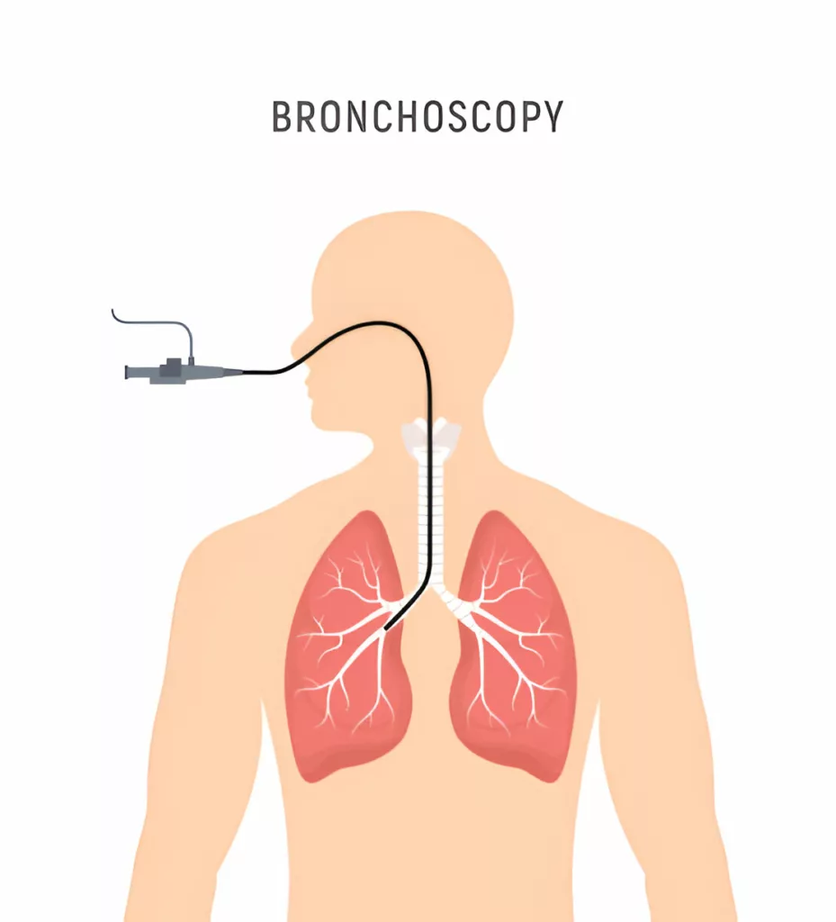
What is Bronchoscopy?
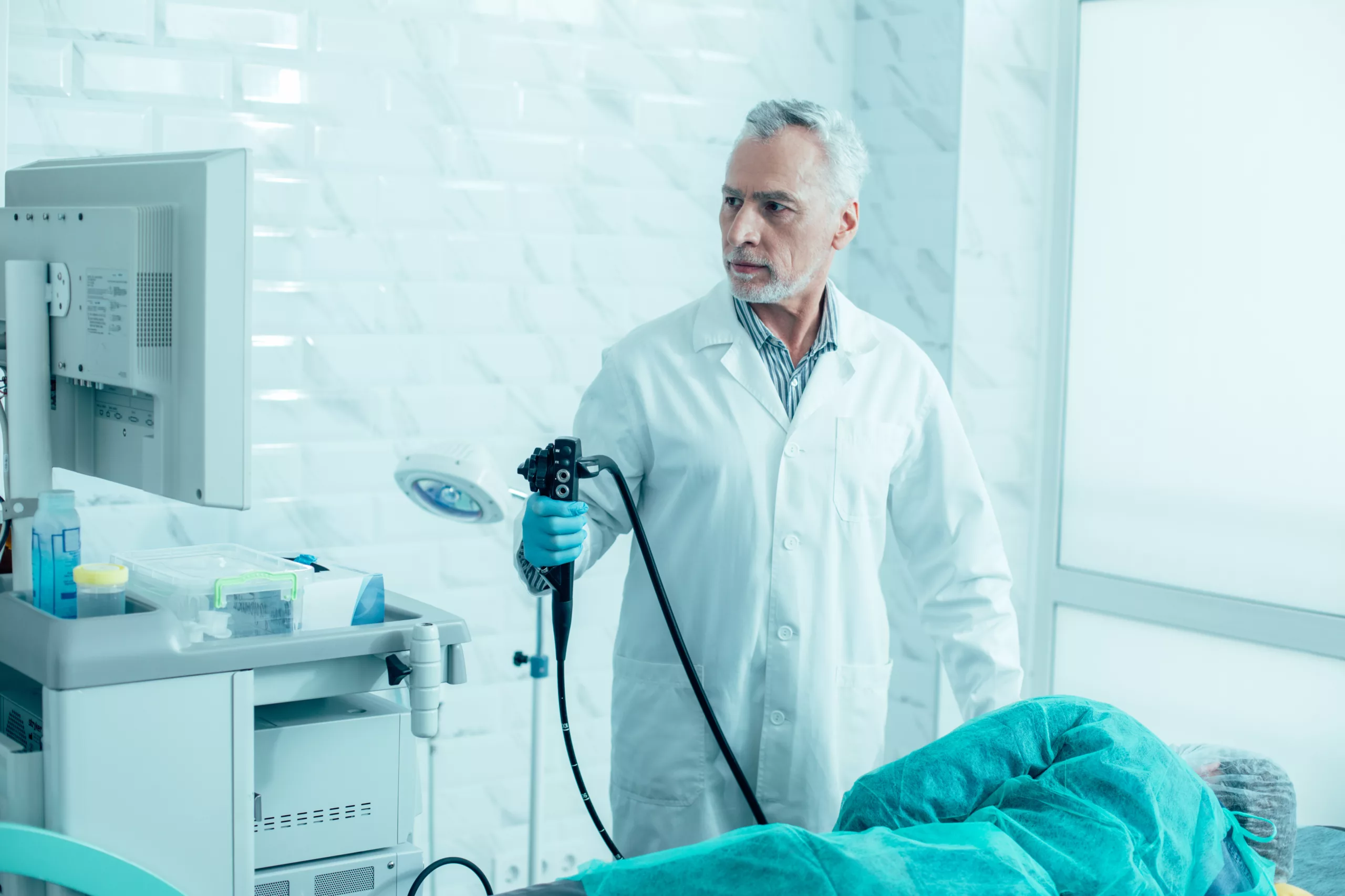
Bronchoscopy is a medical procedure used to diagnose and treat conditions affecting the lungs and airways. It involves inserting a thin, flexible tube called a bronchoscope through the nose or mouth and into the lungs. The bronchoscope has a small camera at its tip, allowing doctors to view and capture detailed images of the lungs and air passages on a monitor.
During the procedure, patients are typically under conscious sedation, meaning they won’t feel pain as the bronchoscope is passed through the mouth or nose, but they can still breathe independently. In certain cases, such as when there is bleeding in the lungs or a foreign object is lodged in the airways, a more rigid bronchoscope may be used to navigate effectively.
Doctors usually recommend bronchoscopy when a patient has a persistent cough, suspected lung infection, or abnormal findings on a chest X-ray or other imaging tests. It can also collect tissue or mucus samples for diagnosing lung cancer, remove foreign bodies or blockages, clear out dead tissue, and plan treatment for various lung conditions.
Types of Bronchoscopes
Medical doctors may use rigid or flexible bronchoscopes, depending on the conditions and requirements of the lungs.
Rigid Bronchoscope:
The rigid bronchoscope consists of a firm tube. It is used to view larger parts of the lungs. The rigid bronchoscope may be used within the bronchi of the lungs to:
- Control lung bleeding
- Remove lung excess secretions and blood
- Eliminate dead lung tissue
- Remove foreign bodies
- Perform lung procedures such as placing stents (to keep your airway open) and other treatments.
Flexible Bronschoscope:
The flexible bronchoscope is used more commonly than the rigid bronchoscope. It can easily go down into bronchioles (smaller air passages).
The following are the uses of a Flexible bronchoscope:
- Pull out lung secretions
- Obtain tissue sample for biopsy
- Directly injecting medicine into the lungs
- Placing an oxygen tube to ease breathing for the patient
Indications
The lung diseases where bronchoscopy is commonly required include:1Mahmoud N, Vashisht R, Sanghavi DK, et al. Bronchoscopy. [Updated 2023 Jul 24]. In: StatPearls [Internet]. Treasure Island (FL): StatPearls Publishing; 2024 Jan-. Available from: https://www.ncbi.nlm.nih.gov/books/NBK448152/
- Pneumonia (lung infection)
- Malignancy
- Presence of any foreign body
- Hemoptysis (blood in the cough)
- Interstitial lung disease
Contraindications
According to the American Thoracic Society, the contraindications for the bronchoscopy include recent medical events like:2Sokolowski Jr., J. W., Burgher, L. W., Jones Jr., F. L., Patterson, J. R., & Selecky, P. A. (Year). Position Paper on Guidelines for Fiberoptic Bronchoscopy in Adults. American Review of Respiratory Disease, 136(4), 1066. https://doi.org/10.1164/ajrccm/136.4.1066
- Recent heart attack
- Heart failure
- Exacerbated asthma symptoms
- Exacerbated COPD symptom
- Low oxygen level
- Abnormal heart rate (arrhythmias) within six weeks
Risks of the Bronchoscopy
Bronchoscopy is considered safe, but medicines used for local sedation (applied on the nose, throat, and mouth) or general anesthesia may have side effects.3Leiten, E. O., Martinsen, E. M., Bakke, P. S., Eagan, T. M., & Grønseth, R. (2016). Complications and discomfort of bronchoscopy: a systematic review. European Clinical Respiratory Journal, 3, 33324. https://doi.org/10.3402/ecrj.v3.33324
The risks may include;
- Abnormal heart rate (arrhythmias)
- Abnormal blood pressure
- Lung infection
- Hypoxia (low oxygen level) during the procedure
- Bleeding, especially after taking a tissue sample (biopsy)
- Pneumonia
- Breathing problem
- Sore throat
- Increased body temperature
- Increased risk of heart attack in patients with pre-existing heart conditions
- During a rigid procedure, there is a small chance that your lungs may collapse (pneumothorax) if your doctor punctures the lung; however, this rarely happens. Your doctor will take a chest x-ray to check for any problem in your lungs after the procedure.
If you undergo general anesthesia for the procedure, you may likely experience;
- Vomiting and Nausea
- Muscle ache
- Fluctuation in blood pressure
- Abnormal heartbeat
Consult your doctor if you find any of these signs
- Chest discomfort or pain
- Blood with cough
- Increased heartbeat
- Problem with breathing
- Elevated body temperature
Complications
The complications of the bronchoscopy are very rare and uncommon; however, it may include:4Stahl, D. L., Richard, K. M., & Papadimos, T. J. (2015). Complications of bronchoscopy: A concise synopsis. International journal of critical illness and injury science, 5(3), 189–195. https://doi.org/10.4103/2229-5151.164995
- Airway trauma
- Vocal cord trauma
- Bronchospasm
- Lung scarring
- Lung puncture (pneumothorax)
- Airway bleeding
- Lung aspiration
- Hypoxemia (reduced oxygen level)
- Arrhythmias
How to prepare for a Bronchoscopy Procedure?
Once your doctor schedules a bronchoscopy, they will provide you with specific instructions to follow before the procedure.5American Lung Association. (n.d.). Bronchoscopy. Retrieved from https://www.lung.org/lung-health-diseases/lung-procedures-and-tests/bronchoscopy
- You will need to avoid foods and drinks for several hours before the procedure.
- Discuss all your medications, including prescription drugs, over-the-counter medications, vitamins, herbal supplements, and other health products. Your doctor may advise you to stop taking certain medications, such as blood thinners, vitamins, and diabetes medications, before the procedure.
- You will sign a consent form that includes the benefits and risks of the bronchoscopy procedure; ensure you get all the answers before signing the consent form.
- The doctor will give you sedatives (numbing medicine) before the procedure, so he may ask you to bring someone with you on the day of the procedure to take you home afterward or make arrangements to get back home.
How is Bronchoscopy Performed?
Bronchoscopy is a quick and painless procedure done as an outpatient if it is a flexible bronchoscopy, which is very common. However, a rigid bronchoscopy procedure requires a longer hospital stay. Normally, bronchoscopy takes 20 minutes to 1 hour, depending on your doctor’s chosen method.
Step-by-Step Procedure
The bronchoscopy procedure follows these steps:6Johns Hopkins Medicine. (n.d.). Bronchoscopy. Johns Hopkins Medicine. Retrieved from https://www.hopkinsmedicine.org/health/treatment-tests-and-therapies/bronchoscopy
- First, you will wear a hospital gown and remove jewelry and other objects, such as dentures and metal objects.
- Afterward, you will lie on the treatment table with your head elevated.
- Your healthcare provider may give you preventative antibiotics before and after the procedure to prevent infections after the bronchoscopy.
- Local sedatives will be given to help you relax, and numbing agents will be applied to your nose, mouth, and throat to anesthetize the airway passages. You will feel drowsy but remain awake during the procedure. If a rigid bronchoscopy is required, you will be given general anesthesia to ensure you are asleep and pain-free throughout the procedure.
- To provide oxygen, a nasal tube, or a face mask will be provided to aid in breathing.
- During the procedure, the medical team will monitor your vitals, such as your heart rate, breathing, and blood pressure.
- Once the numbing agents take effect, the doctor will insert the bronchoscope through your mouth or nose, guiding it down into your lungs until it reaches the bronchi. The bronchoscope may be equipped with additional instruments, such as needles or brushes, to collect lung tissue samples, which can help diagnose various lung conditions.
- In some cases, your doctor may spray saline solution down the lungs with a bronchoscope to wash the airway surface and collect the cells and fluid that are washed. This process is called bronchial washing, and then your doctor may observe these cells under the microscope later.
- Bronchoscopy can help identify issues, such as bleeding, tumors, abscesses, swelling, infections, blockages, or mucus in the lungs. If a blockage is found in the bronchi, the doctor may place a stent (a small tube) to keep the airway open.
- To observe lymph nodes and tissue in the bronchi, a bronchoscope with attached ultrasound is used to guide through the airways.
- After assessing your airway and lungs, the procedure ends
- Your doctor will now remove the bronchoscope.
Recovery after the Procedure
After the procedure, you will stay in the recovery room until the effects of anesthesia are gone. You may feel sleepy and drowsy and experience throat pain, but these symptoms will disappear after a few hours. Meanwhile, the medical team will monitor your blood pressure, heart rate, and breathing.
Your doctor may ask you to cough and spit to check the cough reflex and the mucus to observe the blood in the mucus. He may also advise you to get a chest X-ray after the procedure to examine any signs or problems in your lungs.
You may notice throat swelling, pain, and hoarseness, which are normal and usually go away in a few days with gargles and cough medicine.
If the procedure is outpatient, you can go home on the same day and, if your doctor advises, continue your regular diet and activities after some time.
Result of Bronchoscopy
The bronchoscopy result will be ready after two to three days. Your doctor will discuss the result with you, give details about the findings during the procedure or treatment, and guide you further regarding the condition diagnosed. If a tissue sample was taken during the procedure, a pathologist will review it in 2-3 weeks because tissue samples take time to review.
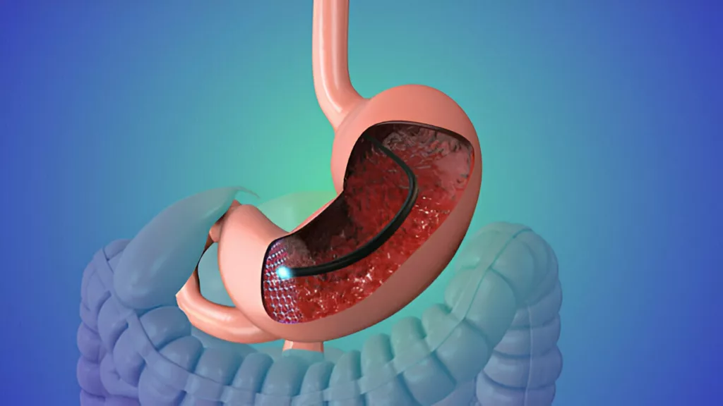
How Biopsy is done using Bronchoscopy
A lung biopsy involves removing tissue or a sample from the bronchi or lung. The procedure of extracting tissue samples during bronchoscopy is called bronchoscopy with lung biopsy, fiber optic bronchoscopy, or trans-bronchial biopsy.
Doctors perform lung biopsy to diagnose lung infection, polyps, tumors, and benign or malignant lung cancer and also to determine the stages of lung cancer.
Biopsy of lung tissue using flexible bronchoscopy helps diagnose several lung conditions and diseases.7Paradis, T. J., Dixon, J., & Tieu, B. H. (2016). The role of bronchoscopy in the diagnosis of airway disease. Journal of Thoracic Disease, 8(12), 3826–3837. https://doi.org/10.21037/jtd.2016.12.68
Some of the airway and lung diseases that require bronchoscopy with lung biopsy are;
- Tumors in the airways of the lungs
- Lung disease causing breathing problem
- Changes in lungs observed in CT scan or any other imaging procedure
- Bronchial infection
- Chronic cough
- Blood in cough
- Lung transplant rejection reactions
How is a tissue sample taken during the Bronchoscopy?
The procedure for trans-bronchial biopsy is similar to a bronchoscopy procedure and takes a few hours or less.
The doctor uses a flexible bronchoscope equipped with a light and camera to collect samples from the bronchi, guided by fluoroscopy or X-ray imaging. Doctors may flush water or normal saline into the bronchi to collect the lung secretions that the doctor will examine later under the microscope. Your vitals during the procedure and recovery period are observed.
The complications of bronchoscopy with lung biopsy are low and are preventable. The most common complications of this procedure include coughing with blood and leaking air from the lungs (pneumothorax).8Jacomelli M, Margotto SS, Demarzo SE, Scordamaglio PR, Cardoso PFG, Palomino ALM, Figueiredo VR. Early complications in flexible bronchoscopy at a university hospital. J Bras Pneumol. 2020;46(4):e20180125. doi: 10.36416/1806-3756/e20180125. Epub 2020 Jun 1. PMID: 32490906; PMCID: PMC7567622.
Bronchoscopy vs. Endoscopy
Endoscopy is a medical technique used to examine the body and diagnose and treat various conditions. Bronchoscopy is a type of endoscopy to examine the airways and lungs.
During endoscopy, a thin tube called a scope enters the organ. The tube or scope has a light and camera attached at the end, which connects to a screen for viewing and taking pictures of the organ.
Doctors can use endoscopy to remove tissue for a sample (biopsy) or perform surgeries for different conditions. Depending on the condition’s requirements, endoscopes can attach different tools for tissue extraction, place a stent, or seal wounds. The procedure of endoscopy is similar to bronchoscopy.
Endoscopy is used to view different organs. The procedure is the same for all organs, and the procedure’s name is based on the organ examined or treated. For example, a colonoscopy is an endoscopy to view and treat colon (intestine) disorders. In laryngoscopy, the scope enters through your nose or mouth and reaches the larynx to view it.
Conclusion
Bronchoscopy is a type of endoscopy where a thin and flexible tube goes inside the organ to check for any disease or condition like tumor, bleeding, or ulcer, or to treat conditions, for example, placing a stent to make the airway open or healing wound. The procedure is safe with lower risks and complications. The procedure usually takes 20 minutes to a few hours, depending on whether it is used for diagnostic or treatment purposes, which could take longer hours.
Refrences
- 1Mahmoud N, Vashisht R, Sanghavi DK, et al. Bronchoscopy. [Updated 2023 Jul 24]. In: StatPearls [Internet]. Treasure Island (FL): StatPearls Publishing; 2024 Jan-. Available from: https://www.ncbi.nlm.nih.gov/books/NBK448152/
- 2Sokolowski Jr., J. W., Burgher, L. W., Jones Jr., F. L., Patterson, J. R., & Selecky, P. A. (Year). Position Paper on Guidelines for Fiberoptic Bronchoscopy in Adults. American Review of Respiratory Disease, 136(4), 1066. https://doi.org/10.1164/ajrccm/136.4.1066
- 3Leiten, E. O., Martinsen, E. M., Bakke, P. S., Eagan, T. M., & Grønseth, R. (2016). Complications and discomfort of bronchoscopy: a systematic review. European Clinical Respiratory Journal, 3, 33324. https://doi.org/10.3402/ecrj.v3.33324
- 4Stahl, D. L., Richard, K. M., & Papadimos, T. J. (2015). Complications of bronchoscopy: A concise synopsis. International journal of critical illness and injury science, 5(3), 189–195. https://doi.org/10.4103/2229-5151.164995
- 5American Lung Association. (n.d.). Bronchoscopy. Retrieved from https://www.lung.org/lung-health-diseases/lung-procedures-and-tests/bronchoscopy
- 6Johns Hopkins Medicine. (n.d.). Bronchoscopy. Johns Hopkins Medicine. Retrieved from https://www.hopkinsmedicine.org/health/treatment-tests-and-therapies/bronchoscopy
- 7Paradis, T. J., Dixon, J., & Tieu, B. H. (2016). The role of bronchoscopy in the diagnosis of airway disease. Journal of Thoracic Disease, 8(12), 3826–3837. https://doi.org/10.21037/jtd.2016.12.68
- 8Jacomelli M, Margotto SS, Demarzo SE, Scordamaglio PR, Cardoso PFG, Palomino ALM, Figueiredo VR. Early complications in flexible bronchoscopy at a university hospital. J Bras Pneumol. 2020;46(4):e20180125. doi: 10.36416/1806-3756/e20180125. Epub 2020 Jun 1. PMID: 32490906; PMCID: PMC7567622.
