Pachyonychia is a rare disease involving your body’s keratin genes. As indicated by its name, it is a congenital condition, meaning it is present from birth. This disease primarily impacts areas of the body that contain keratin, such as the skin and nails. Symptoms of this condition entirely depend upon the type of keratin gene involved.1Agarwal, P., Chhaperwal, M. K., Singh, A., Verma, A., Nijhawan, M., Singh, K., & Mathur, D. (2013). Pachyonychia congenita: A rare genodermatosis. Indian Dermatology Online Journal, 4(3), 225–227. https://doi.org/10.4103/2229-5178.115527
What is Pachyonychia Congenita?
Pachyonychia congenita (PC) is a rare genetic disorder that is inherited in an autosomal dominant pattern, primarily impacting the skin and nails. The condition is marked by severe pain in the soles, palmoplantar keratoderma with blisters underneath, and varying degrees of nail thickening and dystrophy. Other common symptoms include white patches in the mouth (oral leukokeratosis), different types of skin cysts, and excessive skin growth around hair follicles (follicular hyperkeratosis). The disorder is caused by mutations in one of five specific keratin genes: KRT6A, KRT6B, KRT6C, KRT16, or KRT17.2Pachyonychia congenita. (2023). In UpToDate. Retrieved June 26, 2024, from https://www.uptodate.com/contents/pachyonychia-congenita?search=pachyonychia%20congenita&source=search_result&selectedTitle=1%7E12&usage_type=default&display_rank=1
Pathophysiology
The body’s Epithelial cells are made up of keratin filaments, which provide structural support. Two different types of keratin proteins join together to form a structure called a hexamer, and they make additional links to form a large tetramer. Many tetramers then assemble to build the basic structure of keratin, called keratin intermediate filaments. These keratin intermediate filaments express themselves in the keratinocytes of your skin, nails, and mucosa; every epithelial cell in your body represents a different type of keratin gene.
It develops when there is a defect in keratin 6A, keratin 6B, keratin 16, and keratin 17. Keratin 6A and 16A constitute type 1 of pachyonychia congenita, the most common type, and type 2 contains keratin 6B and 17. These mutations prevent the formation of structural solid filaments of keratin, leading to weak skin, nails, and hair epithelial cells and producing symptoms like pain, calluses, or blisters.
Causes of Pachyonychia Congenita
Mutations in any of your keratin genes cause PC. Almost 115 types of mutations have been recorded in keratin genes until now. This condition transmits in an autosomal dominant pattern, which means that only one copy of a mutated gene is enough to cause the disease. Any one of the parents who is affected has a 50% chance of transmitting pachyonychia congenita to the children. Some cases of PC may present without a genetic or family history, and these cases occur due to sporadic (spontaneous )mutation.3Zieman, A. G., & Coulombe, P. A. (2020). Pathophysiology of pachyonychia congenita-associated palmoplantar keratoderma: new insights into skin epithelial homeostasis and avenues for treatment. The British journal of dermatology, 182(3), 564–573. https://doi.org/10.1111/bjd.18033
How Common is this Condition?
Pachyonychia congenita is very infrequent. Only 5000 to 10000 people in the world have suffered from this disease, which equally affects males and females, mostly at birth.
Symptoms of Pachyonychia Congenita
Symptoms of this condition depend upon the type of mutated keratin gene and the location of that gene in your body. Most symptoms are visible when a child reaches the age of ten.
Pachyonychia congenita presents with the following signs and symptoms:
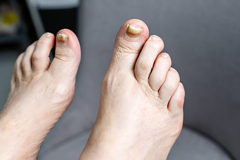
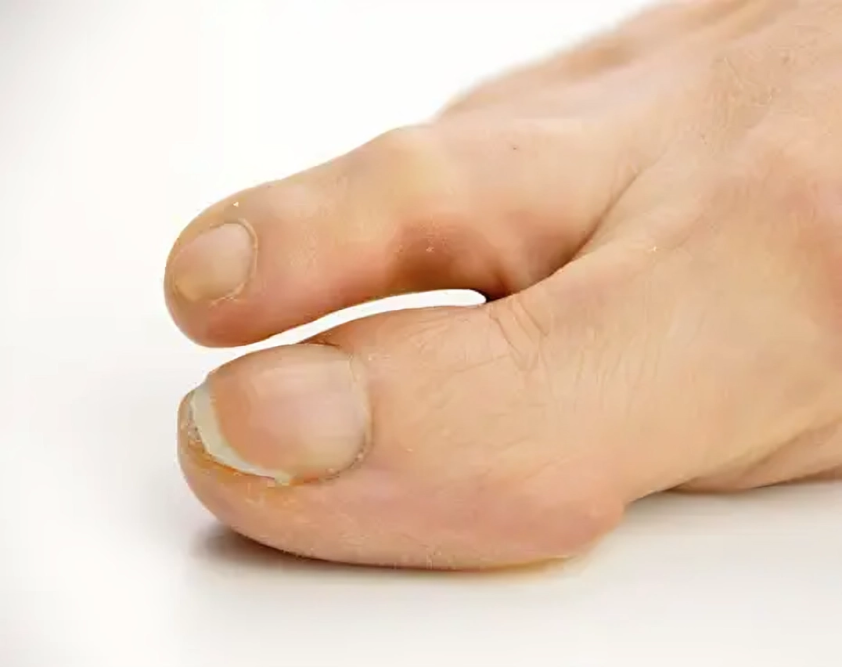
Thickening of Nails
Almost every child with pachyonychia congenita has thickened nails on the toes, which is the most common symptom. Thickening of your fingernail is less common than a toenail. The thickening of nails doesn’t cause pain in the early stages, but as time passes, thickening increases, making it painful. Children with severe thickening of toenails find it challenging to wear shoes and walk. You must carefully tackle this condition by regularly cutting the affected toenail. If not treated correctly in the early stages, it causes an infection of your nails called paronychia, a painful condition. Thickened fingernails and toenails mainly occur when keratin 6A and 17 mutation occurs.
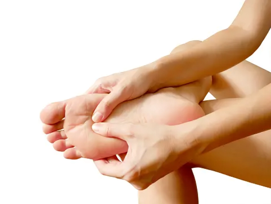
Plantar Keratoderma
More than 95% of patients with pachyonychia congenita have plantar keratoderma, an abnormal thickening of their soles. Plantar keratoderma mainly develops in children when they start walking. Thickening develops in friction and weight-bearing areas, making it painful for the patients. With prolonged walking, there is a risk of fissuring and secondary infection of the affected areas. Some children also develop palmar thickening, but its incidence is less than that of plantar keratoderma.
In a few cases, plantar keratoderma is associated with excessive sweating in your palms and soles (hyperhidrosis).
Plantar Pain
Plantar pain is a common symptom of pachyonychia congenita. Walking and weight-bearing activities increase the intensity of plantar pain, making daily normal activities difficult. The pain doesn’t subside without medications. Some studies show that the origin of plantar pain is neuropathic, which may affect the nerves of the sole.4Krupiczojc, M. A., & O’Toole, E. A. (2018). Plantar pain in pachyonychia congenita. The British journal of dermatology, 179(1), 11–12. https://doi.org/10.1111/bjd.16700
Nail Dystrophy
Pachyonychia congenita also presents as toe and fingernail dystrophy. The nails become discolored in dystrophy and attain an abnormal size and shape. Sometimes, nails grow to an abnormally increased length with a rough surface and upslanting shape. Toenail dystrophy occurs more frequently than fingernail dystrophy. Fingernail dystrophy usually develops due to KRT6A mutation.5Ghosh, R., Chatterjee, K., Barua, J. K., & Roy, A. (2017). Cutaneous Cysts with Nail Dystrophy in a Young Female: A Classical Association. Indian journal of dermatology, 62(6), 661–664. https://doi.org/10.4103/ijd.IJD_473_16
Mucosal Findings
In pachyonychia congenita, plaques form on the buccal surfaces of your mouth and tongue. Although the plaques are benign, they cause difficulty for the child to suck the milk properly. Plaques are primarily associated with KRT6A mutation.
Another mucosal finding in pachyonychia congenita is the formation of white patches on the tongue and buccal mucosa, generally in the areas of trauma, such as bite injury. These white patches resemble a lot with patches of candidiasis and leukoplakia. Therefore, it is crucial to diagnose them properly.
Cysts
Cysts in pachyonychia congenita are pilosebaceous cysts, which are associated with hair follicles and sebaceous glands. The most common type of pilosebaceous cyst in pachyonychia congenita is steatocystomas. These small yellow cysts form on the arms, chest, breast, or armpits. Patients with KRT17 mutations have been found to have these cysts.
Natal Teeth
Children with pachyonychia congenita are often born with natal teeth or develop them in the first year of life. Natal teeth cause problems in sucking milk for the baby and may also cause trauma to the mother’s breast. These teeth are not of serious concern as regular teeth replace them in childhood. Mutations of the KRT17 gene are associated with natal or prenatal teeth.
Follicular Keratoses
Very few cases of pachyonychia congenita have been reported with follicular keratoses on the extremities. Follicular keratosis is the formation of thick bumps or plaques on the skin that may increase in size, causing discomfort and pain.
Calluses & Blisters
When children with pachyonychia congenita start walking, they risk developing calluses (hard skin) and blisters on their soles. These calluses and blisters may lead to secondary infection of their soles.6Mccarthy, R. L., De Brito, M., & O’toole, E. (2023). Pachyonychia Congenita: Clinical Features and Future Treatments. The Keio Journal of Medicine, 10.2302/kjm.2023-0012-IR. Advance online publication. https://doi.org/10.2302/kjm.2023-0012-IR
Diagnosis of Pachyonychia Congenita
95% of the patients with this condition present with typical symptoms of toenail thickening, keratoderma, and plantar pain, which are enough to make a definite diagnosis. However, to differentiate pachyonychia congenita from other similar diseases, it is essential to follow the following protocols:
Family History
The first and crucial step in diagnosing pachyonychia congenita is taking a family history. This is a genetic disease with an autosomal dominant trait, and one of the parents of the affected child also suffers from it.
Molecular DNA Analysis
Molecular DNA analysis is a molecular pathology in which a pathologist takes DNA and RNA samples and recognizes the type of mutations in keratin genes and the type of gene involved. These mutations could be missense, deletion, substitution, or others involving keratin genes like KRT6A, KRT6B, KRT16, and KRT17.
Biopsy
A biopsy is taken by inserting a needle in your affected skin, and a piece of tissue is examined. An oral biopsy of white patches and plaques in your buccal mucosa and tongue differentiates the pachyonychia congenita from the findings of candidiasis and leukoplakia. Taking plantar and skin biopsies is also recommended to make a better diagnosis.
Culture & Microscopy
To differentiate the nail thickening and dystrophy of pachyonychia congenita from other disorders, for instance, candida and fungal infections, the culture of your nail scrapings is taken and examined under a microscope.
Histology
Histological examination reveals the changes in keratin layers of your skin in pachyonychia congenita. It shows increased proliferation of keratinocytes along with parakeratosis (immature epidermal keratinocytes). Histology also indicates a specific basketweave appearance of one of the layers of keratin, a condition called orthokeratosis.
Plaques of your palms and soles are examined under an electron microscope, which reveals the thickening of keratin filaments and the formation of abnormally large granules in keratin layers.7Agarwala, M. K., Schwartz, M. E., & Smith, F. J. (2016). Pachyonychia Congenita: New Classification and Diagnosis. Indian journal of dermatology, 61(5), 567. https://doi.org/10.4103/0019-5154.190110
Prenatal Testing
As pachyonychia congenital is a genetic disease, a pregnant woman must undergo prenatal testing if any of her partners are affected by it. Prenatal testing aids in diagnosing the disease before the baby’s birth and makes it easier for the physician to treat it better.
Treatment of Pachyonychia Congenita
Like any other genetic disease, pachyonychia congenita does not have a specific medication, and its treatment revolves around symptomatic management.
Following are some of the treatment protocols that you can take advantage of to reduce severe symptoms of this rare nail disorder:
- To treat the thickening of your palms and soles, avoid weight-bearing exercises, prolonged walking, and injury, as pressure adversely affects the thickening. Use medicated soft shoes, socks, and gloves to minimize the pressure on palms and soles and avoid trauma.
- Pain associated with plantar keratoderma becomes a significant concern for patients with pachyonychia congenita, as it can become severe if not taken care of. Use prescribed painkillers to decrease the intensity of pain. Walk as little as possible. If the pain restricts regular movement and walking, you can use a wheelchair or crutches.
- Thickened nails on toes and fingers must be trimmed whenever needed using a cutter, rasps, or a filer to avoid secondary infection and trauma. Another option to soften the thick nails is to dip the fingers and toes in water, urea, propylene glycol, or any weak organic acid, such as salicylic acid.8Milstone, L. M., Fleckman, P., Leachman, S. A., Leigh, I. M., Paller, A. S., van Steensel, M. A., & Swartling, C. (2005). Treatment of pachyonychia congenita. The journal of investigative dermatology. Symposium proceedings, 10(1), 18–20. https://doi.org/10.1111/j.1087-0024.2005.10203.x
Some other treatment modalities that your doctor can prescribe are:
Retinoids such as Isotretinoin & Etretinate
Retinoids help alleviate follicular keratosis and the thickening of palms and soles. However, their use is not usually admired due to their unfavorable effects. Retinoids may exacerbate the thickening and tenderness of palms and soles, and with long-term use, they also cause teratogenicity, liver toxicity, hyperlipidemia, and skeletal problems.
Painkillers
Pain in palms and soles becomes a severe concern for patients with pachyonychia congenita. Most of the patients experience a significant reduction in pain by using NSAIDs. Some patients also use narcotic analgesics as well as neuropathic painkillers.
Rapamycin & Statins
Both drugs affect keratin genes by inhibiting the gene itself or inhibiting transcription. In this way, they manage the symptoms of pachyonychia congenita. The statins mainly used are simvastatin and rosuvastatin.
Sirolimus
1% sirolimus is used as an ointment, and studies show that it dramatically reduces the symptoms of plantar keratoderma.
Botulinum Toxin or Aluminium Hydroxide
Excessive sweating of palms and soles in pachyonychia congenita becomes problematic for the patients. Physicians prescribe aluminium hydroxide or injectable botulinum toxin in your soles to treat hyperhidrosis (excessive sweating).9Koren, A., Sprecher, E., Reider, E., & Artzi, O. (2020). A treatment protocol for botulinum toxin injections in the treatment of pachyonychia congenita-associated keratoderma. The British journal of dermatology, 182(3), 671–677. https://doi.org/10.1111/bjd.18169
Surgical Approach
In pachyonychia congenita, surgical options can be used only to treat cysts. Surgeons make incisions, excisions, and drain cysts in pachyonychia congenita like any other cysts. Surgery doesn’t give promising results for problems associated with pachyonychia congenita, such as nail dystrophy or thickening, as the nails can regrow even after the surgery. Similarly, grafting and excision are not advantageous in the case of keratoderma due to its recurrence. Hence, you should treat the symptoms of pachyonychia congenita with helpful remedies and care rather than go for surgery.10Thomsen, R. J., Zuehlke, R. L., & Beckman, B. I. (1982). Pachyonychia congenita: surgical management of the nail changes. The Journal of dermatologic surgery and oncology, 8(1), 24–28. https://doi.org/10.1111/j.1524-4725.1982.tb00229.x
Life Expectancy in Pachyonychia Congenita
This condition doesn’t affect the lifespan of a patient. Hence, its prognosis is satisfactory.
However, due to the unbearable pain in plantar keratoderma, pachyonychia congenita impacts the quality of life in a way that patients cannot walk effortlessly. In that case, using a wheelchair or any other aid makes life a bit manageable.11Wu, A. G., & Lipner, S. R. (2021). Distinctions in the Management, Patient Impact, and Clinical Profiles of Pachyonychia Congenita Subtypes. Skin appendage disorders, 7(3), 194–202. https://doi.org/10.1159/000513340
Conclusion
In conclusion, this condition develops at birth or in childhood due to genetic mutation in keratin genes. These mutated keratin genes exhibit symptoms in your skin, hair follicles, or mucosa. Pachyonychia congenita presents as plantar thickening, nail dystrophy, or pain in your palms and soles. Biopsy of your skin is helpful for the diagnosis of pachyonychia congenita. Your physician may also suggest a culture of nail scraping. Always trim your overgrown nails to avoid any complications. Take proper rest in case of plantar pain. Adequate care of nails and skin promises a reasonable prognosis of pachyonychia congenita.
Refrences
- 1Agarwal, P., Chhaperwal, M. K., Singh, A., Verma, A., Nijhawan, M., Singh, K., & Mathur, D. (2013). Pachyonychia congenita: A rare genodermatosis. Indian Dermatology Online Journal, 4(3), 225–227. https://doi.org/10.4103/2229-5178.115527
- 2Pachyonychia congenita. (2023). In UpToDate. Retrieved June 26, 2024, from https://www.uptodate.com/contents/pachyonychia-congenita?search=pachyonychia%20congenita&source=search_result&selectedTitle=1%7E12&usage_type=default&display_rank=1
- 3Zieman, A. G., & Coulombe, P. A. (2020). Pathophysiology of pachyonychia congenita-associated palmoplantar keratoderma: new insights into skin epithelial homeostasis and avenues for treatment. The British journal of dermatology, 182(3), 564–573. https://doi.org/10.1111/bjd.18033
- 4Krupiczojc, M. A., & O’Toole, E. A. (2018). Plantar pain in pachyonychia congenita. The British journal of dermatology, 179(1), 11–12. https://doi.org/10.1111/bjd.16700
- 5Ghosh, R., Chatterjee, K., Barua, J. K., & Roy, A. (2017). Cutaneous Cysts with Nail Dystrophy in a Young Female: A Classical Association. Indian journal of dermatology, 62(6), 661–664. https://doi.org/10.4103/ijd.IJD_473_16
- 6Mccarthy, R. L., De Brito, M., & O’toole, E. (2023). Pachyonychia Congenita: Clinical Features and Future Treatments. The Keio Journal of Medicine, 10.2302/kjm.2023-0012-IR. Advance online publication. https://doi.org/10.2302/kjm.2023-0012-IR
- 7Agarwala, M. K., Schwartz, M. E., & Smith, F. J. (2016). Pachyonychia Congenita: New Classification and Diagnosis. Indian journal of dermatology, 61(5), 567. https://doi.org/10.4103/0019-5154.190110
- 8Milstone, L. M., Fleckman, P., Leachman, S. A., Leigh, I. M., Paller, A. S., van Steensel, M. A., & Swartling, C. (2005). Treatment of pachyonychia congenita. The journal of investigative dermatology. Symposium proceedings, 10(1), 18–20. https://doi.org/10.1111/j.1087-0024.2005.10203.x
- 9Koren, A., Sprecher, E., Reider, E., & Artzi, O. (2020). A treatment protocol for botulinum toxin injections in the treatment of pachyonychia congenita-associated keratoderma. The British journal of dermatology, 182(3), 671–677. https://doi.org/10.1111/bjd.18169
- 10Thomsen, R. J., Zuehlke, R. L., & Beckman, B. I. (1982). Pachyonychia congenita: surgical management of the nail changes. The Journal of dermatologic surgery and oncology, 8(1), 24–28. https://doi.org/10.1111/j.1524-4725.1982.tb00229.x
- 11Wu, A. G., & Lipner, S. R. (2021). Distinctions in the Management, Patient Impact, and Clinical Profiles of Pachyonychia Congenita Subtypes. Skin appendage disorders, 7(3), 194–202. https://doi.org/10.1159/000513340
