What is Rift Valley Fever?
Rift Valley fever (RVF) is a mosquito-borne viral disease that causes febrile or hemorrhagic illness in ruminants and humans. RVF virus (RVFV) is one of the phleboviruses, first identified in the Rift Valley of Africa in the 1930s.1Daubney, R., & Hudson, J. R. (1931). Enzootic Hepatitis or Rift Valley Fever. An Un-described Virus Disease of Sheep, Cattle and Man from East Africa. RVFV was initially known as a causative agent of abortions and neonatal mortality in sheep. Later, it was identified as a causative agent of febrile illness in humans. Domesticated ruminants such as cattle, goats, camels, and sheep are the most susceptible animals, while wild animals such as giraffes, warthogs, impalas, and pigs can also get this infection.2Nair, N., Osterhaus, A. D., Rimmelzwaan, G. F., & Prajeeth, C. K. (2023). Rift Valley fever virus—Infection, pathogenesis, and host immune responses. Pathogens, 12(9), 1174.
In animals, the prevalence of RVF ranges from 8.8% to 12.9%, while the prevalence of RVF in humans is 5.9%. However, this prevalence rate may vary across geographical regions. 3Clark, M. H., Warimwe, G. M., Di Nardo, A., Lyons, N. A., & Gubbins, S. (2018). Systematic literature review of Rift Valley fever virus seroprevalence in livestock, wildlife, and humans in Africa from 1968 to 2016. PLoS Neglected Tropical Diseases, 12(7), e0006627.
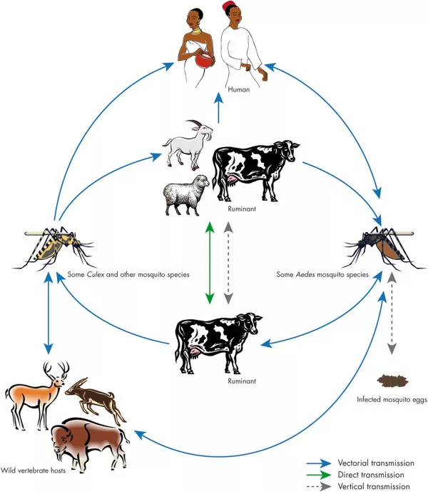
This virus transmission among ruminants is caused by mosquitoes, most commonly Aedes, Culex, and Anopheles species. In humans, direct contact with infected animal blood or tissues, such as during slaughter or handling of carcasses, and the consumption of raw milk from infected animals are the main modes of transmission. Mosquito-borne transmission to humans is rare but possible during outbreaks when mosquito populations surge.4Grossi-Soyster, E. N., Lee, J., King, C. H., & LaBeaud, A. D. (2019). The influence of raw milk exposure on Rift Valley fever virus transmission. PLoS Neglected Tropical Diseases, 13(3), e0007258.
Structure of RVFV
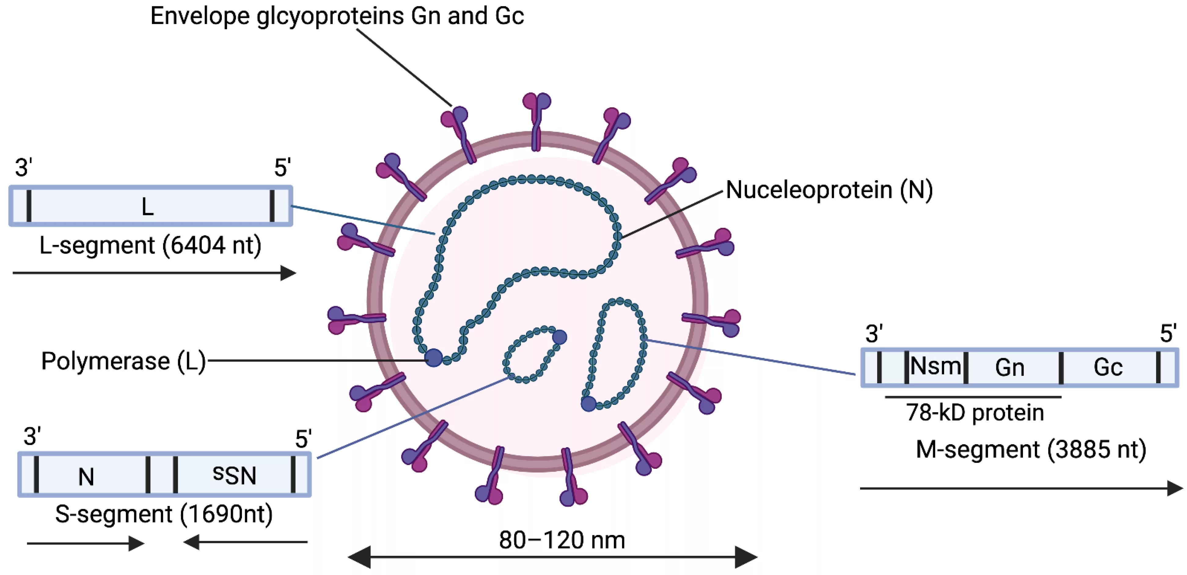
RVFV is an enveloped virus with a diameter of 90–110 nm and a core element of 80–85 nm. It has a segmented RNA genome consisting of three negative-sense RNA segments:
- Small (S) Segment: Encodes the nucleoprotein and non-structural proteins.
- Medium (M) Segment: Encodes two structural glycoproteins involved in host cell entry.
- Large (L) Segment: Encodes RNA-dependent RNA polymerase, essential for viral replication and transcription.
The segmented and complex structure of RVFV enables its efficient replication and adaptability, contributing to its ability to infect diverse hosts.5Liu, L., Celma, C. C., & Roy, P. (2008). Rift Valley fever virus structural proteins: expression, characterization, and assembly of recombinant proteins. Virology journal, 5, 1-13.

Life Cycle of RVFV
The life cycle of RVFV involves a complex interaction between the virus, its mosquito vector, and its susceptible host.
Infection & Transmission
Aedes mosquitoes acquire the virus by feeding on the infected animal.
Once an infected mosquito subsequently bites an animal or human, RVFV enters the host through the bloodstream.
Attachment
The virus attaches to specific receptors on host cells. The receptor cells that facilitate the entry are L-SIGN and DC-SIGN.
Endocytosis
Following attachment, the process of virus internalization is through endocytosis. This process is often pH-dependent. It may involve macropinocytosis or caveolin-mediated endocytosis.
Release of the Viral Genome
The viral envelope fuses with the endosomal membrane. With the help of this fusion, the virus releases the viral RNA genome into the cell cytoplasm of the host.
Replication & Transcription
The process of new RNA synthesis involves using cellular-capped RNA as primers. The negative-sense RNA genome is transcribed into positive-sense RNA. This process takes place through RNA-dependent RNA polymerase. These positive sense RNAs serve as a template for synthesizing viral proteins. The viral proteins include non-structural and structural proteins.
Assemblage
The newly synthesized RNA genomes and viral proteins assemble at specific points in the host cell. Here, they form new virions.
Release
The assembled virions bud off from the host cell membrane, ready to infect neighboring cells or be transmitted back to mosquitoes during feeding.6Nair, N., Osterhaus, A. D., Rimmelzwaan, G. F., & Prajeeth, C. K. (2023). Rift Valley fever virus—Infection, pathogenesis, and host immune responses. Pathogens, 12(9), 1174.
Causes of RVF
As we have discussed earlier, RVFV is the causative agent of Rift Valley Fever (RVF). Environmental factors such as heavy rainfall and flooding create breeding grounds for mosquito populations, significantly increasing the risk of outbreaks. RVFV transmission occurs through two primary cycles:
- Enzootic Cycle: Infected mosquitoes maintain low-level virus circulation among animals.
- Epizootic Cycle: Favorable environmental conditions lead to increased mosquito populations, triggering widespread transmission and outbreaks among livestock and humans.
Humans primarily contract RVF through contact with the blood, tissues, or milk of infected animals. This risk is particularly high for veterinarians, farmers, and workers in the meat industry. While mosquito bites can transmit the virus to humans, this mode of transmission is less common.

Pathogenesis of RVFV
The virus employs several strategies to evade the host’s immune system. The brief overview of the pathogenesis of this virus is as follows:
- The non-structural proteins inhibit the induction of interferon responses. This process allows the virus to replicate without activating significant immune activation.
- The virus manipulates the signaling pathways of the immune system. The disturbance of signaling pathways disrupts cytokine production, leading to a dysregulated immune response.
- These strategies result in unchecked viral replication, contributing to the severity of clinical manifestations.7Ikegami, T., & Makino, S. (2011). The pathogenesis of Rift Valley fever. Viruses, 3(5), 493-519.
Signs & Symptoms of RVF
RVF affects both animals and humans with varying signs and symptoms.
Symptoms in Animals:
The common symptoms of RVF in animals include:
- Fever
- Loss of appetite
- Nasal and ocular discharge
- Abdominal pain, vomiting, and diarrhea (often with blood)
- Lethargy and rapid respiratory distress
- Jaundice and epistaxis (nosebleeds)
- Decreased milk production in lactating animals
In most severe cases, especially in young animals, symptoms progress rapidly, leading to death in just two to three days after the onset of the disease. The mortality ratios among young lambs are high, approximately 90% to 100%. The mortality ratio among adult sheep is 10% to 30%.8Waqar, M. A., Qureshi, A., Ahsan, A., Sadaqat, S., Zulfiqar, H., Razaq, A., … & Ilyas, D. (2023). Epidemiology, clinical manifestations, treatment approaches and future perspectives of Rift Valley fever: epidemiology of Rift Valley fever. Pakistan Journal of Health Sciences, 02-08. Other symptoms include:
- Jaundice
- Rapid respiratory distress
- Prostration
Symptoms In Humans:
After an incubation period of 2–6 days, RVF in humans typically manifests as a self-limiting febrile illness. Early symptoms include:
- Fever, headache, dizziness, and weakness
- Joint stiffness, backache, and muscle pain
- Nasal congestion, sore throat, and fatigue9Bird, B. H., Ksiazek, T. G., Nichol, S. T., & MacLachlan, N. J. (2009). Rift Valley fever virus. Journal of the American Veterinary Medical Association, 234(7), 883-893.
The gastrointestinal symptoms:
- Epigastric distress
- Vomiting
- Nausea
- Diarrhea
- Abdominal pain
- Anorexia
- Dysphagia
The hepatic symptoms due to RVF:
- Hepatomegaly (enlarged liver)
- Jaundice and tenderness in the hypochondriac region
Neurological manifestations:
- Insomnia
- Delirium
- Vertigo
- Confusion
- Disorientation
Obstetric manifestations:
- Preterm delivery
- Miscarriages10Anywaine, Z., Lule, S. A., Hansen, C., Warimwe, G., & Elliott, A. (2022). Clinical manifestations of Rift Valley fever in humans: Systematic review and meta-analysis. PLoS Neglected Tropical Diseases, 16(3), e0010233.
Severe Complications:
In some cases, RVF progresses to severe or life-threatening forms, including:
- Hepatitis or acute hepatic necrosis: Liver damage leading to organ failure
- Retinitis: Vision impairment or blindness due to ocular inflammation
- Encephalitis: Brain inflammation resulting in neurological complications
- Hemorrhagic fever: Excessive bleeding from various sites, such as gums or nose
Severe hemorrhagic syndromes may involve:
- Coagulopathy and disseminated intravascular coagulation (DIC)
- Renal failure and multiple organ dysfunction
The most common causes of death in severe RVF cases include renal or hepatic failure and shock.11Bird, B. H., Ksiazek, T. G., Nichol, S. T., & MacLachlan, N. J. (2009). Rift Valley fever virus. Journal of the American Veterinary Medical Association, 234(7), 883-893.12Anywaine, Z., Lule, S. A., Hansen, C., Warimwe, G., & Elliott, A. (2022). Clinical manifestations of Rift Valley fever in humans: Systematic review and meta-analysis. PLoS Neglected Tropical Diseases, 16(3), e0010233.
Diagnosis of RVF
Accurate diagnosis of RVF relies on a combination of clinical and laboratory evaluations.
Clinical Diagnosis:
Initial diagnosis is made through non-specific symptoms such as fever, myalgia, and gastrointestinal disturbances. The healthcare provider will have a look at the patient’s history regarding exposure to infected animals. However, due to overlapping symptoms with other disorders, clinical evaluation alone is insufficient to confirm the condition.
Laboratory Diagnosis:
Accurate diagnosis of RVF requires specific laboratory tests. Following laboratory tests are employed to diagnose the disease.
Reverse Transcriptase Polymerase Chain Reaction (RT-PCR):
This test is useful during the early stages of infection. This molecular technique detects the viral DNA in the tissue or blood samples. The high sensitivity and specificity of this test allow the detection of viruses even in a very low viral load. Results can be obtained within a few hours. A positive test indicates an active RVF infection.13Petrova, V., Kristiansen, P., Norheim, G., & Yimer, S. A. (2020). Rift Valley fever: diagnostic challenges and investment needs for vaccine development. BMJ Global Health, 5(8), e002694.
Serological Tests:
Serological tests help identify the antibodies produced in response to the infection.
Virus Neutralization Test (VNT)
This test evaluates the ability of the serum to neutralize the virus. Positive results of this test indicate the presence of neutralizing antibodies and a previous exposure to the virus. However, this test requires specialized laboratories for their accomplishment due to biosafety requirements.
Enzyme-Linked Immunosorbent Assay (ELISA)
This test detects Immunoglobulin M (IgM) and Immunoglobulin G (IgG) antibodies against this virus. IgM indicates a recent infection, while IgG indicates a past infection. ELISA is a highly sensitive and specific test used to diagnose RVFV.
Indirect Immunofluorescent Assay (IFA)
IFA uses fluorescently labeled antigens to detect the specific antigens in the serum of the infected individual. IFA provides qualitative results, but this test has lower sensitivity than ELISA.14Lapa, D., Pauciullo, S., Ricci, I., Garbuglia, A. R., Maggi, F., Scicluna, M. T., & Tofani, S. (2024). Rift Valley Fever Virus: An Overview of the Current Status of Diagnostics. Biomedicines, 12(3), 540.
Immunohistochemistry:
It detects viral antigens in infected humans and animals. This test requires stained tissue sections for evaluation. If the section visualizes RVFV antigens, the individual has an active RVF infection. Immunohistochemistry is particularly useful for post-mortem infections.15Petrova, V., Kristiansen, P., Norheim, G., & Yimer, S. A. (2020). Rift Valley fever: diagnostic challenges and investment needs for vaccine development. BMJ Global Health, 5(8), e002694.
Virus Isolation:
During the acute phase of Rift Valley Fever, the virus can be detected using serum or whole blood samples. Virus isolation can be done through cell culture techniques. If cytopathic effects are observed in the cell culture, RVFV is present. It is a time-consuming method and requires several days to confirm the presence of the virus in the cell culture.16Lapa, D., Pauciullo, S., Ricci, I., Garbuglia, A. R., Maggi, F., Scicluna, M. T., & Tofani, S. (2024). Rift Valley Fever Virus: An Overview of the Current Status of Diagnostics. Biomedicines, 12(3), 540.
Treatment & Management of the RVF
Yet, there is no specific treatment for RVF. Most cases resolve on their own within a few days. RVF can be managed through various strategies. Supportive treatment and antiviral medications are the first-line options for managing the symptoms.
Supportive Care:
It is the cornerstone for managing RVF. The supportive care includes:
Coagulation Support
Patients experiencing coagulopathy or excessive bleeding may require transfusions of fresh frozen plasma or platelets to support their coagulation system.
Symptom Specific Treatment Options
These include antiemetics, analgesics, and anxiolytics for managing anxiety, pain, and nausea, respectively.
Monitoring Renal Function
Monitoring of renal function is necessary for patients with severe renal dysfunction. Early diagnosis can prevent severe outcomes in patients with renal failure.
Electrolyte and Fluid Management
Individuals may experience severe dehydration due to gastrointestinal symptoms. Intravenous fluids are needed to maintain electrolyte balance and hydration to manage dehydration.
Antiviral Treatments:
Antiviral treatments can target the process intrinsic to viruses. The healthcare providers are using the following antiviral medications to treat RVF.
Ribavarin
Ribavirin is one of the few drugs that have been permitted to treat viral hemorrhagic fever. The administered dose of ribavirin is 18.8 mg/kg subcutaneously. Now, the use of ribavirin is controversial due to the reports of side effects such as hemolytic anemia and inflammation.17Waqar, M. A., Qureshi, A., Ahsan, A., Sadaqat, S., Zulfiqar, H., Razaq, A., … & Ilyas, D. (2023). Epidemiology, clinical manifestations, treatment approaches and future perspectives of Rift Valley fever: epidemiology of Rift Valley fever. Pakistan Journal of Health Sciences, 02-08.
Favipiravir
Favipiravir can also offer protection against RVFV in animal models. However, more research is required to confirm its effectiveness in humans.
Vaccine for RVF:
Till now, there is no commercially available vaccine for humans against RVF. Scientists have developed an inactivated vaccine for experimental purposes among high-risk groups (veterinarians and laboratory personnel). However, modified live vaccines can help control outbreaks in livestock.18Waqar, M. A., Qureshi, A., Ahsan, A., Sadaqat, S., Zulfiqar, H., Razaq, A., … & Ilyas, D. (2023). Epidemiology, clinical manifestations, treatment approaches and future perspectives of Rift Valley fever: epidemiology of Rift Valley fever. Pakistan Journal of Health Sciences, 02-08.
Prevention of RVF
Preventing RVF requires a combination of vector control, personal protective measures, and public health interventions.
Vector Control
- Regularly clean water containers to prevent mosquito breeding.
- Eliminate standing water, such as in ponds and stock tanks.
- Use insecticides to reduce mosquito populations during outbreak seasons.
- Employ insecticide-treated bed nets (ITNs) and repellents to prevent mosquito bites, especially in endemic areas.
Personal Protection
- Wear long-sleeved clothing and long pants to minimize skin exposure outdoors.
- Avoid handling sick animals or their body fluids without proper protective equipment.
- Practice safe handling of livestock and animal products to minimize exposure.
- Avoid consuming raw milk or undercooked meat.
Livestock Management
- Limit livestock movement during outbreaks to reduce the spread of infection.
- Quarantine new animals during outbreaks to prevent disease transmission.
- Ban the slaughter of sick animals during epidemics to reduce human exposure.
Community and Healthcare Education
- Educate communities about the transmission, symptoms, and prevention of RVF.
- Train healthcare workers to recognize RVF symptoms and implement infection control measures promptly.
- Ensure proper handling of blood and fluids from suspected RVF cases in healthcare and veterinary settings.
Surveillance and Monitoring
- Implement robust surveillance systems for livestock to detect early signs of RVF outbreaks.
- Monitor environmental conditions, such as heavy rainfall that may trigger mosquito population growth and outbreaks.
A Quick Review
RVF is a significant health concern due to its zoonotic nature and potential for major outbreaks. The epidemiology of the disease is closely related to environmental factors which facilitate the breeding of mosquitoes. An early and accurate diagnosis can help in the outbreak management of RF. Effective public health strategies are required to control viral outbreaks effectively. Preventing RVF requires a multifaceted approach involving vector control, education, safe handling practices, and vaccination. Continuous research on the vaccine development for humans against RVF and improved diagnostic methods are crucial to mitigate this viral disease’s effects on humans and livestock.
Refrences
- 1Daubney, R., & Hudson, J. R. (1931). Enzootic Hepatitis or Rift Valley Fever. An Un-described Virus Disease of Sheep, Cattle and Man from East Africa.
- 2Nair, N., Osterhaus, A. D., Rimmelzwaan, G. F., & Prajeeth, C. K. (2023). Rift Valley fever virus—Infection, pathogenesis, and host immune responses. Pathogens, 12(9), 1174.
- 3Clark, M. H., Warimwe, G. M., Di Nardo, A., Lyons, N. A., & Gubbins, S. (2018). Systematic literature review of Rift Valley fever virus seroprevalence in livestock, wildlife, and humans in Africa from 1968 to 2016. PLoS Neglected Tropical Diseases, 12(7), e0006627.
- 4Grossi-Soyster, E. N., Lee, J., King, C. H., & LaBeaud, A. D. (2019). The influence of raw milk exposure on Rift Valley fever virus transmission. PLoS Neglected Tropical Diseases, 13(3), e0007258.
- 5Liu, L., Celma, C. C., & Roy, P. (2008). Rift Valley fever virus structural proteins: expression, characterization, and assembly of recombinant proteins. Virology journal, 5, 1-13.
- 6Nair, N., Osterhaus, A. D., Rimmelzwaan, G. F., & Prajeeth, C. K. (2023). Rift Valley fever virus—Infection, pathogenesis, and host immune responses. Pathogens, 12(9), 1174.
- 7Ikegami, T., & Makino, S. (2011). The pathogenesis of Rift Valley fever. Viruses, 3(5), 493-519.
- 8Waqar, M. A., Qureshi, A., Ahsan, A., Sadaqat, S., Zulfiqar, H., Razaq, A., … & Ilyas, D. (2023). Epidemiology, clinical manifestations, treatment approaches and future perspectives of Rift Valley fever: epidemiology of Rift Valley fever. Pakistan Journal of Health Sciences, 02-08.
- 9Bird, B. H., Ksiazek, T. G., Nichol, S. T., & MacLachlan, N. J. (2009). Rift Valley fever virus. Journal of the American Veterinary Medical Association, 234(7), 883-893.
- 10Anywaine, Z., Lule, S. A., Hansen, C., Warimwe, G., & Elliott, A. (2022). Clinical manifestations of Rift Valley fever in humans: Systematic review and meta-analysis. PLoS Neglected Tropical Diseases, 16(3), e0010233.
- 11Bird, B. H., Ksiazek, T. G., Nichol, S. T., & MacLachlan, N. J. (2009). Rift Valley fever virus. Journal of the American Veterinary Medical Association, 234(7), 883-893.
- 12Anywaine, Z., Lule, S. A., Hansen, C., Warimwe, G., & Elliott, A. (2022). Clinical manifestations of Rift Valley fever in humans: Systematic review and meta-analysis. PLoS Neglected Tropical Diseases, 16(3), e0010233.
- 13Petrova, V., Kristiansen, P., Norheim, G., & Yimer, S. A. (2020). Rift Valley fever: diagnostic challenges and investment needs for vaccine development. BMJ Global Health, 5(8), e002694.
- 14Lapa, D., Pauciullo, S., Ricci, I., Garbuglia, A. R., Maggi, F., Scicluna, M. T., & Tofani, S. (2024). Rift Valley Fever Virus: An Overview of the Current Status of Diagnostics. Biomedicines, 12(3), 540.
- 15Petrova, V., Kristiansen, P., Norheim, G., & Yimer, S. A. (2020). Rift Valley fever: diagnostic challenges and investment needs for vaccine development. BMJ Global Health, 5(8), e002694.
- 16Lapa, D., Pauciullo, S., Ricci, I., Garbuglia, A. R., Maggi, F., Scicluna, M. T., & Tofani, S. (2024). Rift Valley Fever Virus: An Overview of the Current Status of Diagnostics. Biomedicines, 12(3), 540.
- 17Waqar, M. A., Qureshi, A., Ahsan, A., Sadaqat, S., Zulfiqar, H., Razaq, A., … & Ilyas, D. (2023). Epidemiology, clinical manifestations, treatment approaches and future perspectives of Rift Valley fever: epidemiology of Rift Valley fever. Pakistan Journal of Health Sciences, 02-08.
- 18Waqar, M. A., Qureshi, A., Ahsan, A., Sadaqat, S., Zulfiqar, H., Razaq, A., … & Ilyas, D. (2023). Epidemiology, clinical manifestations, treatment approaches and future perspectives of Rift Valley fever: epidemiology of Rift Valley fever. Pakistan Journal of Health Sciences, 02-08.

