What is Tinea Capitis?
Tinea capitis, commonly known as scalp ringworm, is a fungal infection that primarily affects the scalp and hair shafts. It is caused by dermatophytes, specifically species of the genera Microsporum and Trichophyton, which invade the outer layer of the scalp and the hair follicles. This infection is highly contagious and spreads through direct contact with an infected person, animal, or contaminated objects like combs, hats, or pillows. It is most common in children aged 3 to 14 years.
There are two main clinical types:
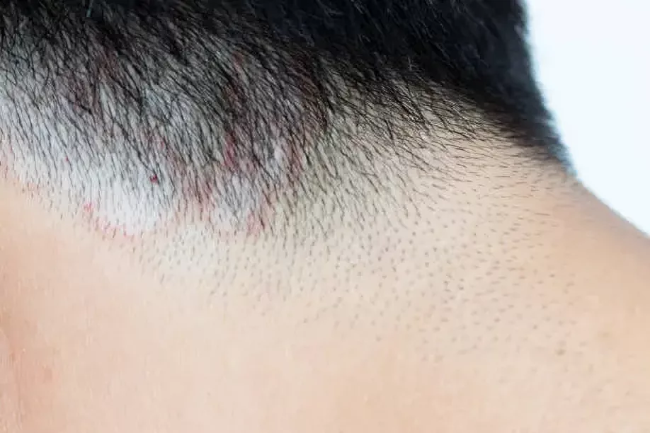
- Non-inflammatory Tinea capitis: Characterized by scaly patches with hair loss, black dots (broken hairs), or gray patches. It is generally less severe and rarely causes permanent damage.
- Inflammatory Tinea capitis: A more aggressive form that may present with kerion (a painful, swollen, pus-filled lesion) or favus (a chronic crusting form). If untreated, it can lead to scarring and permanent alopecia.
In some cases, eyebrows and eyelashes may also be affected, especially in severe or untreated infections. Prompt antifungal treatment is essential to prevent complications, especially with the inflammatory type. Topical treatments are usually ineffective alone—oral antifungal therapy (e.g., griseofulvin or terbinafine) is typically required.1ls. Hay R. J. (2017). Tinea Capitis: Current Status. Mycopathologia, 182(1-2), 87–93. https://doi.org/10.1007/s11046-016-0058-8
Causes of Tinea Capitis
Tinea capitis is a dermatophyte infection, and these are filamentous fungi in the genera Trichophyton, Microsporum, and Epidermophyton. They are commonly known for infecting the keratinised tissue of your skin, hair, and nails. The primary genera responsible for these infections include Trichophyton and Microsporum. Though Epidermophyton is a recognized dermatophyte genus, it rarely causes tinea capitis and more commonly affects other body sites (e.g., tinea cruris or tinea pedis).
Dermatophytes are classified based on their primary habitat and host preference:
- Anthropophilic (human)
- Zoophilic (animal)
- And geophilic (soil) fungi.
Among these, the most common causes of this infection are Anthropophilic and zoophilic dermatophytes. Examples of dermatophyte species associated with tinea capitis include:
Anthropophilic:
These fungi are transmitted from human to human and are the most common cause of tinea capitis worldwide. Common species include:
- Epidermophyton floccosum (very rare cause)
- Trichophyton tonsurans
- Trichophyton violaceum
- Trichophyton soudanens
- Trichophyton schoenleinii (associated with favus)
- Trichophyton interdigitale (formerly T. mentagrophytes var. interdigitale)
- Trichophyton rubrum (rare in tinea capitis)
- Microsporum audouinii
Zoophilic:
These are transmitted from animals and often cause more inflammatory infections:
- Arthroderma benhamiae (a teleomorph of certain Trichophyton species).
- Microsporum canis
- Microsporum nanum
- Trichophyton verrucosum
- Trichophyton equinum
Geophilic:
These species originate from the soil and occasionally cause human infection:
- Microsporum gypseum
- Microsporum fulvum
The prevalence of specific dermatophytes varies by geographic location and population, and may shift over time due to environmental, cultural, or socioeconomic changes.
Pathogenesis of Tinea capitis
Tinea capitis is transmitted through contact of the scalp with the causative dermatophyte. Acquisition of the fungus may occur due to direct contact with an infected individual or animal or by contact with a contaminated object (e.g., comb, brush, or hat). Furthermore, it also spreads among children in crowded places, most commonly through gatherings such as schools, daycare centers, parks, and restaurants.
In some cases, people may get infected by coming into contact with asymptomatic carriers of the fungus. These carriers may not show any visible signs of infection but can still transmit the organism. This mode of transmission is important, especially when considering recurrent tinea capitis in individuals.
When the dermatophyte comes in contact with the stratum corneum of the scalp, it begins to grow downward into this outer layer and invades keratin. Once the hair becomes infected, it weakens, becomes brittle, and eventually breaks off near the scalp surface.2Brasch J. (2010). Pathogenesis of tinea. Journal der Deutschen Dermatologischen Gesellschaft = Journal of the German Society of Dermatology : JDDG, 8(10), 780–786. https://doi.org/10.1111/j.1610-0387.2010.07481.x
Immunosuppression may impair hair shaft growth and strength, leading to easier colonisation. Other associated diseases include:
- Diabetes mellitus
- Prolonged steroid use
- Cancer
- Immunosuppressant medications
- Anemia
Although HIV is a major immunosuppressive condition, the risk of tinea capitis does not appear significantly increased in these patients, possibly due to competing colonisation by Malassezia species. Hair typically gets a fungal infection in one of three principal ways:
Endothrix:
In this pattern, the fungus invades the interior of the hair shaft. Trichophyton tonsurans is a common cause.3Araviysky, A. N., Araviysky, R. A., & Eschkov, G. A. (1975). Deep generalized trichophytosis. (Endothrix in tissues of different origin).Mycopathologia, 56(1), 47–65. https://doi.org/10.1007/BF00493584
Ectothrix:
Here, the fungus coats the outside of the hair shaft. An example is Microsporum canis.
Favus:
In the last category, favus develops, a chronic condition that may continue for several years. It isn’t easy to diagnose favus unless the patient develops alopecia (patchy hair loss ). The main causative organism is Trichophyton schoenleinii.
Common Symptoms of Tinea Capitis
The common symptoms of this infection include the following:4Hill, R. C., Gold, J. A. W., & Lipner, S. R. (2024). Comprehensive Review of Tinea Capitis in Adults: Epidemiology, Risk Factors, Clinical Presentations, and Management. Journal of fungi (Basel, Switzerland), 10(5), 357. https://doi.org/10.3390/jof10050357
- Scaly patches on the scalp
- Hair loss
- Kerion: Inflammatory mass in which remaining hairs are loose. Characterised by boggy, purulent, inflamed nodules and plaques. Usually painful; drains pus from multiple openings, like a honeycomb, thick crusting with matting of adjacent hairs.5Nakagawa, H., Nishihara, M., & Nakamura, T. (2018). Kerion and tinea capitis.IDCases, 14, e00418. https://doi.org/10.1016/j.idcr.2018.e00418
- Swollen lymph nodes in some cases
- Itching on the scalp, also known as scalp pruritus
- Tenderness on the scalp
- Brittle or fragile hair
- Fever (fever as a symptom is rarely not a feature of this disease, but it may occur with kerion or secondary bacterial infection)
- Cervical lymphadenopathy – Palpable cervical lymphadenopathy is a frequent finding in patients with tinea capitis, particularly when clinical signs of inflammation are present.
- Other infrequent symptoms include the occurrence of erythema nodosum in association with kerion.
- Dermatophytid reactions – Autoeczematization reactions (id reactions) are secondary dermatitic eruptions associated with a localised inflammatory skin disorder.
The popular term “dermatophytid reaction” primarily refers to autoeczematization reactions that may occur in association with dermatophyte infections. In this one, the pathogenesis involves an immune reaction to the fungal antigens, and this reaction could be like a delayed-type hypersensitivity response. Dermatophytid reactions often follow the onset of antifungal therapy but may also precede treatment. Most patients can manage dermatophytid reactions if the treatment begins with working on the underlying fungal infection and emollients, oral antihistamines, or topical corticosteroids. Oral corticosteroids, however, are not common drugs to use and refer to, and are mainly useful in severe cases.
How does Tinea Capitis spread?
This is a widespread fungal infection, and it has various ways of transmission. Three modes of transmission are:
- Human-to-human contact
- Animal-to-human contact
- Fomite transmission
Types of Tinea Capitis
Various types of this infection can occur through the three standard modes of transmission mentioned above. Types of tinea capitis based on clinical presentation and pathogenesis are:6Farooqi, M., Tabassum, S., Rizvi, D. A., Rahman, A., Rehanuddin, Awan, S., & Mahar, S. A. (2014). Clinical types of tinea capitis and species identification in children: an experience from tertiary care centres of Karachi, Pakistan. JPMA. The Journal of the Pakistan Medical Association, 64(3), 304–308.
Gray Patch Tinea Capitis:
This type of Tinea Capitis is generally found among children, and the cause is Microsporum.
Black Dot Tinea Capitis:
Trichophyton causes black dot Tinea Capitis. As its name suggests, the hair breaks off at the scalp, leaving black dots on your head.
Kerion:
Kerion refers to an inflammatory mass that sometimes oozes pus, and patients also present with lymphadenopathy and scarring in the case of kerion.
Favus:
This one can be divided into three stages, and the symptoms in each stage vary.7 Ilkit M. (2010). Favus of the scalp: an overview and update. Mycopathologia, 170(3), 143–154. https://doi.org/10.1007/s11046-010-9312-7
- Early cases show perifollicular erythema and matting of hair.
- Later, thick yellow adherent crusts (scutula) are composed of skin debris and hyphae pierced by remaining hair shafts.
- Fetid odor.
- It shows little tendency to clear spontaneously. Often results in scarring alopecia.
Difference between Scalp Psoriasis & Tinea Capitis
Scalp psoriasis is an autoimmune disease that quickly produces new skin cells, while tinea capitis is not an autoimmune disorder; it is a fungal infection of the scalp caused by dermatophytes and spreads through contact with an infected person. People with scalp psoriasis present with white, silvery, scaly patches around the hairline or behind the ears and neck. However, the clinical presentation of Tinea capitis is ring-like, along with hair loss and black dots.
Tinea capitis and scalp psoriasis also differ in their way of spreading, as you can get tinea capitis through physical contact with the person having it. But scalp psoriasis doesn’t spread like that. Scalp psoriasis diagnosis is based on clinical diagnosis; however, in case of any ambiguity, doctors also recommend that the individual go for a biopsy. Tinea capitis can be diagnosed by three standard methods: examining through fungal culture, conducting microscopy, or using a Wood’s lamp.
These two diseases also differ in their therapy. Doctors prescribe topical steroids and medicated shampoos (such as coal tar and salicylic acid) to treat Scalp Psoriasis. In severe cases of Scalp Psoriasis, biological agents play a significant role. In the case of Tinea Capitis, the doctors prescribe Oral antifungals (e.g, griseofulvin, terbinafine) and antifungal shampoos.8Crowley J. (2010). Scalp psoriasis: an overview of the disease and available therapies. Journal of drugs in dermatology : JDD, 9(8), 912–918.

How to diagnose Tinea Capitis?
The diagnosis is usually based on a clinical examination of the patient, which involves a wood lamp examination, microscopy (KOH test), fungal culture, and dermoscopy. 9Wei, L. W., & Qiao, J. J. (2023). Mini-Review: The Diagnostic Methods of Tinea Capitis. Mycopathologia, 188(5), 563–569. https://doi.org/10.1007/s11046-023-00731-3
Physical Examination & Dermoscopy:
The physical examination of Tinea Capitis involves pathological alopecia, cervical lymphadenopathy, scaling regions (“black dots”), and patches that need to be looked for. It is better to see the skin patches with the help of dermoscopy, as it involves the use of a dermatoscope ( a specialised magnifying glass ).
Potassium Hydroxide (KOH) Preparations:
Another standard diagnostic method for this fungal infection is Potassium Hydroxide (KOH) preparations. It helps dissolve the keratin in your skin, hair, and nails, but doesn’t disrupt the fungus in these structures. A sample is taken by scraping the skin of the scalp, and the technician makes a slide of it by putting potassium hydroxide on it. Pathologists see the slide under a microscope and identify the type of fungus. You can get results through KOH preparations within at least 24 hours.10 Levitt, J. O., Levitt, B. H., Akhavan, A., & Yanofsky, H. (2010). The sensitivity and specificity of potassium hydroxide smear and fungal culture relative to clinical assessment in the evaluation of tinea pedis: a pooled analysis. Dermatology research and practice, 2010, 764843. https://doi.org/10.1155/2010/764843
Fungal Culture:
A fungal culture test confirms the diagnosis and determines the species responsible for causing the fungal infection in your scalp. The pathologist takes the sample from the affected body part and lets the fungus grow in a dish with a specific substance. After confirming the presence of dermatophytes, this test will ensure the diagnosis is valid. The only issue with the fungal culture test is that the results may require several weeks.
Wood Lamp Examination:
Many pathologists use wood lamp examination to diagnose Tinea capitis. Wood lamp examination involves a special type of ultraviolet light that helps to identify the fungus by giving it a specific colour like blue, green, or yellow. This process requires the patient to sit in a dark room to make a better diagnosis.11Dyer, J. M., & Foy, V. M. (2022). Revealing The Unseen: A Review of Wood’s Lamp in Dermatology. The Journal of clinical and aesthetic dermatology, 15(6), 25–30.
Treatment of Tinea Capitis
The treatment of this fungal infection includes oral antifungal therapy and adjunctive measures. Besides treatment, it is always essential to maintain your hygiene to prevent the chances of causing tinea capitis. The options for oral antifungal therapy include terbinafine, griseofulvin, fluconazole, and itraconazole. Most doctors recommend using Terbinafine and griseofulvin for initial treatments as they have been reported to be most helpful in treating the infection.12Alkeswani, A., Cantrell, W., & Elewski, B. (2019). Treatment of Tinea Capitis. Skin Appendage Disorders, 5(4), 201–210. https://doi.org/10.1159/000495909
Moreover, in case an infant has developed the disease, they are prescribed Fluconazole by the doctor. The medications prescribed are written below:
A. Griseofulvin (first-line for children):
Children:
- Dose: 20–25 mg/kg/day (microsize) or 0–15 mg/kg/day (ultramicro size) for 6–8 weeks
Adults:
- Dose: 500–1000 mg/day, usually for 6–8 weeks
Take with fatty food to improve absorption
B. Terbinafine (first-line for Trichophyton Infections):
Children: (also depends on the body weight)
- <20 kg: 62.5 mg/day
- 20–40 kg: 125 mg/day
- 40 kg: 250 mg/day•
- Duration: 4 weeks
Adults:
- 250 mg/day for 4–6 weeks
C. Itraconazole (Alternative):
Children:
- 3–5 mg/kg/day, or pulse dosing (e.g., 5 mg/kg/day for 1 week/month)
Adults:
- 100–200 mg/day for 4–6 weeks or in pulse cycles
D. Fluconazole (Alternative):
Children:
- 6 mg/kg/day
Adults:
- 150–300 mg/week, or daily dosing
- Duration: 3–6 weeks
Antifungal shampoos containing fluconazole are also helpful in reducing fungal spores and preventing the spread. Use of these shampoos twice or thrice a week during the shower routine is a recommended dose.
How to prevent Tinea Capitis?
It is often stated that prevention is always better than treatment, and it is true in most skin and scalp-related issues. Keeping your body and skin clean and dirt-free can help avoid many diseases. There are several significant yet simple measures that you need to consider to prevent tinea capitis. These measures and tips include:
- Always try your best to keep your scalp clean and wash your hair regularly.
- Avoid sharing combs, shaving products, straighteners, and other similar personal items, especially if you live in a hostel or shared houses. This will help you keep your scalp free of germs and dirt and reduce the risk of infections.
- If you ever encounter any child or adult affected with Tinea Capitis, keep your distance and altogether avoid direct contact. Since Tinea Capitis is contagious, maintaining distance from the affected individual is highly important.
- For good physical health, maintaining good hygiene is a necessity.
- Keep your hair dry most of the time since fungal infection grows in moist environments.
These preventive measures are essential as they will help you avoid getting infected by Tinea Capitis and reduce the risk of reinfection if you have been infected earlier.13 Chen, X. Q., Zhou, Y. B., Xiao, Y. Y., & Ma, L. (2023). Zhonghua liu xing bing xue za zhi = Zhonghua liuxingbingxue zazhi, 44(12), 1988–1992. https://doi.org/10.3760/cma.j.cn112338-20230613-00373
What are the differentials of Tinea Capitis?
Symptoms of tinea capitis may resemble some other skin diseases. The most common differentials are seborrheic dermatitis and alopecia areata. However, it is easy to rule out Tinea capitis from these differentials based on symptoms. Alopecia areata is characterised by no formation of scales or inflammation, while Tinea capitis patients present with a scaly area along with hair loss and may also experience itching.
Seborrheic dermatitis and tinea capitis both present with scales, so it gets tricky sometimes to differentiate between them. Yet, some differences exist as seborrheic dermatitis presents with diffuse hair loss, and tinea capitis invokes a patchy hair loss. Moreover, seborrheic dermatitis also has greasy yellow scales, unlike Tinea capitis.
Conclusion
Tinea Capitis is a fungal infection; maintaining your physical health and body hygiene is necessary to prevent the disease. If you have developed the disease, you need a proper check-up. Oral fungal therapy is recommended to treat the disease. It is also important to use antifungal shampoos and maintain your overall body hygiene to reduce the risk of developing the disease or its reinfection, or transmission to other individuals in case the disease has already impacted you.
Moreover, follow-up of the patient requires clinical examination to ensure the disease is no longer present, and it is also important for the patient to know when to discontinue oral antifungal treatment. Therefore, Taking care of your physical health and using shampoo prescribed by doctors and dermatologists is key to preventing such diseases.
Refrences
- 1ls. Hay R. J. (2017). Tinea Capitis: Current Status. Mycopathologia, 182(1-2), 87–93. https://doi.org/10.1007/s11046-016-0058-8
- 2Brasch J. (2010). Pathogenesis of tinea. Journal der Deutschen Dermatologischen Gesellschaft = Journal of the German Society of Dermatology : JDDG, 8(10), 780–786. https://doi.org/10.1111/j.1610-0387.2010.07481.x
- 3Araviysky, A. N., Araviysky, R. A., & Eschkov, G. A. (1975). Deep generalized trichophytosis. (Endothrix in tissues of different origin).Mycopathologia, 56(1), 47–65. https://doi.org/10.1007/BF00493584
- 4Hill, R. C., Gold, J. A. W., & Lipner, S. R. (2024). Comprehensive Review of Tinea Capitis in Adults: Epidemiology, Risk Factors, Clinical Presentations, and Management. Journal of fungi (Basel, Switzerland), 10(5), 357. https://doi.org/10.3390/jof10050357
- 5Nakagawa, H., Nishihara, M., & Nakamura, T. (2018). Kerion and tinea capitis.IDCases, 14, e00418. https://doi.org/10.1016/j.idcr.2018.e00418
- 6Farooqi, M., Tabassum, S., Rizvi, D. A., Rahman, A., Rehanuddin, Awan, S., & Mahar, S. A. (2014). Clinical types of tinea capitis and species identification in children: an experience from tertiary care centres of Karachi, Pakistan. JPMA. The Journal of the Pakistan Medical Association, 64(3), 304–308.
- 7Ilkit M. (2010). Favus of the scalp: an overview and update. Mycopathologia, 170(3), 143–154. https://doi.org/10.1007/s11046-010-9312-7
- 8Crowley J. (2010). Scalp psoriasis: an overview of the disease and available therapies. Journal of drugs in dermatology : JDD, 9(8), 912–918.
- 9Wei, L. W., & Qiao, J. J. (2023). Mini-Review: The Diagnostic Methods of Tinea Capitis. Mycopathologia, 188(5), 563–569. https://doi.org/10.1007/s11046-023-00731-3
- 10Levitt, J. O., Levitt, B. H., Akhavan, A., & Yanofsky, H. (2010). The sensitivity and specificity of potassium hydroxide smear and fungal culture relative to clinical assessment in the evaluation of tinea pedis: a pooled analysis. Dermatology research and practice, 2010, 764843. https://doi.org/10.1155/2010/764843
- 11Dyer, J. M., & Foy, V. M. (2022). Revealing The Unseen: A Review of Wood’s Lamp in Dermatology. The Journal of clinical and aesthetic dermatology, 15(6), 25–30.
- 12Alkeswani, A., Cantrell, W., & Elewski, B. (2019). Treatment of Tinea Capitis. Skin Appendage Disorders, 5(4), 201–210. https://doi.org/10.1159/000495909
- 13Chen, X. Q., Zhou, Y. B., Xiao, Y. Y., & Ma, L. (2023). Zhonghua liu xing bing xue za zhi = Zhonghua liuxingbingxue zazhi, 44(12), 1988–1992. https://doi.org/10.3760/cma.j.cn112338-20230613-00373
