What is a Torn Meniscus?
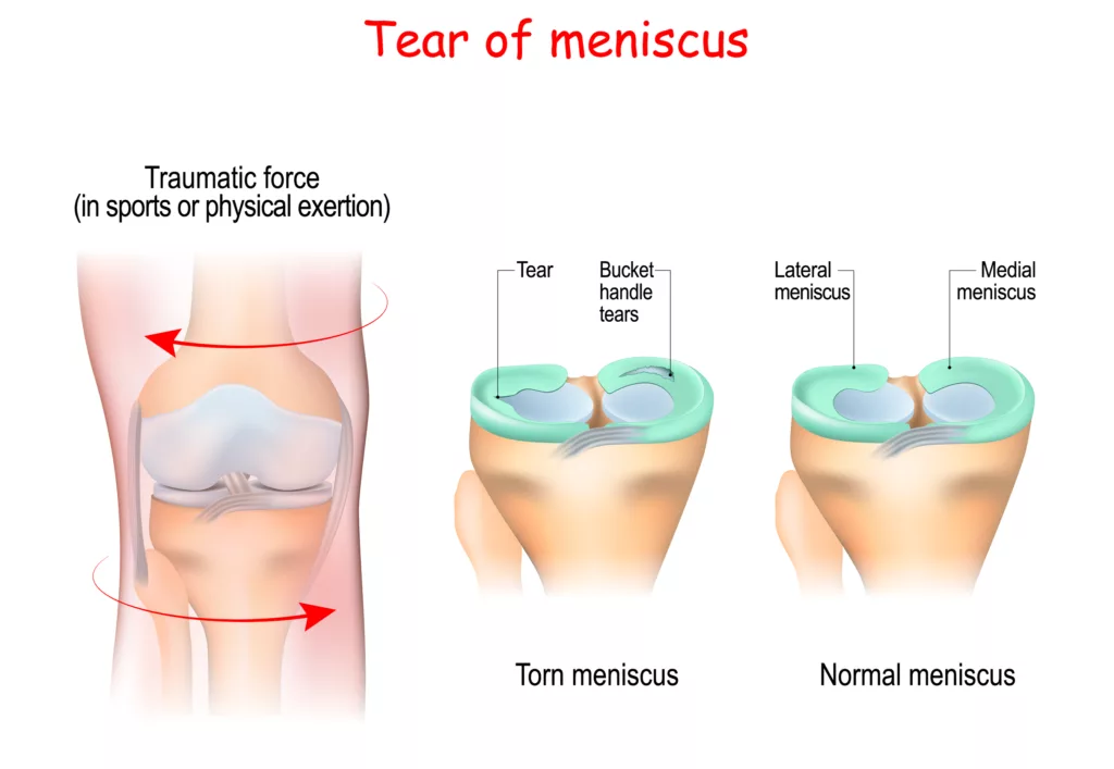
A torn meniscus refers to a tear in the cartilage that cushions and stabilizes the knee joint. The meniscus is a C-shaped cartilage in the knee that stabilizes the joint. The human knee has two menisci. They are located between the femur (upper thigh bone) and the tibia, which is the lower leg bone. The lateral meniscus is located outside the knee, whereas the medial meniscus is on the interior.
The meniscus supports lubricating fluid around the knee to stabilize and improve the fit between the femur and the tibia. It also absorbs stress and distributes weight to the knee.1The role of the meniscus in knee osteoarthritis: a cause or consequence? Englund M, Guermazi A, Lohmander SL. Radiol Clin North Am. 2009;47:703–712. A meniscus tear typically occurs due to a sudden twist or rotation of the knee, often during sports or physical activity. It can cause pain, swelling, stiffness, and difficulty moving the knee and may lead to a feeling of the knee “locking” or “giving way.” Depending on the severity and location of the tear, treatment may range from rest and physical therapy to surgical repair.
Causes of Torn Meniscus
A tear in the meniscus happens when a person abruptly changes his position while jumping or running. Different causes of torn meniscus include traumatic and degenerative changes. A few are mentioned below:
Traumatic Causes:
- Any sudden or brisk movement
- A sudden stop of the knee that stresses the joint with extra pressure may cause a torn meniscus.
- Accidental twisting or catching of the knee.
- A jerky jump or awkward landing.
Degenerative Causes:
- Chronic Arthritis
- Osteoarthritis2Degenerative meniscal tears of the knee: evaluation and management. Matar HE, Duckett SP, Raut V. Br J Hosp Med (Lond) 2019;2:46–50.
- High Obesity Index
- Diabetes > 15 years with HIGH BMI (body mass index)
How it occurs?
The meniscus is a C-type pad of cartilage in the knee that acts as a shock absorber. Meniscus tears frequently occur when an athlete twists or rotates their upper leg while their foot is firmly planted and their knee is bent.3The natural history of meniscus tears. Chambers HG, Chambers RC. J Pediatr Orthop. 2019;39:53–55.
Clinical Manifestations of Tear in Meniscus:
Although each person’s meniscus tear symptoms may vary, some of the most typical ones include:
- Swelling
- Pain and localized discomfort
- Knee catching, locking, and the characteristic ‘giving away’ sensation
- A popping sensation during the injury
- Difficulty in fully bending or extending the leg
- Limited mobility
- Known cases of chronic arthritis will observe an increase in pain in the knee joint.
- Giving way sensation of the knee joint while climbing the stairs or getting out of the car
Who is at risk of Torn meniscus?
A torn meniscus can affect many people, but certain groups are more susceptible to this injury. They are more commonly seen in men than women, with up to 80% of all meniscal tears reported in men.4Bhan K. (2020). Meniscal Tears: Current Understanding, Diagnosis, and Management. Cureus, 12(6), e8590. https://doi.org/10.7759/cureus.8590 Other risk factors are:
- Individuals who frequently play sports such as tennis, football, or baseball.
- Professional athletes who engage in high-impact activities.
- Anyone who experiences a sudden twist of the knee due to a hit or trauma.
- Those suffering from degenerative diseases related to aging.
Types of Meniscal Tears
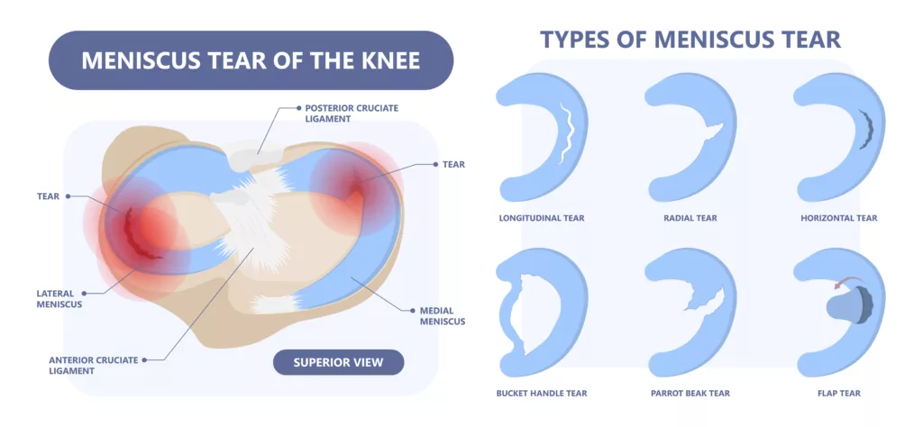
Meniscal tears are classified based on their morphology, guiding diagnosis, and treatment planning. It has the following main types:
Horizontal Tear (Cleavage Tear):
This type runs parallel to the tibial plateau, dividing the meniscus into upper and lower sections. It affects one of the articular surfaces or the free edge of the meniscus.
Vertical Tear:
A tear that runs perpendicular to the tibial plateau and parallel to the meniscus’s long axis, splitting the meniscus into medial and lateral parts. It can be:
- Wrisberg Rip: A specific subtype affecting the lateral meniscus.
- Ramp Lesion: A specific subtype affecting the medial meniscus.
Radial Tear:
This tear is oriented perpendicular to both the tibial plateau and the long axis of the meniscus.
Root Tear:
A specific type of radial tear occurs at the meniscal root.
Complex Tear:
Involves a combination of horizontal, vertical, and radial tears.
Displaced Tear:
Includes tears where a portion is displaced. A displaced tear can be further classified into the following types:
- Flap Tear: A displaced horizontal or vertical tear.
- Bucket-Handle Tear: A displaced vertical tear resembling a bucket handle.
- Parrot Beak Tear: An oblique radial tear.
How Is A Torn Meniscus Diagnosed?
If a meniscal tear is suspected, your orthopedist will begin with a detailed medical history and a comprehensive knee examination. Initially, your healthcare provider will ask about your symptoms and how the injury occurred, followed by a physical exam where they may perform specific tests to check for pain, swelling, or limited knee movement.
One common test is the McMurray test, where the doctor will bend, rotate, and straighten your knee to see if it causes a clicking sound or pain.5Hing, W., White, S., Reid, D., & Marshall, R. (2009). Validity of the McMurray’s Test and Modified Versions of the Test: A Systematic Literature Review. The Journal of manual & manipulative therapy, 17(1), 22–35. https://doi.org/10.1179/106698109790818250 To confirm the diagnosis and assess the extent of the injury, they may also order X-rays and an MRI for a closer evaluation of the knee joint.
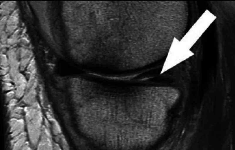
MRI:
An MRI is a diagnostic method that creates comprehensive images of the knee and its relative structure using a combination of giant magnets, radiofrequency technology, and a computer. It is a diagnostic test that depicts arterial supply compromise, the exact location of a torn meniscus, and the extent of injury or disease in an adjacent ligament or muscle.6Meniscal tears. Maffulli N, Longo UG, Campi S, Denaro V. Open Access J Sports Med. 2010;26:45–54.
XRAY:
X-rays are diagnostic procedures that use invisible electromagnetic energy to create images of inside organs, bones, and tissues on film. When a regular X-ray is not precise enough, a joint X-ray with contrast dye may also be used to examine joints like the knee or hip.
Additionally, doctors may suggest arthroscopy and arthrograms for a torn meniscus. Arthroscopy involves inserting a camera into the knee to visualize and potentially treat the tear directly. An arthrogram uses contrast dye injected into the joint to enhance the visibility of the meniscus on X-rays or MRIs, providing clearer images for diagnosis. However, diagnostic arthroscopy is not recommended without the therapeutic benefit.
Treatment for Meniscal Tear:
The treatment for a torn meniscus depends on the tear’s type, size, and location. Conservative management is recommended for meniscus tears located in the highly vascularized areas of the meniscus, specifically the red/red zone. This includes the peripheral 30% of the medial meniscus and 25% of the lateral meniscus. Tears in these areas tend to heal better with non-surgical treatment, especially if smaller than 5 mm, as these are generally considered stable and more likely to respond well to conservative care.7Treatment of meniscal injuries in young athletes. Giuliani JR, Burns TC, Svoboda SJ, Cameron KL, Owens BD. J Knee Surg. 2011;24:93–100.
Conservative management:
Surgery is typically not necessary since tears associated with arthritis often improve over time with arthritis medication. Many other tears that aren’t connected to locking or a block to knee mobility won’t worsen over time. Therefore, they don’t need to be surgically repaired.
Your physician might advise:
Rest:
Avoid doing anything that makes your knee hurt more, especially if it involves twisting, rotating, or pivoting.
Ice:
Knee discomfort and edema can be reduced with ice. Use a cold pack, a bag of frozen veggies, or a towel filled with
Medications:
Over-the-counter medicines like NSAIDs are recommended for pain. The most common are ibuprofen and naproxen for relieving pain and inflammation.
Physiotherapy:
After thoroughly examining your knee, your physiotherapist will create a unique exercise-based therapy program. The exercises will progressively strengthen your knee and leg muscles, improving your knee’s range of motion, strength, and stability.
Surgical Management:
When conservative measures are inadequate, or for tears in less vascularized areas, surgical intervention may be necessary:
Arthroscopic Partial Meniscectomy (APM):
This procedure involves removing damaged tissue via small incisions. While common, recent studies suggest its effectiveness may not exceed that of placebo treatments.8Arthroscopic partial meniscectomy in young patients with symptomatic discoid lateral meniscus: an average 10-year follow-up study. Lee CR, Bin SI, Kim JM, Lee BS, Kim NK. Arch Orthop Trauma Surg. 2018;138:369–376.
In this minimally invasive procedure, an arthroscope (a surgical instrument with a camera) removes the torn meniscus fragment. Instead of one huge open incision in the knee, arthroscopic surgery requires two to three tiny incisions. The procedure takes 30 minutes to an hour, depending on the tear size.
Post-surgical healing:
After surgery, crutches are recommended for up to six weeks. Physical therapy speeds up the healing process in 2-3 months.
Meniscal Repair:
This aims to heal the meniscus while preserving its function. Various techniques are used, such as inside-out, all-inside, and outside-in repairs. Short-term success is good, but long-term outcomes can vary, with failure rates up to 30% at five years.9Meniscal repair outcomes at greater than five years: a systematic literature review and meta-analysis. Nepple JJ, Dunn WR, Wright RW. J Bone Joint Surg Am. 2012;19:2222–2227.
Meniscus Replacement:
If repairing the meniscus isn’t possible or if a meniscectomy has already been performed, meniscus replacement has proven useful.
-
Meniscal Allografting:
Involves replacing the damaged meniscus with a donor meniscus. This complex procedure is called Meniscal Allograft Transplantation (MAT) and has a 10-year survival rate of about 89.2%.10Midterm and long-term results of medial versus lateral meniscal allograft transplantation: a meta-analysis. Bin SI, Nha KW, Cheong JY, Shin YS. Am J Sports Med. 2018;46:1243–1250.
-
Meniscal Scaffolds:
These FDA-approved, minimally invasive options allow tissue to grow into a scaffold that mimics meniscal function for partial meniscus replacement.
Conclusion
A torn meniscus is a trauma. Although painful, the best thing is that it recovers to its full if treated carefully. The patient should follow the self-care advice of his doctor for a good prognosis. Yet, it is time-consuming but manageable.
Refrences
- 1The role of the meniscus in knee osteoarthritis: a cause or consequence? Englund M, Guermazi A, Lohmander SL. Radiol Clin North Am. 2009;47:703–712.
- 2Degenerative meniscal tears of the knee: evaluation and management. Matar HE, Duckett SP, Raut V. Br J Hosp Med (Lond) 2019;2:46–50.
- 3The natural history of meniscus tears. Chambers HG, Chambers RC. J Pediatr Orthop. 2019;39:53–55.
- 4Bhan K. (2020). Meniscal Tears: Current Understanding, Diagnosis, and Management. Cureus, 12(6), e8590. https://doi.org/10.7759/cureus.8590
- 5Hing, W., White, S., Reid, D., & Marshall, R. (2009). Validity of the McMurray’s Test and Modified Versions of the Test: A Systematic Literature Review. The Journal of manual & manipulative therapy, 17(1), 22–35. https://doi.org/10.1179/106698109790818250
- 6Meniscal tears. Maffulli N, Longo UG, Campi S, Denaro V. Open Access J Sports Med. 2010;26:45–54.
- 7Treatment of meniscal injuries in young athletes. Giuliani JR, Burns TC, Svoboda SJ, Cameron KL, Owens BD. J Knee Surg. 2011;24:93–100.
- 8Arthroscopic partial meniscectomy in young patients with symptomatic discoid lateral meniscus: an average 10-year follow-up study. Lee CR, Bin SI, Kim JM, Lee BS, Kim NK. Arch Orthop Trauma Surg. 2018;138:369–376.
- 9Meniscal repair outcomes at greater than five years: a systematic literature review and meta-analysis. Nepple JJ, Dunn WR, Wright RW. J Bone Joint Surg Am. 2012;19:2222–2227.
- 10Midterm and long-term results of medial versus lateral meniscal allograft transplantation: a meta-analysis. Bin SI, Nha KW, Cheong JY, Shin YS. Am J Sports Med. 2018;46:1243–1250.
