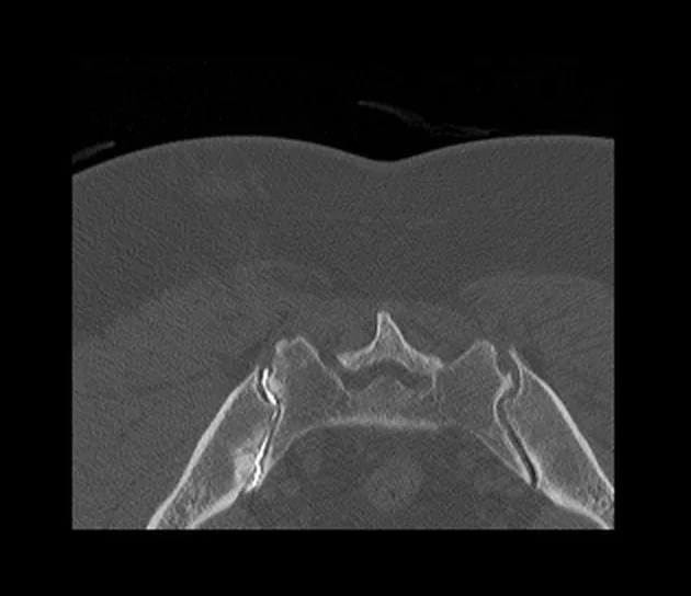
An Arthrogram, also called arthrography, is an invasive imaging test that aims to find out the cause of pain or joint problems, which is not very clear when using regular imaging tests. This test uses contrast dye to get a detailed image of a joint. Arthrogram is commonly performed to assess knee and shoulder joints. However, it is also performed to evaluate other joints as well, including wrist, elbow, ankle, hip, and other joints.1Berquist T. H. (1997). Imaging of articular pathology: MRI, CT, arthrography. Clinical anatomy (New York, N.Y.), 10(1), 1–13. https://doi.org/10.1002/(SICI)1098-2353(1997)10:1<1::AID-CA1>3.0.CO;2-#
What is an Arthrogram?
An arthrogram is a medical imaging test that involves injecting contrast dye into the joint before taking X-rays, CT scans, fluoroscopy, or MRIs to get a clearer picture of the joint. The arthrogram allows distention of the joint and makes structures visible and distinguishable from the soft tissues. A radiologist performs the procedure, and a technologist assists by taking images of the joint that your doctor will later check and assess. The test can help identify tears in ligaments, tendons, and joint capsules, and it can also be helpful in finding dislocated joints or bone fragments that might cause pain. It is also useful after joint replacement surgery to ensure the joint is properly placed and functioning correctly.2Zhu, Q., & Nobuhara, K. (2001). The role of radiocarpal injection arthrography and magnetic resonance imaging in the diagnosis of triangular fibrocartilage complex injuries. Chinese journal of traumatology = Zhonghua chuang shang za zhi, 4(2), 78–81.
Arthrography is considered safe yet not recommended if you are pregnant or have a joint infection or arthritis.3Berquist T. H. (1997). Imaging of articular pathology: MRI, CT, arthrography. Clinical anatomy (New York, N.Y.), 10(1), 1–13. https://doi.org/10.1002/(SICI)1098-2353(1997)10:1<1::AID-CA1>3.0.CO;2-#
Types of Arthrogram
An arthrogram is an effective imaging tool for identifying diseases and conditions of ligaments, tendons, cartilage, synovial fluid, and other joint structures. There are two types of arthrogram: direct and indirect.
Direct & Indirect Arthrogram
Indirect arthrography involves injecting contrast material into the bloodstream, eventually reaching the joint. Conversely, contrast material is directly injected into the joint in direct arthrography. Both direct and indirect methods improve the visualization of the joint area after injecting the contrast.
The imaging techniques used to capture the joint images include Computed Tomography (CT) scans, Magnetic Resonance Imaging (MRI), and fluoroscopy (a real-time X-ray) and X-ray.
There are two ways for Direct Arthrography:
- Conventional Direct Arthrography: It uses iodine contrast dye injected into the joint, guided by fluoroscopy, to enhance the visualization of joint structures.
- MRI or CT Direct Arthrography: It uses MRI or CT to guide the injection of contrast material into the joint. The contrast material used in MRI arthrography is gadolinium. MRI uses strong magnetic and radiofrequency waves and a computer to get detailed pictures of organs, soft tissues, bones, and other internal structures of the body. CT direct arthrography uses the same iodine contrast as conventional direct arthrography. CT provides cross-sectional images of the soft tissues and internal body structures using X-rays.
Direct arthrography is preferred over indirect arthrography as it improves the view of small internal structures by enlarging the joint space. This helps in better evaluating joint diseases and conditions.
When do I need an Arthrogram?
Your doctor will recommend an arthrogram to get detailed images of the joint for making an accurate diagnosis and treatment plan for your condition. Your doctor might recommend an arthrogram if:
- Regular Imaging tests are not helpful: When regular X-rays, MRI scans, and CT scans don’t clearly show the root cause of the joint pain or dysfunction.
- Suspected Joint Tissue Damage: When your doctor suspects tears in ligaments, capsules, tendons, or cartilage.
- Joint Displacement: To identify joint dislocation or displacement.
- Post-Operative Assessment: After joint replacement surgery to ensure the joint is placed properly and functioning correctly.
- Chronic Pain: To find the root cause of chronic pain that has not yet been diagnosed.
- Inject Medicine: To inject cortisone, a pain-relieving medicine, into painful joints.

Contraindications for Arthrogram
The arthrogram procedure is a safe method with minimal risks to contrast dye, but there are some contraindications for the procedure, and those are;
- Anaphylaxis (severe allergic reaction) to contrast dye
- Contraindications for MRI including cochlear and dental implants4Ghadimi M, Sapra A. Magnetic Resonance Imaging Contraindications. [Updated 2023 May 1]. In: StatPearls [Internet]. Treasure Island (FL): StatPearls Publishing; 2024 Jan-. Available from: https://www.ncbi.nlm.nih.gov/books/NBK551669/
- Pregnancy5Garcia-Bournissen, F., Shrim, A., & Koren, G. (2006). Safety of gadolinium during pregnancy. Canadian family physician Medecin de famille canadien, 52(3), 309–310.
- Infective joint condition
- Recent local or systemic infection
- Avascular necrosis of a joint
- Recent MR arthrogram or CT
- Not able to stay still for the procedure
- Active arthritis6Johns Hopkins Medicine. (n.d.). Arthrography. Retrieved from https://www.hopkinsmedicine.org/health/treatment-tests-and-therapies/arthrography#:~:text=Arthrography%20is%20not%20recommended%20for,may%20lead%20to%20birth%20defects.
How Do I Prepare Myself for the Test?
You don’t need to do anything special before undergoing the arthrogram procedure, but wearing comfortable clothes and removing anything metal, including jewelry, is essential. Avoid eating and drinking right before the procedure to prevent discomfort like nausea and vomiting. If you fear tight or closed spaces, they will give you medicine to help you stay calm. Before going for the procedure, inform your doctor about:
- All medical devices you use, including pacemakers, hearing aids, and cochlear implants.
- Any allergies to dyes and medicines.
- The medicines you usually take.
- If you are pregnant or have a joint infection.
The Procedure of the Arthrogram
The arthrogram procedure is done as an outpatient or as a part of a hospital stay. The procedure differs depending on the area/joint undergoing the procedure. However, the procedure will generally look like this;
- You will change into a hospital gown and lie on the procedure table to allow the radiologist to perform the arthrogram.
- He may then take images of the joint before injecting contrast dye to compare them with images after injecting the dye.
- Before beginning the procedure, the radiologist/technician will clean the procedure area with antiseptic.
- Next, using anesthesia, he will numb the procedure site that might feel painful.
- If the joint has swelling or fluid, your technician will remove it using a syringe. The fluid may then be sent to the laboratory for diagnostic purposes.
- Now, he will inject contrast dye into the joint using ultrasound, fluoroscopy, CT, or MRI. Usually, doctors inject gadolinium into the joint for an MRI arthrography. If the patient cannot undergo an MRI, they use an iodine-containing contrast and perform a CT scan instead. If the procedure is for therapeutic purposes, then medication or anesthesia is injected in this step to reduce pain from the joint.7Crossan K, Rawson D. Shoulder Arthrogram. [Updated 2023 Apr 3]. In: StatPearls [Internet]. Treasure Island (FL): StatPearls Publishing; 2024 Jan-. Available from: https://www.ncbi.nlm.nih.gov/books/NBK580562/
- Next, he will ask you to move the joint to spread the dye effectively and get images of the joint and tissues.
- He may now take images in different positions and angles using MRI, CT, Fluoroscopy, Ultrasound, or X-ray, depending on the doctor’s recommendation.
Result of Arthrogram
The technician will review the images from your arthrogram and send them to your doctor within a day or two. Your doctor will then discuss the results with you and plan possible treatments, such as surgery or joint replacement, to resolve your pain and condition. If needed, your doctor may also request further tests.
What can I expect after the Arthrogram?
After an arthrogram, your joint may swell, feel tender, and make popping sounds. These are normal and should go away in a few hours or days. Your doctor will suggest ways to manage these symptoms. They may recommend using cold packs to reduce swelling and taking medicine for tenderness and pain. Avoid intense physical activity for a while.
Contact your doctor if you notice:
- A high temperature
- Swelling, tenderness, and severe pain lasting more than 2-3 days
- Fluid draining from the procedure site
Risks from Arthrogram
Arthrogram is a safe procedure with low risks, but you may experience:
- Allergic reaction to the contrast material, which may cause hives, stomach upset, or dizziness.
- Infection or bleeding at the injection site, though this is a rare complication.
- Radiation exposure – while single radiation doses are generally not harmful, repeated exposure may pose health risks like cancer. Be sure to inform your doctor if you are pregnant, as it may affect the baby.
- Pain from the needle at the injection site
If you experience severe pain, prolonged swelling, fever, or any signs of infection, contact your doctor immediately.
MRI vs. Arthrogram
MRI (Magnetic Resonance Imaging) is a non-invasive technique that uses strong magnetic and radio waves, along with a computer, to provide detailed images of soft tissues like the spleen, liver, breast tissue, kidneys, and joints.8Li, Q., Amano, K., Link, T. M., & Ma, C. B. (2016). Advanced Imaging in Osteoarthritis. Sports health, 8(5), 418–428. https://doi.org/10.1177/1941738116663922
In contrast, an arthrogram is an invasive imaging method that injects contrast dye directly into the joint or indirectly through the bloodstream to get detailed images of soft tissues and joints.
Both procedures are safe methods for evaluating joint and other conditions, as they don’t use ionizing radiation. They help identify conditions that are otherwise difficult to diagnose. MRI gives cross-sectional images of body parts, while an arthrogram uses dye injected into the joint and then employs regular imaging techniques like X-ray, MRI, fluoroscopy, or CT scan to get clear images.
Conclusion
An arthrogram, or arthrography, is a procedure where contrast material is injected into a joint for clearer imaging. It’s useful for detecting joint diseases, drawing diagnostic fluid, and injecting anesthesia. Common joints include the shoulder, knee, wrist, elbow, hip, and ankle.
The procedure, which uses X-rays, fluoroscopy, MRI, or CT scans, takes about two hours. There are two types of arthrography: indirect and direct, with direct being preferred for better clarity. Risks are minimal but include a slight chance of allergic reaction. It’s not recommended for infected joints or during pregnancy. Post-procedure soreness or swelling usually resolves within two days.
Refrences
- 1Berquist T. H. (1997). Imaging of articular pathology: MRI, CT, arthrography. Clinical anatomy (New York, N.Y.), 10(1), 1–13. https://doi.org/10.1002/(SICI)1098-2353(1997)10:1<1::AID-CA1>3.0.CO;2-#
- 2Zhu, Q., & Nobuhara, K. (2001). The role of radiocarpal injection arthrography and magnetic resonance imaging in the diagnosis of triangular fibrocartilage complex injuries. Chinese journal of traumatology = Zhonghua chuang shang za zhi, 4(2), 78–81.
- 3Berquist T. H. (1997). Imaging of articular pathology: MRI, CT, arthrography. Clinical anatomy (New York, N.Y.), 10(1), 1–13. https://doi.org/10.1002/(SICI)1098-2353(1997)10:1<1::AID-CA1>3.0.CO;2-#
- 4Ghadimi M, Sapra A. Magnetic Resonance Imaging Contraindications. [Updated 2023 May 1]. In: StatPearls [Internet]. Treasure Island (FL): StatPearls Publishing; 2024 Jan-. Available from: https://www.ncbi.nlm.nih.gov/books/NBK551669/
- 5Garcia-Bournissen, F., Shrim, A., & Koren, G. (2006). Safety of gadolinium during pregnancy. Canadian family physician Medecin de famille canadien, 52(3), 309–310.
- 6Johns Hopkins Medicine. (n.d.). Arthrography. Retrieved from https://www.hopkinsmedicine.org/health/treatment-tests-and-therapies/arthrography#:~:text=Arthrography%20is%20not%20recommended%20for,may%20lead%20to%20birth%20defects.
- 7Crossan K, Rawson D. Shoulder Arthrogram. [Updated 2023 Apr 3]. In: StatPearls [Internet]. Treasure Island (FL): StatPearls Publishing; 2024 Jan-. Available from: https://www.ncbi.nlm.nih.gov/books/NBK580562/
- 8Li, Q., Amano, K., Link, T. M., & Ma, C. B. (2016). Advanced Imaging in Osteoarthritis. Sports health, 8(5), 418–428. https://doi.org/10.1177/1941738116663922

