Vulvar Intraepithelial Neoplasia is a non-invasive squamous cell lesion that affects your vulvar skin. It manifests as abnormal growth and discoloration of your vulvar skin. It can occur anywhere on the skin of the vulva. This neoplasm is limited to the skin of the vulva. Therefore, malignancy is rare but can occur if it remains undiagnosed for long.
What is Vulvar Intraepithelial Neoplasia?
Vulvar intraepithelial neoplasia is a squamous cell lesion that affects the female genital area, specifically the vulva. It is a precursor to squamous cell carcinoma and mainly targets adult females. It is a premalignant condition (condition before cancer) and is limited to the skin of the vulva but can be malignant if it remains neglected for a long period. There is no screening test available for this neoplasia. A careful clinical examination and biopsy are helpful in the diagnosis.
Vulvar intraepithelial neoplasia most commonly targets females in their forties. It is more common in white females than in black ones. Undifferentiated vulvar intraepithelial neoplasia (VIN) is more common in adult females (in the fourth decade of life), while differentiated vulvar intraepithelial neoplasia is more common in old ages (in the sixth decade of life).1Ayala, M. (2023, October 18). Vulvar intraepithelial neoplasia. StatPearls – NCBI Bookshelf. https://www.ncbi.nlm.nih.gov/books/NBK540982/
Causes of Vulvar Intraepithelial Neoplasia
There is no known cause of vulvar intraepithelial neoplasia. However, this is more common in white females than in blacks. People with vulvar intraepithelial neoplasia also have a high-risk form of human papillomavirus (HPV).
HPV is a highly contagious and sexually transmitted disease. The transmission rate is higher in sexually active patients. The incidence and prevalence of HPV in vulvar intraepithelial neoplasia is 72-100%. Out of all HPV viruses, HPV16 is the most common form, which is linked with vulvar intraepithelial neoplasia.
Classification of Vulvar Intraepithelial Neoplasia
Vulvar epithelial neoplasia (VIN) is classified into three types. There are three grades of VIN (VIN1, VIN2, and VIN3). The higher the grade, the more severe the disease.
High-Grade Squamous Cell Intraepithelial Lesion (HSIL):
This is the most common form of VIN. People with this type of VIN are at greater risk of having malignancy than others. This lesion, previously referred to as VIN2 and VIN3, is characterized by the presence of undifferentiated vulvar intraepithelial neoplasia.
It most commonly targets females of 40 years of age. It also occurs in patients who have a history of smoking and are immunocompromised. Early diagnosis and treatment are necessary because this type of VIN is premalignant and can develop into a malignant form if it remains untreated for a long duration. However, the risk of conversion into a malignant form is low.
Low-Grade Squamous Cell Intraepithelial Lesion (LSIL):
This less severe form of VIN manifests as flat condyloma (warts) on the vulvar skin, previously referred to as VIN1. These condylomas are non-cancerous and shed away on their own without any treatment. However, regular follow-up is necessary for evaluation.
Differentiated Vulvar Intraepithelial Neoplasia (VIN):
It is a less common type of vulvar intraepithelial neoplasia, accounting for only 5% of all VIN cases. It is not caused by human papillomavirus (HPV) but is called VIN simplex disease. Differentiated valvular intraepithelial neoplasia occurs in patients with a positive history of lichen sclerosus (an inflammatory disease that causes itching and the formation of white patches). Additionally, this type of VIN most commonly affects women aged between 50 and 60 years.
This lesion is more dangerous than HISL because the risk of malignancy is higher. Therefore, surgery is indicated to treat this type of VIN. 2Vulval intraepithelial neoplasia (VIN). Squamous intraepithelial lesion (SIL) | DermNet. (n.d.). https://dermnetnz.org/topics/vulval-intraepithelial-neoplasia
Symptoms of Vulvar Intraepithelial Neoplasia
Symptoms vary from person to person depending upon the immune status of the person and the type of vulvar intraepithelial neoplasia. However, some people remain asymptomatic and have only patchy discoloration on the vulvar skin on examination. While some people experience symptoms. These symptoms are the following:
- Severe itching that does not settle down
- Patch of discoloration around the skin or thickened skin
- Mild to moderate burning sensation
- The presence of warts on the skin (flat or raised)
- Dyspareunia (pain during intercourse)
Lesions of vulvar intraepithelial neoplasia can develop on any part of the vulva. But are most commonly found in hair-bearing parts (labia majora), non-hairy parts (labia minora), and posterior fourchettes. The differentiated vulvar intraepithelial neoplasia most commonly develops on non-hairy skin. 3Vulval intraepithelial neoplasia (VIN). (n.d.). Cancer Research UK. https://www.cancerresearchuk.org/about-cancer/vulval-cancer/vulval-intraepithelial-neoplasia
Which form of HPV causes VIN?
VIN is most commonly linked with HPV infection. There are different types of HPV. But HPV16, HPV18, and HPV23 cause vulvar intraepithelial neoplasia. Out of these, HPV 16 is the most common.4Management of vulvar intraepithelial neoplasia. (n.d.). ACOG. https://www.acog.org/clinical/clinical-guidance/committee-opinion/articles/2016/10/management-of-vulvar-intraepithelial-neoplasia
Who is at risk of having Vulvar Intraepithelial Neoplasia?
Some factors increase the risk of having vulvar intraepithelial neoplasia. However, these factors include:
- Positive history of HPV infection
- Positive history of herpes simplex virus
- Smoking
- History of human immunodeficiency virus (HIV)
- Immunocompromised
- People who are taking immunosuppressive medications after organ transplant
- Lichen sclerosis
- Chronic vulvar infection
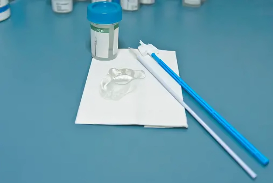
How to Diagnose Intraepithelial Neoplasia?
Diagnosing this condition is challenging because no screening test is available for VIN. However, it is diagnosed by taking history, clinical examination, laboratory investigations, and histopathology.
History:
Your doctor will ask some questions regarding the disease. Your history will help your doctor make a diagnosis. He may ask about your age, profession, marital status, history of itching, discharge, diabetes, AIDS, HPV, HSV, Organ transplant, smoking, alcohol, and sexual history. He will also inquire about past medical history.
Physical Examination:
During a physical examination, your doctor will examine your vulvar skin to see if it has warts, patchy discoloration, tenderness, or discharge. However, all parts of the vulva should be examined, including the labia majora, labia minora, clitoris, and perineal area. In case of any suspicion during the examination, cytology is necessary.
The anal canal should also be examined because HSIL may include the anal portion. If there is any lesion, a biopsy is helpful to confirm the diagnosis.
Pap Smear
This test is necessary for screening for cervical carcinoma. In this test, your doctor will take a sample from the cervix, and a pathologist will examine it for any pathology. It is not a painful procedure.
Colposcopy
In colposcopy, your doctor will examine your vulva, cervix, and vagina with the help of a camera.
Laboratory Investigations:
Laboratory investigations are rarely advised if the diagnosis is confirmed. However, in suspicious cases, laboratory tests are advised to rule out other causes. These tests include Complete blood count (CBC) with ESR and white blood cell count.
Biopsy:
It is a confirmatory test to diagnose VIN. In this test, a part of the vulva is taken and sent for histopathology to look for abnormalities.
Staging of Vulvar Cancer
VIN is not a cancer but can develop into cancer if it remains undiagnosed. However, staging helps to know the carcinoma’s severity and spread. The staging of vulvar cancer is as follows:
5Facs, E. L. J. M. M. F. (n.d.). Vulvar Cancer Staging: Overview. https://emedicine.medscape.com/article/2156984-overview?scode=msp&st=fpf&socialSite=google&icd=login_success_gg_match_fpf&form=fpf
Primary Tumor (T)
| Tx | Tumor can not be assessed |
| T0 | There is no evidence of a tumor |
| Tis | Carcinoma in situ |
| T1A | The lesion is less than 2 cm and limited to the vulva, with a stromal invasion of less than 1 mm. |
| T1B | The lesion is greater than 2 cm and confined to the vulva, with a stromal invasion of more than 1 mm. |
| T2 | Tumors extend into adjacent structures, including the distal part of the urethra, the distal part of the vagina, and the anal canal |
| T3 | Tumors involve the proximal part of the urethra, vagina, bladder, and rectal mucosa. |
Regional Lymph Nodes (N)
| Nx | Regional lymph nodes can not be assessed |
| N0 | no regional lymph node metastasis |
| N1 | one or two regional lymph nodes are involved |
| N1a (3A) | one or two regional lymph node metastasis, each less than 5 mm |
| N1B (3A) | One regional lymph node metastasis of more than 5 mm |
| N2 | Three or more regional lymph nodes |
| N2a (3b) | Three or more lymph node metastases less than 5 mm |
| N2B (3b) | Two or more regional lymph node metastasis greater than 5 mm |
| N2c (3c) | extracapsular spread |
| N3 (4A) | ulcerated lymph nodes |
Distant Metastasis (M):
M0= No distant metastasis
M1= Distant metastasis (4B)
How to treat Intraepithelial Neoplasia?
The treatment of VIN depends upon the site of lesion, invasion, or spread and the associated symptoms. The main goal of treatment is to remove the lesion and restore normal functioning of the vulva. However, the treatment options include the following:
Watchful Monitoring:
If your lesion is low-grade and noninvasive, monitoring and regular follow-up are advised. Your doctor will evaluate you closely and treat you if the disease becomes symptomatic.
Surgical Excision:
Surgeons perform vulvectomy, an invasive procedure involving excision of the vulva. They remove the abnormal part along with some normal areas, then send the specimen for histopathology.
Laser Ablation:
It is an alternative treatment for VIN in which a CO2 laser is used for ablation.6Penna C, Fallani MG, Fambrini M, Zipoli E, Marchionni M. CO2 laser surgery for vulvar intraepithelial neoplasia. Excisional, destructive and combined techniques. J Reprod Med. 2002 Nov;47(11):913-8. PMID: 12497680. This procedure has some advantages and disadvantages.
The disadvantage is that laser ablation destroys the affected part, producing a pathologic specimen that may harm the body. Therefore, this procedure has a high recurrence rate.
On the other hand, the advantages of this procedure include less scarring and preservation of the normal structure of the vulva.
Medical Management:
Some trial medications are used to treat VIN. Imiquimod is an effective drug to treat VIN. This drug has multiple properties (immunomodulator, antiviral, and antitumor). It also boosts your immunity against HPV infection. However, this drug is under trial and has the same response as surgical excision therapy. It may have some side effects that are:7Martin, L. (2023, November 29). What to know about vulvar intraepithelial neoplasia. https://www.medicalnewstoday.com/articles/vulvar-intraepithelial-neoplasia#symptoms
- Local irritation
- Redness
- Erosion
How can I prevent Vulvar Intraepithelial Neoplasia?
There are no known preventive measures that can prevent you from having VIN. However, some preventative methods can lower the risk of having VIN. These preventative methods include:
Smoking Cessation:
Smoking may increase the risk of having VIN. Therefore, by stopping smoking, you can lower your risk of having this lesion.
Vaccination of HPV:
HPV is most commonly linked with VIN. Therefore, vaccination of HPV has reduced the frequency of having VIN. Moreover, HPV vaccination is done prophylactically.
Safe Sex:
This disease spreads by direct skin-to-skin contact and also by sexual intercourse. Therefore, by limiting your number of sexual partners and using barrier methods, you can lower your risk of having VIN.
By treating Lichen Sclerosis:
The symptoms of lichen sclerosis include (itching, white patches, and raised papules). This disease has a most common link with differentiated VIN. Therefore, treating lichen sclerosis can reduce your risk of having VIN.
Regular Follow-Up:
Regular doctor visits may also reduce your risk of having VIN because your doctor will perform a PAP smear test. These visits will allow your doctors to diagnose and treat early.
Moreover, doctors recommend a follow-up every 6-12 months (5 years for undifferentiated VIL and whole-life differentiated VIL) because of the higher recurrence rate.
Prognosis
The prognosis of the disease varies depending on the diagnosis and treatment. People who are diagnosed early and treated show a better prognosis than others who remain undiagnosed. Without treatment, the non-invasive undifferentiated VIL can progress into invasive undifferentiated vulvar intraepithelial neoplasia in 6-7 years. However, differentiated vulvar intraepithelial neoplasia is more dangerous than undifferentiated vulvar intraepithelial neoplasia because, if left untreated, it progresses into a malignant form in 2-3 years.
Difference between Vulvar & Vaginal Intraepithelial Neoplasia
VIN is a premalignant lesion that affects your vulva. It presents with the formation of warts, patchy discoloration, and ongoing itching. Moreover, it only involves the vulva and has a link with HPV infection.
In comparison, vaginal intraepithelial neoplasia affects the inner lining of your vagina. Like VIN, this is also not a cancer but can develop into cancer. However, it most commonly develops between 40-60 years. It has a link to the HPV infections. 8Vulvar intraepithelial neoplasia (VIN): Symptoms & treatment. (n.d.). City of Hope. https://www.cancercenter.com/risk-factors/vulvar-intraepithelial-neoplasia
Conclusion
To conclude, It is a condition that affects genital areas, especially vulva in females. It is not a cancerous condition, but it can lead to cancer if it remains untreated. It presents as continuous itching in the genital area, patches of discoloration, a burning sensation, and pain during intercourse. Watchful monitoring, surgical treatment, laser ablation, and medical treatment are treatment options for vulvar intraepithelial neoplasia. Differentiated vulvar intraepithelial neoplasia has a higher chance of developing into cancer.
Refrences
- 1Ayala, M. (2023, October 18). Vulvar intraepithelial neoplasia. StatPearls – NCBI Bookshelf. https://www.ncbi.nlm.nih.gov/books/NBK540982/
- 2Vulval intraepithelial neoplasia (VIN). Squamous intraepithelial lesion (SIL) | DermNet. (n.d.). https://dermnetnz.org/topics/vulval-intraepithelial-neoplasia
- 3Vulval intraepithelial neoplasia (VIN). (n.d.). Cancer Research UK. https://www.cancerresearchuk.org/about-cancer/vulval-cancer/vulval-intraepithelial-neoplasia
- 4Management of vulvar intraepithelial neoplasia. (n.d.). ACOG. https://www.acog.org/clinical/clinical-guidance/committee-opinion/articles/2016/10/management-of-vulvar-intraepithelial-neoplasia
- 5Facs, E. L. J. M. M. F. (n.d.). Vulvar Cancer Staging: Overview. https://emedicine.medscape.com/article/2156984-overview?scode=msp&st=fpf&socialSite=google&icd=login_success_gg_match_fpf&form=fpf
- 6Penna C, Fallani MG, Fambrini M, Zipoli E, Marchionni M. CO2 laser surgery for vulvar intraepithelial neoplasia. Excisional, destructive and combined techniques. J Reprod Med. 2002 Nov;47(11):913-8. PMID: 12497680.
- 7Martin, L. (2023, November 29). What to know about vulvar intraepithelial neoplasia. https://www.medicalnewstoday.com/articles/vulvar-intraepithelial-neoplasia#symptoms
- 8Vulvar intraepithelial neoplasia (VIN): Symptoms & treatment. (n.d.). City of Hope. https://www.cancercenter.com/risk-factors/vulvar-intraepithelial-neoplasia
