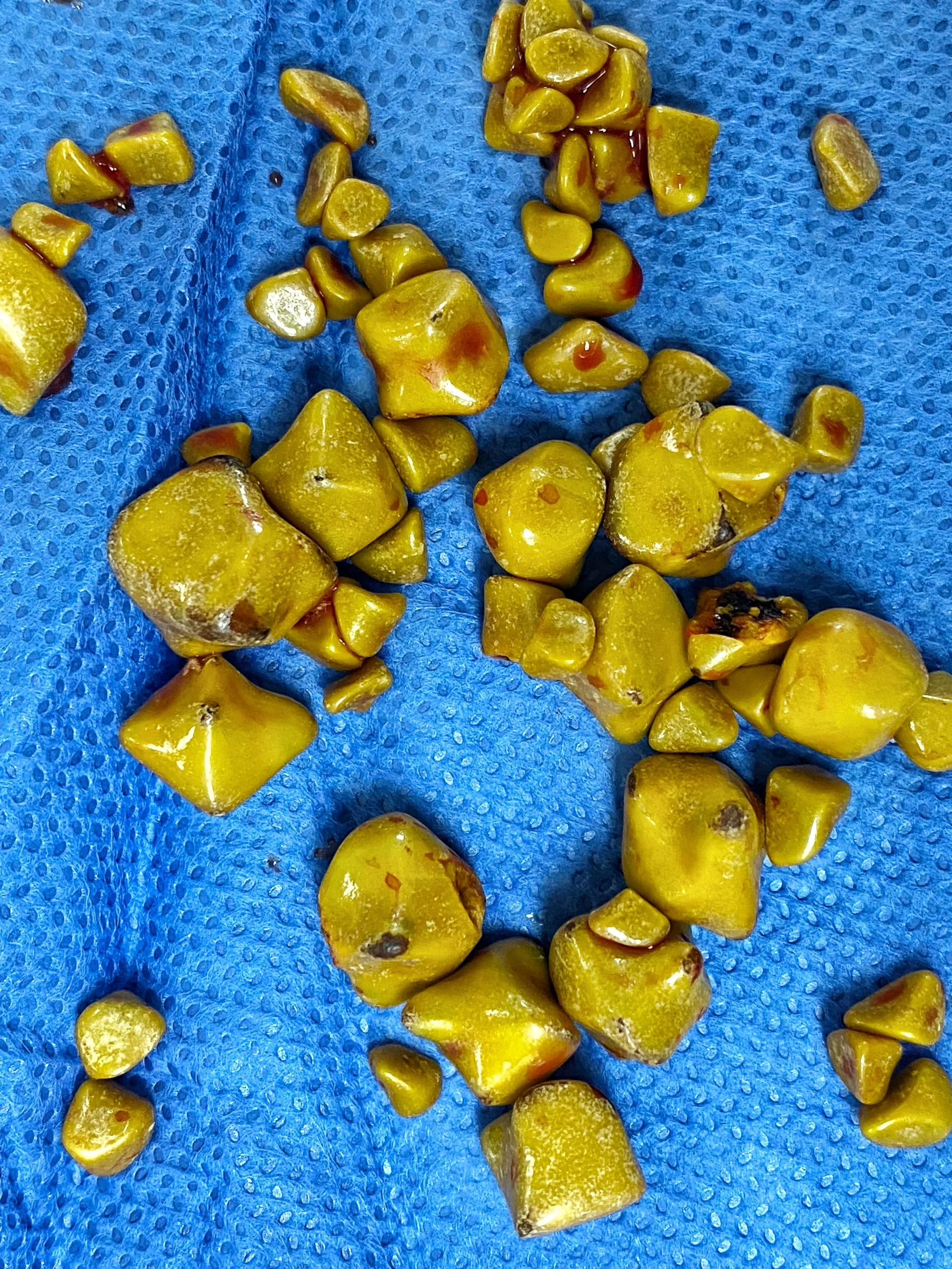
Biliary Disease, also known as bile duct disease, includes a variety of disorders involving the bile duct. They all cause blockage of bile ducts in different ways, manifesting the same symptoms. Typically, individuals in mid-adulthood, most prominently women, are at a greater risk of developing biliary disease. The most common are gallstones, cholangiocarcinoma, strictures, and primary biliary cholangitis, all readily treatable. However, the rarest one, biliary atresia, can become fatal if not treated with surgery.
What is Biliary Disease?
Any structural, functional, or metabolic abnormality of the biliary system is called biliary disease. The biliary system is an essential portion of the associative digestive tract that is involved in producing, reserving, and transferring bile. It consists of the gallbladder and the hepatic biliary tree, including hepatic, bile, and cystic ducts and bile canaliculi.
Bile is necessary to integrate fats and absorb fat-soluble soluble vitamins, which serve as co-enzymes in various biochemical processes. It is the hydrophilic portion of the bile that increases the permeability of the lipid core of the vitamins K, A, D, and E, which prevents their excretion1Ren, Xiaoxue, Yifan Wu, Mingle Huang, Yubin Xie, Ming Kuang, Xiaoxing Li, and Lixia Xu. “IDDF2022-ABS-0194 Patient-derived organoids predict chemotherapy response in biliary tract cancer.” In Abstracts of the International Digestive Disease Forum (IDDF), Hong Kong, 2–4 September 2022. BMJ Publishing Group Ltd and British Society of Gastroenterology, 2022. .
The liver produces the bile and gallbladder stores and releases it into the small intestine (duodenum) via the common bile duct. The bile consists of a mixture of cholesterol, conjugated bilirubin, bile acids/ salts, phospholipids, and water.
All types of bile diseases block bile ducts, affecting the overall health of the biliary system. Acute or chronic biliary diseases may result from any injury or infections in associated biliary organs, leading to inflammation.
Symptoms of Bile Duct Disease
Symptoms of a clogged bile duct may be instantaneous or may present gradually for several years after swelling in the bile duct. Biliary disease mostly manifests symptoms associated with the liver products’ excretion and their release into the blood flow. Several other symptoms result from bile ducts’ poor performance in transferring certain digestive secretions (bile acids) to the small intestine.2Luchini, Claudio, Michele Simbolo, and Aldo Scarpa. “Pathology of Biliary Tract Cancers.” In The IASGO Textbook of Multi-Disciplinary Management of Hepato-Pancreato-Biliary Diseases, 65–70. Singapore: Springer Nature Singapore, 2022.
Common signs and symptoms associated with blocked bile duct are as follows:
- Change of skin color, like the yellowish appearance of skin (jaundice) or the white part of eyes (icterus), due to piling up of an excreting component called bilirubin
- High body temperature or night sweating
- Skin irritation and allergies (not confined to one site; may be severe at night time or in hot temperature places)
- Tiredness/Dizziness
- Light brown urine
- Abnormal body weight loss
- Loss of appetite
- Abdominal pain, localized under the right side of the rib cage
- Greasy or light mud-coloured stools
- Nausea and vomiting
What Causes Biliary Disease?
Common causes of biliary disease include:
- Genetics
- Increasing age
- Overweight
- Obesity
- High Fat-diet
- Specific gastrointestinal disorders (GERD, celiac disease)
- Certain prescription medications
- Opportunistic infections (like HIV, Chinese liver fluke)
Risk Factors of Biliary Disease – The Five Fs
The pneumonic ‘five Fs’ is a good way to remember the common risk factors associated with biliary disease, particularly gallstones. The five Fs include the following3Levi, Brittany E. Choledochal Cysts: In Brief with Dr. Alexander Bondoc. Stay Current, May 2022. http://dx.doi.org/10.47465/sc1.:
- Fair (White-western population)
- Female (twice as likely as men)
- Fat (individuals with high cholesterol diet or obesity)
- Fertile (associated with high estrogen levels)
- Forty
Although statistical evidence proves, the five Fs4Bass G, Gilani SN, Walsh TN, Leader F. Validating the 5Fs mnemonic for cholelithiasis: time to include family history. Postgraduate medical journal. 2013 Nov;89(1057):638-41., it is not a law and does not exclude other causes like family history, alcohol or drugs.
Types of Biliary Disease
Gallstones
The most common biliary disease is gallstones (cholelithiasis). When the bile produces excessive calcium salt, cholesterol components, or bilirubin, it leads to gallstone formation. Their size may range from the size of a small salt grain to that of a golf ball.
Acute cholangitis
Gallstones can also lead to acute cholangitis, which is a bacterial infection of the biliary system outside the liver. It commonly presents with the Charcot triad: right upper quadrant abdominal pain, jaundice, and fever. Acute cholangitis prompts immediate treatment and is a medical emergency as it can lead to hepatic abscess, septicemia, or infective endocarditis.

Choledocholithiasis
Among common sources of extrahepatic biliary disorders is choledocholithiasis. This condition involves the presence of one or more stones in the bile or hepatic duct leading to biliary obstruction.5Cianci, Pasquale, and Enrico Restini. “Management of cholelithiasis with choledocholithiasis: Endoscopic and surgical approaches.” World Journal of Gastroenterology 27, no. 28 (2021): 4536.
It commonly co-exists with gallstones. These stones can lead to inflammation in the bile duct, developing a condition called bacterial cholangitis. If left untreated, it can convert into secondary bile cirrhosis or gallstone pancreatitis. In some individuals, choledocholithiasis remains asymptomatic, but most patients complain about upper abdominal pain with episodic jaundice.
Primary Biliary Cirrhosis
The inflammation and destruction of bile ducts in the liver due to the body’s immune response (auto-immune) is called primary biliary cirrhosis (PBC). Primary biliary cirrhosis often leads to liver cirrhosis due to cholestasis (reduced bile flow). It is common in women, and a key to diagnosing it is no extra-hepatic involvement. Some cases of primary biliary cirrhosis are also associated with pernicious anemia.6Chung, Chen-Shuan, Yao-Chun Hsu, Shang-Yi Huang, Yung-Ming Jeng, and Chien-Hung Chen. “Primary biliary cirrhosis associated with pernicious anemia.” Canadian Family Physician 56, no. 9 (2010): 889-891.
Cholangiocarcinoma
Cholangiocarcinoma, also known as bile duct cancer, is common in the elderly. It stays silent and becomes symptomatic when terminal with symptoms of itching, pain, dark urine, jaundice, and weight loss. Hepatitis B and C, HIV, liver cirrhosis, fatty liver, and bile duct cysts can result in this cancer. Although cholangiocarcinoma is rare, it is extremely fatal and has a poor prognosis.
Biliary Dyskinesia:
A condition in which the gallbladder has abnormal motility, leading to symptoms like pain and nausea. Biliary dyskinesia is often diagnosed through a HIDA scan, which assesses the gallbladder’s ability to contract and release bile properly.
Billary Atresia
Biliary atresia presents with long-standing jaundice, dark urine, and pale stools in term infants. It happens due to inflammation and fibrosis in some or all of the bile ducts, which blocks them. Kasai procedure, a surgical operation that connects the liver to the intestine, is the only treatment available.7Vij, Mukul, and Mohamed Rela. “Biliary atresia: pathology, aetiology and pathogenesis.” Future science OA 6, no. 5 (2020): FSO466. Additionally, patients need a liver transplant to survive, as biliary atresia ultimately causes cirrhosis and liver failure.
Other miscellaneous types of biliary tract diseases are:
- Pancreatic disorders-bile duct obstruction due to pancreatic head cancer
- Benign and ampullary tumors-Benign or malignant lesions leading to bile obstruction
- Morizzi’s syndrome developed due to cystic duct syndrome
Diagnosing Bile Duct Diseases
Your healthcare provider may suspect a bile duct disorder if you experience typical symptoms or if blood tests reveal elevated levels of bilirubin. Your medical history will be reviewed, and a physical examination will be conducted to identify any signs of bile duct impairment. To distinguish between conditions like hepatitis and cirrhosis, which share similar symptoms, your healthcare provider will also inquire about your medication history and alcohol consumption, both of which can contribute to liver diseases.8Wang, Wei, Dongfeng Chen, Jun Wang, and Liangzhi Wen. “Cellular homeostasis and repair in the biliary tree.” In Seminars in Liver Disease, vol. 42, no. 03, pp. 271-282. Thieme Medical Publishers, Inc., 2022.
If you possess gallstones or other disorders like pancreatitis and abdominal surgery or experienced signs of an autoimmune disorder (such as osteoarthritis, dry mouth or infected eyes, skin redness, or diarrhea with blood), immediately inform your doctor. You will do so as some medications can decrease the rate of drainage occurring through the bile ducts, which requires a medicine review to avoid further complications.9Maev, I. V., D. S. Bordin, T. A. Ilchishina, and Yu A. Kucheryavyy. “The biliary continuum: an up-to-date look at biliary tract diseases.” Meditsinskiy sovet = Medical Council, no. 15 (October 19, 2021): 122–34
Blood Tests:
Blood tests are first-line and are usually as follows:
Liver Profile Tests
This assesses the liver functioning by measuring the number of liver enzymes (ALT, AST, GGT, ALP) and the waste produced called bilirubin (conjugated plus unconjugated). They help diagnose obstructive diseases.
Antibody Tests
These include anti-mitochondrial antibodies, also known as AMAs positive in primary biliary cholangitis, and p-ANCA and anti-SMA antibodies positive in primary sclerosing cholangitis.
Lipid Profile
Cholesterol levels are usually high in gallstones and primary biliary cirrhosis.
Serum Tumor Markers
The tumor marker CA-19-9 is high in cholangiocarcinoma.
Imaging:
Abdominal Ultrasound (USG)
This test provides images of the liver, gallbladder, and associated bile duct using high-frequency sound waves. Abdominal USG is the gold standard for gallstones. Ultrasound of the right upper quadrant is of choice. USG also helps to diagnose cholecystitis (thickened head of gall bladder)
Computed Tomography (CT)
CT scans help diagnose cancers, particularly cholangiocarcinoma. Billary duct cysts can also be viewed.
Cholangiography
It involves ERCP and MRCP to view the biliary tract.10Nayab, Seema, Ameet Jesrani, Riaz Hussain Awan, and Kosar Magsi. “Diagnostic accuracy of MRCP in obstructive biliopathy taking ERCP as a gold standard. Experience at tertiary care hospital of a developing country.” The Professional Medical Journal 29, no. 03 (2022): 285-290.
- Endoscopic retrograde cholangiopancreatography (ERCP)
ERCP uses a small camera device on the front of a flexible cord. The procedure involves adding radioactive dye into the biliary system by passing the camera tube from the mouth to the duodenum. X-ray images follow this and visualize the biliary tree. Since ERCP is invasive, it allows biopsy and block relieving procedures.

Source: Salerno, Raffaele, Nicolò Mezzina, and Sandro Ardizzone. “Endoscopic retrograde cholangiopancreatography, lights and shadows: Handle with care.” World Journal of Gastrointestinal Endoscopy 11, no. 3 (2019): 219.
- Magnetic resonance cholangiopancreatography (MRCP)
This diagnostic test allows an examination that resembles the endoscopic exam mentioned above. The most prominent advantage of this test includes MRI images that can be collected without inserting an endoscopic cord into the stomach. However, it also has a disadvantage: it does not allow biopsy examination.
Liver Biopsy
It involves obtaining a sample by inserting a needle that passes through the skin. The sampled tissues of the liver are observed for the presence of swelling or tumors.
Biliary Disease Treatment
Based on the specifications of biliary disorder, your healthcare providers will recommend a customized treatment plan, which may consist of all conventional and surgical options, including prescription medications, invasive procedures, or chemotherapy.
Surgical options:
Obstructive diseases with stones require surgery.
Laparoscopic Cholecystectomy (Gall bladder Removal)
Laparoscopic cholecystectomy is an elective (planned) surgical operation to remove the gall bladder. Previously, open cholecystectomy was the only option where a large abdominal scar opened the abdominal region. A laparoscope is a camera device; four ports are inserted into the abdomen, and the gallbladder is taken out by visualizing through a screen. Moreover, the abdomen is filled with carbon dioxide beforehand. The procedure allows quick recovery, minimal scarring, and low risk of complications.11Kamarajah, Sivesh K., Santhosh Karri, James R. Bundred, Richard PT Evans, Aaron Lin, Tania Kew, Chinenye Ekeozor, Susan L. Powell, Pritam Singh, and Ewen A. Griffiths. “Perioperative outcomes after laparoscopic cholecystectomy in elderly patients: a systematic review and meta-analysis.” Surgical endoscopy 34 (2020): 4727-4740. However, in the case of complicated cholelithiasis, open surgery is the main choice.

Independent Stone Removal
Most gastroenterologists accomplish stone extraction with endoscopic retrograde cholangiopancreatography (ERCP), often guided by sphincterotomy. However, placement of an endoscopic biliary stent is also helpful in conditions including bacterial cholangitis. It causes decompression of biliary ducts when a larger stone is difficult to remove.
Percutaneous Transhepatic Cholangiography (PTHC)
A substitute for ERCP for the therapy of choledocholithiasis involves the application of percutaneous transhepatic cholangiography (PTHC).12Alotaibi, Khalid M., and Hanan M. Alghamdi. “Percutaneous endoscopic biliary exploration in complex biliary stone disease: Case series study.” International Journal of Surgery Open 24 (2020): 73-78. This procedure can be used to enhance bile drainage in cases of cholangitis. Additionally, by inserting a guidewire through the duodenum via a percutaneous approach, endoscopists may be able to perform ERCP for stone removal, even after previous unsuccessful attempts.
Chemo-Radio Therapy:
Non-surgical treatment approaches may involve radiation and chemotherapy that lessen the size of tumors. Cholangiocarcinoma is usually managed with this.
Medication:
Drug management involves the following drugs:
Ursodiol
Ursodiol, also known as ursodeoxycholic acid, is the drug of choice for primary biliary cirrhosis. It helps improve bile flow by decreasing cholesterol and bile salt production.13Ghonem, Nisanne S., Adam M. Auclair, Christopher L. Hemme, Gina M. Gallucci, Randolph de la Rosa Rodriguez, James L. Boyer, and David N. Assis. “Fenofibrate improves liver function and reduces the toxicity of the bile acid pool in patients with primary biliary cholangitis and primary sclerosing cholangitis who are partial responders to ursodiol.” Clinical Pharmacology & Therapeutics 108, no. 6 (2020): 1213-1223.
Cholestyramine
Cholestyramine reduces cholesterol levels by binding with bile salts to prevent them from reabsorbing the gut. It can be used in conditions with excess bile production due to blockage.14Nevens, Frederik, Michael Trauner, and Michael P. Manns. “Primary biliary cholangitis as a roadmap for the development of novel treatments for cholestatic liver diseases.” Journal of Hepatology 78, no. 2 (2023): 430-441.
Some healthcare practitioners may also suggest lifestyle modifications focusing on diet management. It may involve a low-fat diet or nutritional supplementations for improved nutrient absorption, particularly fat-soluble vitamins.
Prevention of Biliary Disease
To prevent biliary diseases, the following considerations must be followed:
- Biliary duct disorders may arise due to obesity or high cholesterol levels in the body. Hence, try to work and achieve a healthy body mass. It can be made possible through the incorporation of physical activities and diet. Eating a well-balanced diet with fiber, water, and five portions of fruit and vegetables is beneficial.
- Avoid smoking to prevent cholangiocarcinoma.
- Avoid eating undercooked fish if you are traveling to Asia. Eating undercooked fish is associated with certain parasitic infections (Chinese Liver fluke) that lead to biliary disorders like cholangitis and cholangiocarcinoma.15Espinoza, J. Luis. “Fluke-Related Cholangiocarcinoma: Challenges and Opportunities.” Pathogens 12, no. 12 (2023): 1429.
Conclusion
Biliary diseases can progress towards complicated ailments. It may have touched serious levels by the time you observe symptoms. Fortunately, most biliary diseases can be treated with medication or surgery. The noticeable point is to observe the symptoms seriously. You may not consider them severe, such as acute biliary pain that recovers on its own, but if left untreated, it can develop into a persistent and prolonged disorder. Don’t wait for severe warnings; visit your nearest healthcare provider.
Refrences
- 1Ren, Xiaoxue, Yifan Wu, Mingle Huang, Yubin Xie, Ming Kuang, Xiaoxing Li, and Lixia Xu. “IDDF2022-ABS-0194 Patient-derived organoids predict chemotherapy response in biliary tract cancer.” In Abstracts of the International Digestive Disease Forum (IDDF), Hong Kong, 2–4 September 2022. BMJ Publishing Group Ltd and British Society of Gastroenterology, 2022.
- 2Luchini, Claudio, Michele Simbolo, and Aldo Scarpa. “Pathology of Biliary Tract Cancers.” In The IASGO Textbook of Multi-Disciplinary Management of Hepato-Pancreato-Biliary Diseases, 65–70. Singapore: Springer Nature Singapore, 2022.
- 3Levi, Brittany E. Choledochal Cysts: In Brief with Dr. Alexander Bondoc. Stay Current, May 2022. http://dx.doi.org/10.47465/sc1.
- 4Bass G, Gilani SN, Walsh TN, Leader F. Validating the 5Fs mnemonic for cholelithiasis: time to include family history. Postgraduate medical journal. 2013 Nov;89(1057):638-41.
- 5Cianci, Pasquale, and Enrico Restini. “Management of cholelithiasis with choledocholithiasis: Endoscopic and surgical approaches.” World Journal of Gastroenterology 27, no. 28 (2021): 4536.
- 6Chung, Chen-Shuan, Yao-Chun Hsu, Shang-Yi Huang, Yung-Ming Jeng, and Chien-Hung Chen. “Primary biliary cirrhosis associated with pernicious anemia.” Canadian Family Physician 56, no. 9 (2010): 889-891.
- 7Vij, Mukul, and Mohamed Rela. “Biliary atresia: pathology, aetiology and pathogenesis.” Future science OA 6, no. 5 (2020): FSO466.
- 8Wang, Wei, Dongfeng Chen, Jun Wang, and Liangzhi Wen. “Cellular homeostasis and repair in the biliary tree.” In Seminars in Liver Disease, vol. 42, no. 03, pp. 271-282. Thieme Medical Publishers, Inc., 2022.
- 9Maev, I. V., D. S. Bordin, T. A. Ilchishina, and Yu A. Kucheryavyy. “The biliary continuum: an up-to-date look at biliary tract diseases.” Meditsinskiy sovet = Medical Council, no. 15 (October 19, 2021): 122–34
- 10Nayab, Seema, Ameet Jesrani, Riaz Hussain Awan, and Kosar Magsi. “Diagnostic accuracy of MRCP in obstructive biliopathy taking ERCP as a gold standard. Experience at tertiary care hospital of a developing country.” The Professional Medical Journal 29, no. 03 (2022): 285-290.
- 11Kamarajah, Sivesh K., Santhosh Karri, James R. Bundred, Richard PT Evans, Aaron Lin, Tania Kew, Chinenye Ekeozor, Susan L. Powell, Pritam Singh, and Ewen A. Griffiths. “Perioperative outcomes after laparoscopic cholecystectomy in elderly patients: a systematic review and meta-analysis.” Surgical endoscopy 34 (2020): 4727-4740.
- 12Alotaibi, Khalid M., and Hanan M. Alghamdi. “Percutaneous endoscopic biliary exploration in complex biliary stone disease: Case series study.” International Journal of Surgery Open 24 (2020): 73-78.
- 13Ghonem, Nisanne S., Adam M. Auclair, Christopher L. Hemme, Gina M. Gallucci, Randolph de la Rosa Rodriguez, James L. Boyer, and David N. Assis. “Fenofibrate improves liver function and reduces the toxicity of the bile acid pool in patients with primary biliary cholangitis and primary sclerosing cholangitis who are partial responders to ursodiol.” Clinical Pharmacology & Therapeutics 108, no. 6 (2020): 1213-1223.
- 14Nevens, Frederik, Michael Trauner, and Michael P. Manns. “Primary biliary cholangitis as a roadmap for the development of novel treatments for cholestatic liver diseases.” Journal of Hepatology 78, no. 2 (2023): 430-441.
- 15Espinoza, J. Luis. “Fluke-Related Cholangiocarcinoma: Challenges and Opportunities.” Pathogens 12, no. 12 (2023): 1429.

