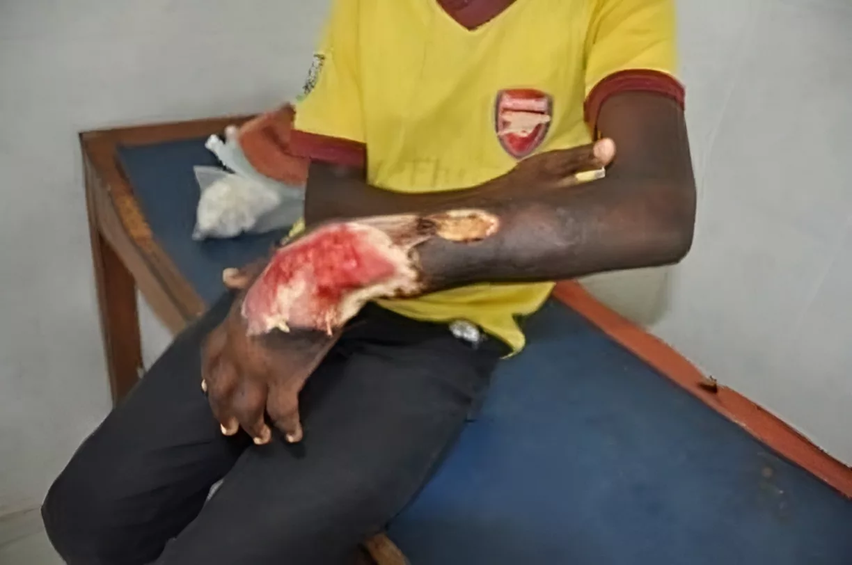
Buruli ulcer is a serious skin infection caused by the bacteria Mycobacterium ulcerans. It begins slowly with a painless swelling or lump on the skin, which can gradually turn into an open wound. The bacteria release a harmful toxin that destroys the skin and underlying tissues.
What is a Buruli Ulcer?
Buruli ulcer was first identified in Australia in the late 1800s and was later named after Buruli County in Uganda, where large outbreaks occurred. Caused by Mycobacterium ulcerans, a bacterium related to those responsible for tuberculosis and leprosy, the infection begins as a painless lump on the skin. Over time, this lump grows, eventually breaking down into a large open sore. If left untreated, the ulcer can spread deeper, damaging the skin, muscles, and even bones, leading to severe tissue destruction and long-term disability.
Cases of Buruli ulcer have been reported in at least 33 countries, with over 2700 cases being reported in 2018, mainly in rural areas across Africa, Australia, South America, and Asia. In these regions, limited access to proper healthcare often delays treatment, increasing the risk of complications.
Other names of Buruli ulcer include:
- Bairnsdale ulcer
- Daintree ulcer
- Mossman ulcer
- Kumasi ulcer
- Searls ulcer1Simpson, H., Kebede Deribe, Tabah, E. N., Peters, A., Maman, I., Frimpong, M., Ampadu, E., Phillips, R., Saunderson, P., Pullan, R. L., & Cano, J. (2019). Mapping the global distribution of Buruli ulcer: a systematic review with evidence consensus. The Lancet Global Health, 7(7), e912–e922. https://doi.org/10.1016/s2214-109x(19)30171-8
Causes of Buruli Ulcer
Buruli ulcer is caused by Mycobacterium ulcerans, a bacterium that enters the body through small cuts, insect bites, or direct contact with dirty water and soil.
Once inside, Mycobacterium ulcerans releases a toxin called mycolactone. This toxin weakens the immune system, kills skin cells, and damages deeper tissues, with nerves being especially affected. Because of this, the infection doesn’t cause much pain or inflammation, but it slowly destroys the skin and fat underneath. These bacteria reproduce inside your body for weeks or months before symptoms appear.2Zingue, D., Bouam, A., Tian, R. B. D., & Drancourt, M. (2018). Buruli Ulcer, a Prototype for Ecosystem-Related Infection, Caused by Mycobacterium ulcerans. Clinical Microbiology Reviews, 31(1). https://doi.org/10.1128/cmr.00045-17
Risk Factors for Buruli Ulcer
The exact modes of transmission of Buruli ulcer are still being researched. However, a few risk factors have been identified that considerably increase your chances of contracting the infection.
Limited Education:
Limited education can contribute to a lack of awareness about Buruli ulcer, including how it spreads and how to recognize early symptoms. This may result in delays in seeking proper medical care. Individuals with less formal education might also have reduced access to healthcare services or may rely on traditional remedies that are ineffective against the disease.
Environmental Exposure:
The bacteria that cause Buruli ulcers thrive in swampy areas, muddy water, and moist soil. If you frequently wade through swamps, fetch water from contaminated sources, or work near cocoa plantations or forests, you have a higher chance of coming into contact with the bacteria. Even tiny scratches or insect bites can allow the bacteria to enter your skin. People who live in these areas may also have homes close to these water sources, increasing daily exposure.
Wearing Short Clothes:
If you work on a farm or in fields while wearing shorts, skirts, or other clothing that leaves your legs uncovered, you are at a greater risk. Farming often involves brushing against rough plants, getting small cuts, or being bitten by insects. These tiny wounds can serve as entry points for the bacteria.
Poor Wound Care Practices:
If you get a cut, scrape, or insect bite and don’t clean it properly, the bacteria can enter and start spreading under your skin. Using adhesive bandages without washing the wound first can trap dirt and moisture, which creates a perfect environment for a buruli ulcer to develop. Some people may not take minor wounds seriously, thinking they will heal on their own, but even small injuries can become dangerous if exposed to contaminated water or soil.
Protection Against Insect Bites:
Insect bites are strongly suspected as culprits in the transmission of Mycobacterium ulcerans. If you don’t sleep under a bed net, you’re exposed to more insect bites, which could increase your risk. Mosquito coils are sometimes used to prevent bites, but they may not be effective enough. People who live in areas with a high number of biting insects might face a greater risk of exposure to the bacteria.
Compromising on Cleanliness & Home Remedies:
Regularly washing your clothes helps remove bacteria from fabric, reducing the chances of it coming into contact with your skin. In some communities, people use traditional treatments like crushed leaves or rubbing alcohol on wounds. These may help kill germs if applied immediately, but they are not a replacement for proper medical care. When these precautions aren’t taken, the risk of Buruli ulcer in endemic communities increases.
Living Conditions:
If you live in a household where hygiene is not a priority, the risk of infection increases. In addition, if your home is near contaminated water, where people often get minor injuries, or where healthcare is not easily available, the chances of Buruli ulcers are much higher.3Pouillot, R., Matias, G., Wondje, C. M., Portaels, F., Valin, N., Ngos, F., Njikap, A., Marsollier, L., Fontanet, A., & Eyangoh, S. (2007). Risk Factors for Buruli Ulcer: A Case Control Study in Cameroon. PLoS Neglected Tropical Diseases, 1(3), e101. https://doi.org/10.1371/journal.pntd.0000101
Is Buruli ulcer Contagious?
Buruli ulcer is not a contagious condition. Unlike tuberculosis or leprosy, it does not spread from person to person. Instead, it is said to be transmitted through environmental exposure and insect vectors.
Symptoms of Buruli Ulcer
Unlike other skin infections, Buruli ulcer is typically painless and does not cause systemic symptoms like fever or redness. It is usually located on your legs (over 60% of cases) and arms (around 25% of cases). Its clinical presentation includes:
- A small, firm, painless nodule or swelling that often gets mistaken for an insect bite.
- A non-healing scab
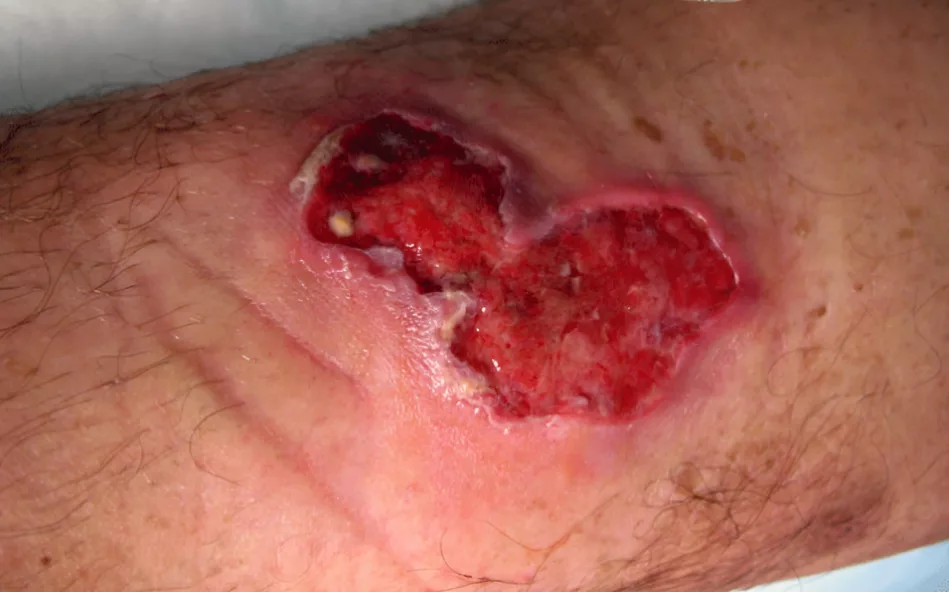
- A deep, expanding ulcer with undermined edges
- Swelling
- Raised lumps
- Thickened skin patches
- Localized pain without an open ulcer4Brown, S. C. (2024, August 26). Buruli Ulcer Clinical Presentation: History, Physical Examination. Medscape.com; Medscape. https://emedicine.medscape.com/article/1104891-clinical
What are the stages of Buruli Ulcer?
Buruli ulcer develops slowly. It starts with mild symptoms before causing serious skin damage. Buruli ulcer is classified into three stages based on how big and severe the infection is. Knowing these stages of ulceration can help you understand the disease and why early treatment is important.
Category I:
Category I is the mildest stage of a buruli ulcer. If you have a small, single lesion less than 5 cm in size, it falls into this category. These early sores are usually painless, so you might not notice them right away. But if treated early with antibiotics, they heal well without causing serious damage. About 32% of Buruli ulcer cases are in this stage.
Category II:
Category II includes larger ulcers or swollen skin patches between 5–15 cm. At this stage, the infection is more noticeable and can cause discomfort or swelling. If left untreated, the skin can break down, forming deep ulcers. Moving around may become harder, especially if the ulcer is near a joint. About 35% of cases fall into this category.
Category III:
Category III is the most severe stage. If your ulcer is larger than 15 cm, involves multiple sores, or spreads to your bones and joints, it’s in this category. The infection can cause deformities and long-term disability. This stage makes up about 33% of cases and often requires surgery.5World. (2019, August 7). Buruli ulcer (Mycobacterium ulcerans infection). Archive.org; World Health Organization: WHO. https://web.archive.org/web/20200606231902/https://www.who.int/health-topics/buruli-ulcer
What are the complications of Buruli Ulcer?
Without treatment, Buruli ulcers get complicated and cause distressing symptoms, such as:
- Deep tissue damage affecting the underlying fat and muscle
- Large ulcers cause severe scarring and contractures that restrict movement
- Infection reaching the bone leads to osteomyelitis with chronic pain
- Permanent disability occurs when joints are destroyed
- Secondary infections that increase the risk of systemic illness
- Extensive tissue loss, which requires skin grafting or reconstructive surgery6Adu, E. J. K. (2013). Management of complications of Mycobacterium ulcerans disease: A three-year review. International Journal of Mycobacteriology, 2(4), 206–210. https://doi.org/10.1016/j.ijmyco.2013.08.003
How is a Buruli ulcer diagnosis established?
To diagnose a Buruli ulcer, doctors rely on your history, physical exam, and lab tests.
History & Physical Exam:
When you see your doctor, they’ll start by asking about your medical history, especially if you’ve recently traveled to areas where Buruli ulcer is more common — places like rural parts of West and Central Africa. They’ll also want to know about the lesions you’ve noticed, such as how long they’ve been there, their appearance, and if you’ve felt any pain or swelling.
During the physical exam, your doctor will carefully look at the affected area. Usually, a Buruli ulcer shows up as a painless, swollen patch of skin that eventually becomes an ulcer with raised edges and a necrotic center. They’ll also check for other signs of infection, like redness or scaling, and might look for any involvement of nearby lymph nodes. Sometimes, these things can be a bit tricky to spot or might be mistaken for other skin conditions, so the doctor will take their time to make a thorough assessment.
Lab Investigations:
To confirm a Buruli ulcer diagnosis, doctors use specific laboratory tests to identify Mycobacterium ulcerans, the bacteria that cause the infection. The key tests are:
- A Polymerase Chain Reaction/ PCR test can detect even very small amounts of the bacteria. It is one of the most sensitive and reliable methods, especially useful in the early stages of infection.
- Histopathology is often done in cases of tissue biopsy for Buruli ulcers. A tissue sample is obtained surgically and checked under a microscope to see the extent of the infection. The test reveals the typical tissue damage caused by Mycobacterium ulcerans, including the absence of inflammation and the necrosis that is characteristic of Buruli ulcer.
- Bacterial cultures involve swabs taken from the ulcer and cultured at lower temperatures (between 29 and 33°C) with low oxygen levels (around 2.5%). If the bacteria are found in blood cultures, it indicates a more serious form of the infection, as the bacteria may have spread beyond the skin.7Röltgen, K., Cruz, I., Ndung’u, J. M., & Gerd Pluschke. (2019). Laboratory Diagnosis of Buruli Ulcer: Challenges and Future Perspectives. Springer EBooks, 183–202. https://doi.org/10.1007/978-3-030-11114-4_10
Is there any treatment for Buruli Ulcer?
Buruli ulcers can be completely cured with treatment if diagnosed early. Management for Buruli ulcer usually involves:
Antibiotics:
Buruli ulcer is treated primarily with antibiotics to kill Mycobacterium ulcerans and stop the disease from progressing. You’ll have to take them for at least eight weeks. Healing takes half a year. A combination regimen of rifampicin and streptomycin is used first.
- Rifampicin blocks bacterial RNA synthesis and prevents bacteria from multiplying.
- Streptomycin is an aminoglycoside that disrupts protein production in bacteria and kills them.
- In cases where these cannot be used, drugs like moxifloxacin, amikacin, and clarithromycin are being studied for their potential role.
Surgery:
Sometimes, antibiotics aren’t enough to treat Buruli ulcers. That’s when surgery comes in. It’s especially needed when the ulcer is deep, widespread, or not responding to medication. In these cases, surgery prevents further damage.
During surgery for a Buruli ulcer, doctors remove infected tissue from your body in a procedure called debridement, which is sometimes followed by skin grafting to cover the wound. You’re given antibiotics to clear the infection completely.
Supportive Care:
While antibiotics and surgery are the most important treatment options for curing Buruli ulcers, supportive therapies can speed up your recovery from the infection. These include:
- You may require analgesics such as NSAIDs or, in severe cases, opioids to manage pain.
- Anti-inflammatory medications or steroids are sometimes given to reduce excessive immune responses.
- Malnutrition can delay wound healing, so you’re advised to consume a diet rich in protein, vitamins, especially vitamin C and zinc, because of their healing properties.
- If ulcers affect your mobility, physical therapy helps restore joint movement and prevent stiffness.
- Options like hyperthermia, hyperbaric oxygen therapy, and ozone therapy are being researched for their benefits in managing Buruli ulcers.
Wound Care:
Wound care for Buruli ulcers starts with cleaning the ulcer using saline or mild antiseptic solutions to remove dirt and prevent infection. You’ll be prescribed special hydrocolloid or silver-infused dressings to help your wounds heal faster while lowering further risk. Since the mycolactone toxin affects nerves, dressing changes can be painful, so painkillers or local anesthetics can help make wound Care easier. If the ulcer produces too much fluid, absorbent dressings and regular changes keep the skin dry and prevent further infection.8Popa, G. L., Muntean, A. A., & Popa, M. I. (2023). Recent Advances in the Management Strategies for Buruli Ulcers. Pathogens, 12(9), 1088–1088. https://doi.org/10.3390/pathogens12091088
What to do for Buruli Ulcer Prevention?
The best way to manage Buruli ulcer is to prevent it in the first place. Since the infection progresses silently and painlessly, a diagnosis might be made too late only when significant tissue damage has already occurred. You can take simple preventive measures that can help reduce both the disease and its complications, such as:
- Avoid contact with stagnant or slow-moving water, as these environments are associated with the bacteria that cause Buruli ulcers
- Wear protective clothing, including long sleeves, trousers, gloves, and boots, especially if you are working in waterlogged areas or fields.
- Use insect repellent on exposed skin to minimize the risk of insect bites, which are said to play a role in disease transmission.
- Sleep under insecticide-treated mosquito nets to reduce exposure to potential vectors, especially if you’re a resident of an endemic area.
- Maintain proper hygiene by regularly washing the body and clothes and avoiding direct contact with contaminated water or soil.
- Clean any cuts, wounds, or insect bites immediately with soap and water, and cover them properly to prevent infection.
- Partake in awareness campaigns, which should emphasize early symptom recognition and seeking medical care without delay.9Pasteur, I. (2007, December 18). Buruli Ulcer: New possibilities for the prevention of the disease. Institut Pasteur. https://www.pasteur.fr/en/buruli-ulcer-new-possibilities-prevention-disease?language=fr
Buruli Ulcer vs. Necrotizing Fasciitis
Both Buruli ulcer and necrotizing fasciitis can lead to significant tissue destruction, but they differ greatly in terms of cause, progression, symptoms, and treatment. Recognizing these differences is critical for timely and effective management.10Phanzu, Delphin & Bafende, Aombe & Imposo, Bofunga & Meyers, Wayne & Portaels, Françoise. (2010). Short Report: Under Treated Necrotizing Fasciitis Masquerading as Ulcerated Edematous Mycobacterium ulcerans Infection (Buruli Ulcer). The American journal of tropical medicine and hygiene. 82. 478-81. 10.4269/ajtmh.2010.09-0256.
| Feature | Buruli Ulcer | Necrotizing Fasciitis |
|---|---|---|
| Cause | Mycobacterium ulcerans | Group A Streptococcus or mixed bacteria |
| Progression | Slow (weeks to months) | Rapid (hours to days) |
| Pain | Usually painless | Extremely painful |
| Ulcer | Deep with undermined edges | Rapidly spreading necrosis, blisters, discoloration |
| Systemic Symptoms | Rare | Common and severe (fever, shock) |
| Treatment | Oral antibiotics; surgery if needed | Emergency surgery + IV antibiotics |
| Mortality Risk | Low with treatment | High without urgent care |
Conclusion
Buruli ulcer is an infectious skin condition caused by Mycobacterium ulcerans. It starts as a painless bump but can grow into a deep, open wound if not treated. The infection damages tissues under the skin and can lead to disability. Treatment lasts eight weeks with antibiotics like rifampicin and streptomycin to kill the bacteria. Severe cases may need surgery to remove dead tissue or close large wounds. Prevention includes avoiding dirty water, wearing protective clothing, and using insect repellent. Keeping wounds clean and using mosquito nets can also help.
Refrences
- 1Simpson, H., Kebede Deribe, Tabah, E. N., Peters, A., Maman, I., Frimpong, M., Ampadu, E., Phillips, R., Saunderson, P., Pullan, R. L., & Cano, J. (2019). Mapping the global distribution of Buruli ulcer: a systematic review with evidence consensus. The Lancet Global Health, 7(7), e912–e922. https://doi.org/10.1016/s2214-109x(19)30171-8
- 2Zingue, D., Bouam, A., Tian, R. B. D., & Drancourt, M. (2018). Buruli Ulcer, a Prototype for Ecosystem-Related Infection, Caused by Mycobacterium ulcerans. Clinical Microbiology Reviews, 31(1). https://doi.org/10.1128/cmr.00045-17
- 3Pouillot, R., Matias, G., Wondje, C. M., Portaels, F., Valin, N., Ngos, F., Njikap, A., Marsollier, L., Fontanet, A., & Eyangoh, S. (2007). Risk Factors for Buruli Ulcer: A Case Control Study in Cameroon. PLoS Neglected Tropical Diseases, 1(3), e101. https://doi.org/10.1371/journal.pntd.0000101
- 4Brown, S. C. (2024, August 26). Buruli Ulcer Clinical Presentation: History, Physical Examination. Medscape.com; Medscape. https://emedicine.medscape.com/article/1104891-clinical
- 5World. (2019, August 7). Buruli ulcer (Mycobacterium ulcerans infection). Archive.org; World Health Organization: WHO. https://web.archive.org/web/20200606231902/https://www.who.int/health-topics/buruli-ulcer
- 6Adu, E. J. K. (2013). Management of complications of Mycobacterium ulcerans disease: A three-year review. International Journal of Mycobacteriology, 2(4), 206–210. https://doi.org/10.1016/j.ijmyco.2013.08.003
- 7Röltgen, K., Cruz, I., Ndung’u, J. M., & Gerd Pluschke. (2019). Laboratory Diagnosis of Buruli Ulcer: Challenges and Future Perspectives. Springer EBooks, 183–202. https://doi.org/10.1007/978-3-030-11114-4_10
- 8Popa, G. L., Muntean, A. A., & Popa, M. I. (2023). Recent Advances in the Management Strategies for Buruli Ulcers. Pathogens, 12(9), 1088–1088. https://doi.org/10.3390/pathogens12091088
- 9Pasteur, I. (2007, December 18). Buruli Ulcer: New possibilities for the prevention of the disease. Institut Pasteur. https://www.pasteur.fr/en/buruli-ulcer-new-possibilities-prevention-disease?language=fr
- 10Phanzu, Delphin & Bafende, Aombe & Imposo, Bofunga & Meyers, Wayne & Portaels, Françoise. (2010). Short Report: Under Treated Necrotizing Fasciitis Masquerading as Ulcerated Edematous Mycobacterium ulcerans Infection (Buruli Ulcer). The American journal of tropical medicine and hygiene. 82. 478-81. 10.4269/ajtmh.2010.09-0256.
