Necrotizing Fasciitis is a severe infection in your skin and soft tissues that occurs due to the introduction of bacteria into your skin. It spreads very quickly and aggressively and becomes deadly if you don’t treat it earlier. This is why It is also called flesh-eating disease. It is most common in diabetic people and alcoholics.
What Is Necrotizing Fasciitis?
Necrotizing fasciitis, or flesh-eating disease, is an inflammatory condition of the skin that causes necrosis of the muscle fascia and soft tissues, which have a poor blood supply. The inflammation spreads gradually, affecting the fascias, perifascial planes, skin, soft tissues, and muscles. It can also be post-surgical or post-trauma.1Hite, M., McCrae, A. L., & Privette, A. (2018). Fungal Necrotizing Fasciitis after Penetrating Trauma. The American Surgeon, 84(8), e302–e304.
It is an acute pathology. It occurs when your skin barrier is damaged, introducing many bacteria into your skin, including Staphylococcus aureus and Streptococci. These bacteria are responsible for most skin infections. However, polymicrobial infection can also occur, which includes a combination of gram-positive and gram-negative bacteria.
How Common is Necrotizing Fasciitis?
This rare skin condition affects 0.4 in 100,000 people in the United States annually. However, in some areas of the world, it affects 1 in 100,000 people annually. Moreover, among these people, Methicillin-resistant strains of Staphylococcus aureus (MRSA) mostly affect children.2Khalid, M., Junejo, S., & Mir, F. (2018). Invasive Community-Acquired Methicillin-Resistant Staphylococcal Aureus (CA-MRSA) Infections in Children. Journal of the College of Physicians and Surgeons–Pakistan: JCPSP, 28(9), S174–S177. https://doi.org/10.29271/jcpsp.2018.09.S174
Types Of Necrotizing Fasciitis
There are the following types of necrotizing fasciitis:
Type I:
It is a polymicrobial necrotizing fasciitis in which more than one bacteria is involved. These include Haemophilus influenza, Escherichia coli, streptococcus, and Bacteroides fragilis. It most commonly occurs in elders, diabetic patients, or people with any systemic disease.
Type II:
It occurs due to group A alpha-hemolytic Streptococci or staphylococci. Many of these are Methicillin-Resistant Strains of Staphylococcus aureus (MRSA). It is called flesh-eating disease and affects all age groups. Healthy people are also at risk of developing this type of necrotizing fasciitis.
Type III:
This type of necrotizing fasciitis occurs due to Clostridium perfringens and causes gas gangrene. It mostly occurs after some trauma or injury. It forms gas under the skin, and your doctor can easily hear a crepitus in the skin. People who inject black tar (heroin) are also at risk of this type of necrotizing fasciitis.
Other Types Of Necrotizing Fasciitis:
Some studies also consider marine organisms like vibrio and Aeromonas hydrophila to be part of type III necrotizing fasciitis. Type IV necrotizing fasciitis involves fungal infections like zygomycetes and candida.3Hite, M., McCrae, A. L., & Privette, A. (2018). Fungal Necrotizing Fasciitis after Penetrating Trauma. The American Surgeon, 84(8), e302–e304.
The necrotizing fasciitis due to marine organisms occurs if a fish bites you or you eat raw seafood. If you have liver disease, you are more prone to this infection. Moreover, it becomes fatal if you don’t consult your doctor in forty-eight hours.
Fungal Necrotizing Fasciitis:
Fungal necrotizing fasciitis occurs in immunocompromised people when they get traumatic wounds.
Causes Of Necrotizing Fasciitis
Bacteria like streptococcus, staphylococcus aureus, vibrio vulnificus, clostridium perfringens, some other types of bacteria, and some fungi mostly cause Necrotizing Fasciitis. They enter from any break in your skin, infecting your superficial fascia and spreading gradually. Bacteria can enter your skin due to the following conditions and cause this flesh-eating infection:
- Surgical wounds
- Abrasions
- Puncture wounds
- Cuts
- Insect bites
- Scrapes
- Needles injury
- Poor wound care
- Poor hand washing
- Aspirin or Nonsteroidal anti-inflammatory drugs (NSAIDs)
How Do Cervicofacial Necrotizing Fasciitis Occur?
Cervicofacial necrotizing fasciitis occurs after cervical trauma or dental infection.
Risk Factors for Necrotizing Fasciitis
This infection is common in people with these conditions:
- Diabetics
- People with peripheral artery disease
- Alcoholics
- Immunocompromised people
- Obese people
- Intravenous drug users
- People having cancer
- Smokers
- People with systemic diseases
Symptoms Of Necrotizing Fasciitis
There are the following symptoms:
Initial Symptoms:
This infection starts with severe pain that worsens over time. It first affects the lower limbs, then the perineum, buttocks, neck, and head.
You may experience symptoms like nausea, fever, vomiting, diarrhea, and malaise, along with the development of lesions. Intense thirst and dehydration may also occur.
Symptoms After Three To Four Days:
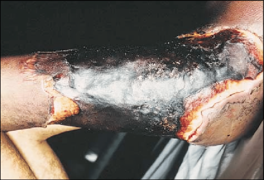
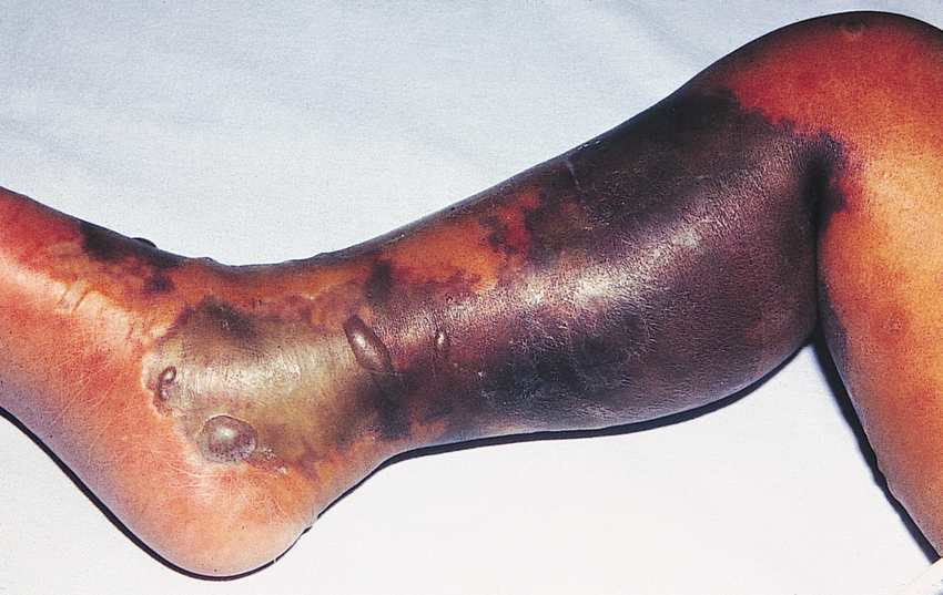
In three to four days, the affected area starts swelling and develops purple rashes, which turn into black blisters on your skin. These black blisters are filled with gas or dark fluid. Your doctor can feel a crepitus in your skin. Later, the wound starts becoming edematous and extremely painful.
Symptoms After Five To Six Days:
After five to six days, blood pressure falls, body temperature rises, and toxemia (toxins in the blood) may occur, leading to toxic shock. Moreover, metastasis to the liver, brain, kidneys, and pericardium can also occur.
Stages Of Necrotizing Fasciitis
Based on appearance of symptoms, this infection is divided into the following stages:
Stage 1: Incubation:
In incubation, the bacteria enter the wound, and there are mild symptoms such as pain, redness, and swelling. These symptoms last for almost the first four days.
Stage 2: Infection:
The bacteria start to multiply and infect you, causing pain, redness, fever, chills and vomiting. It can last for one to three days.
Stage 3: Fascial Involvement:
In this stage, the infection spreads to the fascias (tissue surrounding your muscles), swelling and bruising your skin. This turns your skin greyish. This stage lasts for one to two days.
Stage 4: Muscle Involvement:
In this stage, the infection spreads to your muscles and causes muscle necrosis. Patients can have septic shock during this stage. It also lasts for one to two days.
Stage 5: Sepsis And Multi-Organ Failure:
In stage five, the bacteria enter systemic circulation and cause sepsis. The patient may develop multiorgan failure, such as kidney failure or respiratory failure. This is a life-threatening stage, and its duration is variable.
Stage 6: Recovery Or Death:
If doctors start early treatment, the patient can recover in this stage. However, if left untreated, the infection worsens, leading to death. The duration of this stage is variable, depending on the patient and the severity of the disease.4Fais, P., Viero, A., Viel, G., Giordano, R., Raniero, D., Kusstatscher, S., Giraudo, C., Cecchetto, G., & Montisci, M. (2018). Necrotizing fasciitis: case series and review of the literature on clinical and medico-legal diagnostic challenges. International journal of legal medicine, 132(5), 1357–1366. https://doi.org/10.1007/s00414-018-1838-0
How Does Your Doctor Diagnose Necrotizing Fasciitis?
The patient with this infection presents with severe pain and vomiting. It is an emergency condition, and early diagnosis and treatment is necessary. Your doctor diagnoses necrotizing fasciitis by proper history, examination, and laboratory investigations.5Erichsen Andersson, A., Egerod, I., Knudsen, V. E., & Fagerdahl, A. M. (2018). Signs, symptoms and diagnosis of this infection experienced by survivors and family: a qualitative Nordic multi-center study. BMC infectious diseases, 18(1), 429. https://doi.org/10.1186/s12879-018-3355-7.
History:
The doctors ask these questions for diagnosis:
- History of any trauma, cut, needle injury wound, recent surgery, or abrasion.
- History of any systemic disease, diabetes, alcohol intake, smoking, or intravenous drug intake.
- History of use of non-steroidal anti-inflammatory drugs (NSAIDs) or contact with any person having the same disease.
Examination:
After history, your doctor performs a complete physical examination along with a systemic examination and thorough skin examination to check for initial lesions, gangrene, or crepitus. They examine your skin, head, neck, legs, arms, and buttocks for any signs of gangrene. Moreover, they also check for crepitus in the affected area of your skin. The positive findings your doctor can find include crepitus, edema, abscess, cellulitis, bullae, and anesthesia of the affected skin area.
Finger Test:
A positive finger test is diagnostic for necrotizing fasciitis. Your doctor makes a two-centimeter vertical incision in your skin and pushes the index finger into the tissues. The test is positive if the finger passes the tissue without any resistance.
Laboratory Investigations:
Laboratory investigations are important for the diagnosis of necrotizing fasciitis. Your doctor predicts the severity of disease and motility by using scales like the Laboratory Risk Indicator for Necrotizing fasciitis scale (LRINEC). This score consists of six variables, including:
Total White Blood Cells (Leucocytes) Count (TLC) (cells per millimeter)
Less than 15 (0)
15 to 25 (1)
More than 25 (2)
C Reactive Protein (CRP)
Less than 150 (0)
More than 150 (4).
Hemoglobin (gram/decilitre)
More than 13.5 (0)
11 to 13.5 (1)
Less than 11 (2)
Sodium (millimole / litre)
135 or greater (0)
Less than 135 (2)
Glucose (milligrams/deciliter)
180 or less (0)
More than 180 (1)
Creatinine (milligrams/deciliters)
1.6 or less (0)
More than 1.6 (2)6Ballesteros-Betancourt, J. R., García-Tarriño, R., Ríos-Guillermo, J., Rodriguez-Roiz, J. M., Camacho, P., Zumbado-Dijeres, A., Domingo-Trepat, A., Llusá-Pérez, M., Combalia-Aleu, A., García-Ramiro, S., & Soriano-Viladomiu, A. (2017). Necrotizing fasciitis attended in the Emergency Department in a tertiary Hospital: Evaluation of the LRINEC scale. Infecciones necrosantes de partes blandas atendidas en un servicio de urgencias de tercer nivel: evolución y correlación con la escala laboratory risk indicator for necrotizing fasciitis (LRINEC). Revista espanola de cirugia ortopedica y traumatologia, 61(4), 265–272. https://doi.org/10.1016/j.recot.2017.04.003
Interpretation
A score of six has a positive predictive value of 92 percent, and if you have a score of 8, you are at more than 75 percent risk of necrotizing fasciitis infection.
Imaging Studies:
Imaging studies like computed tomography and magnetic resonance imaging are important for diagnosing the infection. Plain X-rays have no role in the diagnosis. Your doctor may probe the skin area under local anesthesia.
Gram Staining:
Aspiration and gram staining of the soft tissue content are also helpful for diagnosing various strains of bacteria that cause necrotizing fasciitis, such as Streptococci, staphylococci, clostridium perfringens, and others.
B Mode Colour Doppler Ultrasound:
Your doctor may perform a B mode color Doppler Ultrasound on the bedside to diagnose the necrotizing fasciitis on the bedside.
Biopsy:
Tissue biopsy can also help diagnose this flesh-eating infection.
Management Of Necrotizing Fasciitis
This infection is an emergency condition, and its early recognition and treatment are important for the survival of patients. If there is any delay in the treatment, the patient may go into shock and die ultimately. There are following treatment options for necrotizing fasciitis after hospitalization and admission of patients in the intensive care unit (ICU):
Resuscitation:
Mostly, patients with necrotizing fasciitis are in shock, so early resuscitation is necessary. They are in hypotension, so the doctor gives them intravenous fluids and inotropic support to maintain their blood pressure. The doctors also start enteral feeding as soon as the patient is hemodynamically stable. Enteral feeding is effective against negative protein catabolism in the body. Moreover, the doctors give artificial ventilation if the patient is not maintaining saturation.
Antibiotics:
Your doctor gives you a first dose of intravenous antibiotics against the causative organism. Mostly, they give antibiotics like amoxicillin, clindamycin, cephalosporins, metronidazole, linezolid, and vancomycin.
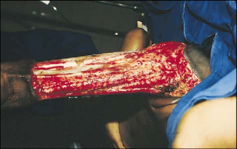
Debridement & Surgery:
The next step in treating necrotizing fasciitis is surgery. The doctors do not delay surgery in surgical consultation; they debride the wound extensively and remove the affected area. Early surgery reduces the chances of limb amputation. Moreover, a second surgery might be needed. The doctors keep the wound open and pack it with wet gauze. They change the dressing daily and perform debridement daily, keeping in view the hemodynamic stability. 7Kim, Y. H., Ha, J. H., Kim, J. T., & Kim, S. W. (2018). Managing necrotizing fasciitis to reduce mortality and increase limb salvage. Journal of wound care, 27(Sup9a), S20–S27. https://doi.org/10.12968/jowc.2018.27.Sup9a.S20
Immunoglobulins:
The doctors give intravenous immunoglobulins to immunocompromised people to improve their immune system.
Soft Tissue Reconstruction:
After your doctor removes all the necrotic tissue, they consult a plastic surgeon for the reconstruction of the soft tissues. The plastic surgeon tries to cover the wound area by primary closure. However, if primary closure is impossible, they close it using a muscle flap. They may use artificial skin if reconstruction by natural tissue is not possible.
Hyperbaric Oxygenation:
Doctors may give hyperbaric oxygen (Oxygen at high pressure) to the patients during their stay in the intensive care unit. However, it is effective for small wounds, has no effect on large wounds, and does not prolong life.8Bonne, S. L., & Kadri, S. S. (2017). Evaluation and Management of Necrotizing Soft Tissue Infections. Infectious disease Clinics of North America, 31(3), 497–511. https://doi.org/10.1016/j.idc.2017.05.011
Necrotizing Fasciitis Vs. Gangrene
| Necrotizing Fasciitis | Gangrene | |
| Definition | Necrotizing fasciitis is an inflammation of the skin’s soft tissues, affecting the fascias, soft tissues, and muscles and gradually causing their necrosis. | Gangrene is a condition in which the death of tissues occurs due to lack of blood supply, infection, or trauma. |
| Pathogenesis
Progression |
It occurs due to any break in the skin’s integrity that introduces bacteria to the skin. The inflammation starts, spreading gradually, making the prognosis worse.
It spreads rapidly within hours or days. |
Gangrene occurs due to a lack of blood supply to the tissues, which causes hypoxia and cell death.
It progresses slowly, spreading in days or weeks. |
| Cause | Necrotizing fasciitis occurs due to some bacteria like Streptococci, staphylococci, and Clostridium perfringens.
Necrotizing fasciitis gradually affects your fascias, para fascias, muscles, and soft tissues. |
Gangrene occurs due to reduced blood supply (ischemia) or infection, such as in diabetic foot.
Gangrene affects your skin, muscles, and organs due to reduced blood supply. |
| Pain | There is severe, excruciating pain in necrotizing fasciitis. | The pain ranges from moderate to severe, depending on location and severity. |
Is Necrotizing Fasciitis Contagious?
It is not directly contagious. However, the bacteria causing this infection can be transferred from one person to another through secretions, fluids, wounds, and common things like razors, towels, and medical equipment. To prevent this, you need to be very careful. Take care of hygiene, and do not use common towels, bedsheets, razors, and other equipment. Moreover, keep the patient’s wounds clean and seek proper medical care if you contact the affected individual.
Conclusions
This infection is an inflammatory condition that affects fascia, soft tissues, and muscles. Its symptoms include pain, vomiting, redness, swelling, purple rashes, and bullae formation in the affected area of the skin. It is common in diabetics, obese, alcoholics, and immunocompromised people. Early recognition and treatment are important for the better prognosis of necrotizing fasciitis. If left untreated, it can lead to sepsis, multi-organ failure, and death.
Refrences
- 1Hite, M., McCrae, A. L., & Privette, A. (2018). Fungal Necrotizing Fasciitis after Penetrating Trauma. The American Surgeon, 84(8), e302–e304.
- 2Khalid, M., Junejo, S., & Mir, F. (2018). Invasive Community-Acquired Methicillin-Resistant Staphylococcal Aureus (CA-MRSA) Infections in Children. Journal of the College of Physicians and Surgeons–Pakistan: JCPSP, 28(9), S174–S177. https://doi.org/10.29271/jcpsp.2018.09.S174
- 3Hite, M., McCrae, A. L., & Privette, A. (2018). Fungal Necrotizing Fasciitis after Penetrating Trauma. The American Surgeon, 84(8), e302–e304.
- 4Fais, P., Viero, A., Viel, G., Giordano, R., Raniero, D., Kusstatscher, S., Giraudo, C., Cecchetto, G., & Montisci, M. (2018). Necrotizing fasciitis: case series and review of the literature on clinical and medico-legal diagnostic challenges. International journal of legal medicine, 132(5), 1357–1366. https://doi.org/10.1007/s00414-018-1838-0
- 5Erichsen Andersson, A., Egerod, I., Knudsen, V. E., & Fagerdahl, A. M. (2018). Signs, symptoms and diagnosis of this infection experienced by survivors and family: a qualitative Nordic multi-center study. BMC infectious diseases, 18(1), 429. https://doi.org/10.1186/s12879-018-3355-7.
- 6Ballesteros-Betancourt, J. R., García-Tarriño, R., Ríos-Guillermo, J., Rodriguez-Roiz, J. M., Camacho, P., Zumbado-Dijeres, A., Domingo-Trepat, A., Llusá-Pérez, M., Combalia-Aleu, A., García-Ramiro, S., & Soriano-Viladomiu, A. (2017). Necrotizing fasciitis attended in the Emergency Department in a tertiary Hospital: Evaluation of the LRINEC scale. Infecciones necrosantes de partes blandas atendidas en un servicio de urgencias de tercer nivel: evolución y correlación con la escala laboratory risk indicator for necrotizing fasciitis (LRINEC). Revista espanola de cirugia ortopedica y traumatologia, 61(4), 265–272. https://doi.org/10.1016/j.recot.2017.04.003
- 7Kim, Y. H., Ha, J. H., Kim, J. T., & Kim, S. W. (2018). Managing necrotizing fasciitis to reduce mortality and increase limb salvage. Journal of wound care, 27(Sup9a), S20–S27. https://doi.org/10.12968/jowc.2018.27.Sup9a.S20
- 8Bonne, S. L., & Kadri, S. S. (2017). Evaluation and Management of Necrotizing Soft Tissue Infections. Infectious disease Clinics of North America, 31(3), 497–511. https://doi.org/10.1016/j.idc.2017.05.011
