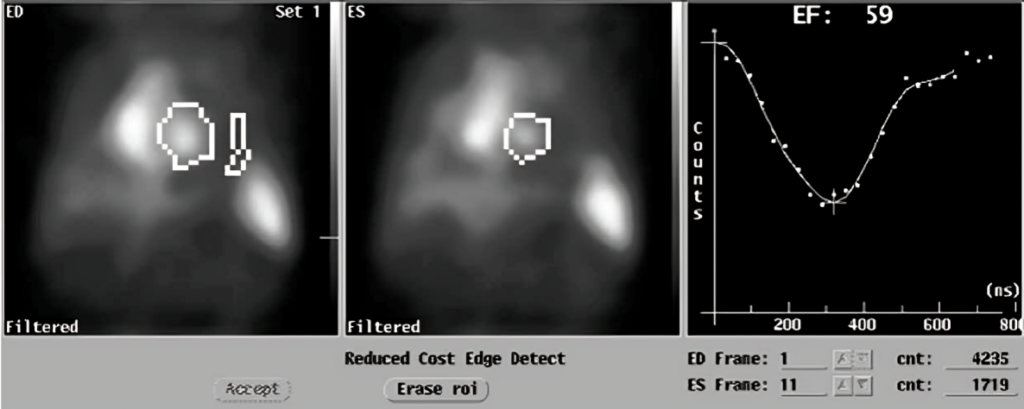
A MUGA scan is a medical procedure that helps doctors better examine your heart and its functioning. Since its results are much more accurate than those of other tests, it is a frequently used imaging modality in healthcare.
What is a MUGA scan?
A MUGA scan is an imaging procedure in nuclear medicine. It involves inserting a radioactive tracer into your blood and using a gamma camera to take photos of your heart and record a parameter known as ejection fraction. Ejection fraction is the force with which the ventricular chambers of your heart pump blood all over the body.
“MUGA” stands for “multiple-gated acquisition” — the term “multiple-gated” refers to the various phases or gates of the cardiac cycle, and “acquisition” refers to the images captured in these different “gates”. Other names for the MUGA scan include:
- Radionuclide ventriculography
- Nuclear ventriculography
- Gated blood pool scan
- Equilibrium radionuclide angiography1Mihir Odak, & Kayani, W. T. (2023, July 25). MUGA Scan. Nih.gov; StatPearls Publishing. https://www.ncbi.nlm.nih.gov/books/NBK564365/
Indications for MUGA Scan
MUGA scans are an accurate and non-invasive method of investigating various heart functions, such as:
- If different chambers of the heart are contracting with enough force during each cycle
- The blood volume in the ventricles just before they contract
- The blood volume in the ventricles just after they contract and have pumped all the blood out
- The speed at which the ventricles fill with and then eject blood
- The total volume of blood pumped out per contraction
- The total volume of blood pumped out each minute
- If there is any backflow of the blood through the heart’s valves
- If the patterns of blood flow between different chambers are normal or have “shunted” in an abnormal direction
Such functional parameters need inspection in a number of heart diseases, as well as conditions where doctors need to rule out damage to cardiac structures. Examples include:
Monitoring Cardiotoxicity
The most important use of MUGA scans is periodically examining cancer patients for cardiac damage. Cancer patients need to undergo rigorous chemotherapy regimens, and many of the drugs included in them can cause cardiotoxicity. The scan is mainly used to record the left ventricular ejection fraction and volumes.2D’Amore, C. (2014). Nuclear imaging in detection and monitoring of cardiotoxicity. World Journal of Radiology, 6(7), 486–486. https://doi.org/10.4329/wjr.v6.i7.486
Cardiac Disease
Even though it has now been replaced by other modalities that are more readily available, the MUGA scan is still used whenever highly accurate information about ventricular function is required to diagnose or assess cardiac disorders. Examples include:
- Heart failure
- Congenital heart disease
- Ischemic heart disease
- Pulmonary hypertension
- Cardiomyopathy
- Arrhythmia
- Postop evaluation after cardiac bypass surgery3Iqbal, S. (2021, October 27). Cardiac blood pool scan. Radiopaedia; Radiopaedia.org. https://radiopaedia.org/articles/cardiac-blood-pool-scan
Contraindications of the MUGA scan
The MUGA scan is safe and is often used in situations where the patient cannot undergo invasive procedures like angiography and angioplasty. However, because of the use of a radio topic tracer (Techtenium-99), it has specific contraindications. These include:
- Allergy to the tracer
- Pregnancy
- Breastfeeding mothers
- Small children
- Uncontrolled medical conditions or unstable patient
- Severe renal disease
- Inability to lie still during the scan or to establish IV access4Henzlova, M. J., W. Lane Duvall, Einstein, A. J., Travin, M. I., & Verberne, H. J. (2016). ASNC imaging guidelines for SPECT nuclear cardiology procedures: Stress, protocols, and tracers. Journal of Nuclear Cardiology, 23(3), 606–639. https://doi.org/10.1007/s12350-015-0387-x

What can I do to prepare for the MUGA scan?
There isn’t a lot to prepare for when it comes to a MUGA scan because of its safety and noninvasiveness. Preparation consists of a medical exam and a few do’s and don’ts, as your doctor advises.5Mary Beth Farrell, Galt, J. R., Panagiotis Georgoulias, Malhotra, S., Pagnanelli, R., Christoph Rischpler, & Bital Savir-Baruch. (2020). SNMMI Procedure Standard/EANM Guideline for Gated Equilibrium Radionuclide Angiography*. Journal of Nuclear Medicine Technology, 48(2), 126–135. https://doi.org/10.2967/jnmt.120.246405
History & Examination
Your doctor will take a comprehensive cardiac health history before your MUGA scan. They will ask you if you have already been diagnosed with any heart condition or have been experiencing any cardiac symptoms such as:
- Chest pain and discomfort
- Dizziness
- Breathlessness
- Fatigue
- Swelling in your feet and legs
They will also ask you about the medications you are taking. In addition, they will ensure you don’t have a history of allergic reactions to radioactive isotopes that are used in nuclear medicine.
In addition to a detailed history, your doctor will examine you and ensure you are medically fit for the MUGA scan. They will also run a series of tests, including:
- Complete blood count
- Renal function tests
- Electrocardiogram
- Serum electrolytes
- Cardiac stress test
- Liver function tests
Informed Consent
Once you have been declared medically fit for a MUGA scan, you will be educated about the steps, risks, and outcomes of the procedure. You will then be asked to sign a document as proof of your informed consent.
Do’s & Don’ts before MUGA Scan
There are several best practices to take up before your MUGA scan. These activities improve patient experiences during the procedure, as well as improve your chances of getting more accurate results. Examples include:
- Drink plenty of water to ensure your kidneys are in perfect health and your body can easily eliminate the radiotracer you will be injected with.
- Take your prescribed medications on time unless your doctor advises you against doing so.
- Wear comfortable, loose-fitting clothing that can easily be unbuttoned. This will allow your medical team to perform the MUGA scan without compromising on modesty.
- Let your doctor know immediately if there’s any chance you could be pregnant or are currently a lactating mother.
- Avoid consuming substances that can affect the function of your heart, such as caffeine and nicotine.
- Avoid strenuous activity, which can make your heart pump more blood than it usually does.
- Before your scan, undergo the complete panel of tests your doctor has advised so that no medical issue is missed.
Step-by-step procedure for a MUGA scan
A MUGA scan procedure takes place in the nuclear medicine department of a hospital or medical center. It is carried out by a team of professionals trained in radiology or nuclear medicine. A technologist performs the procedure, a radiologist interprets it, and the results are sent to your doctor.
During the MUGA Scan:
At the start of your MUGA scan, you will be requested to lie on your back on a hospital bed, and the medical team will prepare you for the procedure.
- Your chest will be exposed, and electrodes from an ECG machine will be placed across it to track your heart’s activity.
- A technician or nurse will pass an IV line into your arm. This will draw blood and inject you with the tracer.
- They will draw your blood and mix it with a radioactive tracer. The most commonly used tracer for the MUGA scan is Technetium-99m, which is used because of its ability to tag red blood cells.
- Your blood will be injected back into your body. Bound to your blood cells, the tracer will travel to your heart, providing a clear picture of its inner workings.
- During regular “rest” scans, you are required to lie down while a gamma camera fixed above takes various photos. “Exercise” scans are different as you need to participate in physical activity, e.g., running or walking on a treadmill machine or pedaling on a bicycle until you reach the peak activity of your heart. Afterward, you will be asked to lie down again while the camera takes more photos of your heart.
- The entire medical procedure lasts no more than two hours.6American Heart Association. (n.d.). Radionuclide ventriculography or radionuclide angiography (MUGA scan). Retrieved from https://www.heart.org/en/health-topics/heart-attack/diagnosing-a-heart-attack/radionuclide-ventriculography-or-radionuclide-angiography-muga-scan
What happens after a MUGA Scan?
Once the images have been captured, a doctor analyzes them and interprets the results. Meanwhile, you are free to leave the hospital. You are usually notified about the results in a matter of weeks and need to return to your primary care physician for follow-up.
Possible results of a MUGA scan
MUGA scans can be normal or abnormal. Normal scans are “negative” for abnormalities, whereas abnormal tests are “positive” for abnormalities in your heart’s structure and function. The MUGA scan results are shown as a percentage called the left ventricle ejection fraction or LVEF.
Normal or Negative MUGA Scan Results
Normal or negative MUGA scans showcase findings that are consistent with those found in healthy populations. An ejection fraction between 50 and 70 percent is considered normal. This means that the heart is pumping an adequate flow of blood to the body’s organs.
Abnormal or Positive MUGA Scan Results
Abnormal or positive MUGA scans have findings with abnormalities in the way your heart pumps blood. Examples include:7Clinic, C. (2022). Multigated Acquisition Scan (MUGA). Cleveland Clinic. https://my.clevelandclinic.org/health/diagnostics/17247-multigated-acquisition-scan-muga
| Left Ventricle Ejection Fraction | Interpretation | Causes |
| <40% | The left ventricle’s ability to pump blood has become greatly reduced |
|
| <40 to 55% | There has been damage to the heart’s muscle, which affects its ability to pump blood well |
|
| >75% | The heart’s musculature has become abnormally thickened, limiting the amount of blood the heart can take in and pump out. | Hypertrophic cardiomyopathy |
Follow-up to the MUGA scan
Your doctor will tell you about your results as soon as they become available. If the findings are negative, your doctor will continue the same treatment plan you already have or make minor changes based on your results.
In case your results are positive, your doctor may recommend some additional tests, such as:
- Echocardiogram
- Cardiac MRI
- Stress test
- Coronary angiography
Depending on your condition, they may also add or remove drugs from your regimen. In addition, you will be scheduled for more MUGA scans in your follow-up appointments if you have a chronic condition like cancer and/or are taking drugs that cause cardiotoxicity. These drugs include:
- Anthracyclines
o Doxorubicin
o Daunorubicin - Trastuzumab
o Herceptin - Cyclophosphamide
- 5-Fluorouracil
- Tyrosine kinase inhibitors
o Sunitinib
o Imatinib - Proteasome inhibitors
o Bortezomib8Kelleni, M. T., & Mahrous Abdelbasset. (2018). Drug-Induced Cardiotoxicity: Mechanism, Prevention, and Management. InTech EBooks. https://doi.org/10.5772/intechopen.79611
Side Effects of the MUGA Scan
MUGA scans are not associated with side effects the way invasive procedures like cardiac catheterization are. However, they can still cause a few complications, such as allergies and radiation exposure.
Allergies
Researchers have noted 2.1 to 6 cases of allergic reactions per 100,000 injections of radioactive substances. Similarly, Technetium-99m can also cause allergic reactions in vulnerable individuals. Signs and symptoms of these reactions include:
- Skin rash
- Itching
- Hives
- Swelling seen on the face or lips
- Lightheadedness
- Nausea
- Difficulty breathing
- Swelling of the throat or tongue
The most severe form of these allergic reactions is anaphylaxis, which can result in death. Therefore, it is important to make sure people who are allergic to radioactive pharmaceuticals do not undergo radioactive scans like the MUGA scan.9Schreuder, N., Quincy de Hoog, van, & Puijenbroek, van. (2019). Anaphylactic Reaction to Tc-99m Macrosalb. Drug Safety – Case Reports, 6(1). https://doi.org/10.1007/s40800-019-0097-4
Radiation Exposure
MUGA scans only use a low dose of radiation and does not have any long-term or serious consequences. However, they must be avoided in populations that are more sensitive to radiation exposure than the average adult, such as pregnant women, breastfeeding mothers, and young children whose bones are still developing.
Others
Other than allergic reactions and radiation exposure, there are a few side effects that you might experience with a MUGA scan, such as:
- Pain or discomfort at the injection site
- Swelling or bruising
- Infection at the injection site
- Back pain and discomfort after lying on your back for long periods, especially if you have joint issues
- False positives/ false negatives during the interpretation of your test results
MUGA Scan vs Echocardiogram
Even though both MUGA scans and echocardiograms assess the heart’s functioning, MUGA scans are more accurate when measuring the ejection fraction of your ventricles. Some of the differences between the two modalities are listed below:
| MUGA Scan | Echocardiogram |
| Employs a radioactive tracer | Employs ultrasound waves |
| Quantitative measurement of the ejection fraction of ventricles | No quantitative measurement of the ejection fraction of ventricles |
| The procedure is longer and takes 1 to 2 hours | The procedure is shorter and takes ½ to 1 hour |
| Unsafe for pregnant women | Safe for pregnant women |
Conclusion
The MUGA scan is an imaging modality in nuclear medicine. It is important in assessing cardiotoxicity caused by drugs, e.g., chemotherapeutic agents. MUGA Scan uses a Technetium-99m tracer and a gamma camera to help medical professionals visualize your heart and take various photos to precisely measure your ventricles’ ejection fraction and other functional parameters. It is a safe procedure with few side effects. Patients don’t need to undergo extensive preparation or aftercare and are discharged right after the procedure.
Refrences
- 1Mihir Odak, & Kayani, W. T. (2023, July 25). MUGA Scan. Nih.gov; StatPearls Publishing. https://www.ncbi.nlm.nih.gov/books/NBK564365/
- 2D’Amore, C. (2014). Nuclear imaging in detection and monitoring of cardiotoxicity. World Journal of Radiology, 6(7), 486–486. https://doi.org/10.4329/wjr.v6.i7.486
- 3Iqbal, S. (2021, October 27). Cardiac blood pool scan. Radiopaedia; Radiopaedia.org. https://radiopaedia.org/articles/cardiac-blood-pool-scan
- 4Henzlova, M. J., W. Lane Duvall, Einstein, A. J., Travin, M. I., & Verberne, H. J. (2016). ASNC imaging guidelines for SPECT nuclear cardiology procedures: Stress, protocols, and tracers. Journal of Nuclear Cardiology, 23(3), 606–639. https://doi.org/10.1007/s12350-015-0387-x
- 5Mary Beth Farrell, Galt, J. R., Panagiotis Georgoulias, Malhotra, S., Pagnanelli, R., Christoph Rischpler, & Bital Savir-Baruch. (2020). SNMMI Procedure Standard/EANM Guideline for Gated Equilibrium Radionuclide Angiography*. Journal of Nuclear Medicine Technology, 48(2), 126–135. https://doi.org/10.2967/jnmt.120.246405
- 6American Heart Association. (n.d.). Radionuclide ventriculography or radionuclide angiography (MUGA scan). Retrieved from https://www.heart.org/en/health-topics/heart-attack/diagnosing-a-heart-attack/radionuclide-ventriculography-or-radionuclide-angiography-muga-scan
- 7Clinic, C. (2022). Multigated Acquisition Scan (MUGA). Cleveland Clinic. https://my.clevelandclinic.org/health/diagnostics/17247-multigated-acquisition-scan-muga
- 8Kelleni, M. T., & Mahrous Abdelbasset. (2018). Drug-Induced Cardiotoxicity: Mechanism, Prevention, and Management. InTech EBooks. https://doi.org/10.5772/intechopen.79611
- 9Schreuder, N., Quincy de Hoog, van, & Puijenbroek, van. (2019). Anaphylactic Reaction to Tc-99m Macrosalb. Drug Safety – Case Reports, 6(1). https://doi.org/10.1007/s40800-019-0097-4

