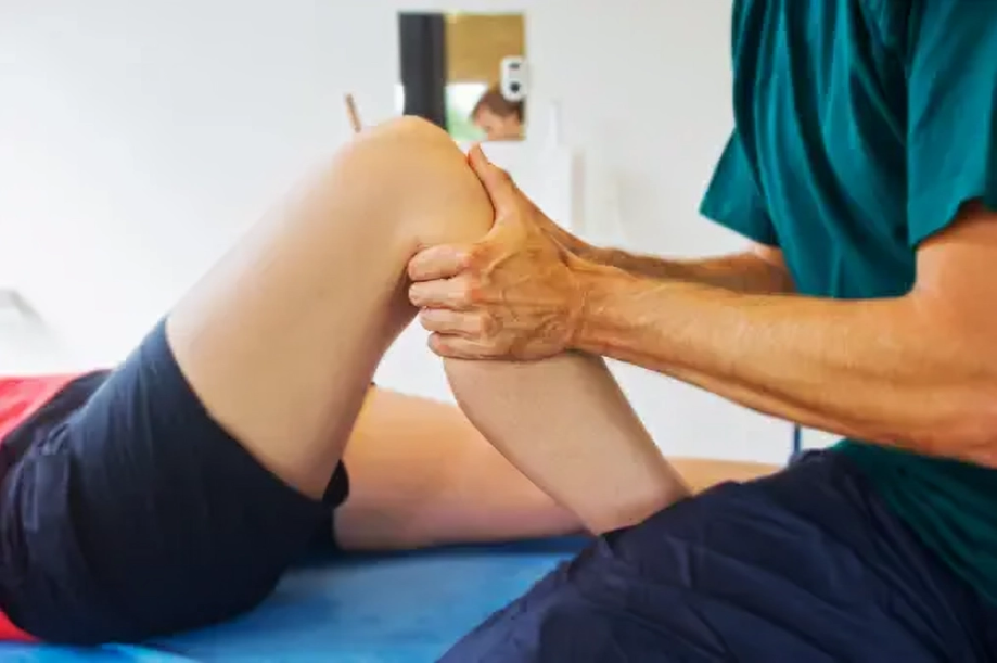
Medial Tibial Stress Syndrome is an overuse injury that mostly affects athletes’ lower extremities. The pain develops in any area between the knee and ankle on the anterior tibia. It is not a serious condition, but precautions must be taken to avoid serious consequences.
Medial Tibial Stress Syndrome
Medial Tibial Stress Syndrome (MTSS), commonly known as shin splints, is an overuse injury that affects the lower legs, particularly the inner edge of the tibia. It is a frequent condition among athletes, runners, and military personnel, causing pain along the posteromedial border of the tibia between the knee and ankle. While MTSS is not typically a severe condition, early management is crucial to prevent disease progression to stress fractures or other complications.1Deshmukh, N. S., & Phansopkar, P. (2022). Medial Tibial Stress Syndrome: A Review Article. Cureus, 14(7). https://doi.org/10.7759/cureus.26641
Causes of Medial Tibial Stress Syndrome
MTSS occurs due to repetitive stress on the tibia and its surrounding tissues, often resulting from physical activities that involve high-impact forces. Key contributing factors include:
- Repetitive Stress: Overuse injury from activities such as running, jumping, or dancing creates microtrauma in the tibial bone and associated muscles.
- Periosteal Inflammation: Irritation of the periosteum (the thin layer covering the bone) is a common mechanism behind MTSS.
- Biomechanical Issues: Flat feet, overpronation, and improper lower limb alignment increase stress on the tibia.
- Training Errors: Sudden increases in activity intensity, frequency, or duration can overload the tibial structure.
- Inappropriate Footwear: Ill-fitted or worn-out shoes fail to provide adequate support, exacerbating the impact on the tibia.
Strenuous activities involving frequent starts and stops, such as basketball or tennis, are particularly likely to cause MTSS. Early recognition and modification of contributing factors are essential to alleviate symptoms and prevent progression.2Franklyn, M., & Oakes, B. (2015). Aetiology and mechanisms of injury in medial tibial stress syndrome: Current and future developments. World journal of orthopedics, 6(8), 577–589. https://doi.org/10.5312/wjo.v6.i8.577
Epidemiology
This syndrome occurs more in military people as compared to athletes and runners. Military recruiters have an incidence of up to 35%, and runners have an incidence between 13.6% and 20% of developing this syndrome. The incidence increases when you increase exercise intensity, load, and frequency.3Bliekendaal, S., Moen, M., Fokker, Y., Stubbe, J. H., Twisk, J., & Verhagen, E. (2018). Incidence and risk factors: a prospective study in Physical Education Teacher Education students. BMJ open sport & exercise medicine, 4(1), e000421. https://doi.org/10.1136/bmjsem-2018-000421
Symptoms
The main complaint of patients with this syndrome is pain. The nature of pain is either dull ache or sharp. Pain is typically localized to the medial side of the tibia, particularly in the middle or lower third, though it can occasionally be more generalized. In the early stages, pain often occurs at the start of physical activity and subsides with continued movement. As the condition progresses, pain may persist during and after activity, potentially causing significant discomfort. Swelling at the site of pain may be observed in some cases, but it is not a universal symptom. Tenderness of the Achilles tendon is not characteristic of MTSS and is more commonly associated with other conditions, such as Achilles tendinopathy.4Bhusari, N., & Deshmukh, M. (2023). Shin Splint: A Review. Cureus, 15(1), e33905. https://doi.org/10.7759/cureus.33905
How to diagnose Medial Tibial Stress Syndrome?
This is typically a clinical disease that is easily diagnosed by:
History:
Diagnosis of MTSS is largely clinical and relies on a detailed history of the patient’s symptoms. Your doctor will inquire about the intensity, location, and nature of the pain, as well as any history of repetitive activity or exercise. A hallmark feature of MTSS is pain in the lower extremities, strongly associated with exercise or strenuous activity. The pain typically starts at the onset of activity and may subside with continued movement in the early stages.
Physical Examination:
Physical examination plays a crucial role in diagnosing medial tibial stress syndrome. Your doctor carefully examines your lower extremities, including the shin, ankle, and knee. People with this condition may experience swelling and tenderness at the site of pain.

Imaging Modalities:
Imaging is not routinely required but may be used to confirm the diagnosis or rule out other conditions like tibial stress fractures or compartment syndrome.
- X-rays: Typically normal in MTSS. The presence of a “dreaded black line” suggests a stress fracture, not MTSS.
- MRI: The preferred imaging modality for severe cases showing periosteal or bone marrow edema. MRI can also identify other injuries, such as stress fractures.
- Nuclear Scans: These are useful but less specific than MRI, as they may show a “double stripe” pattern in the cortical bone, indicating stress injury.
- CT Scans: Not commonly recommended due to low specificity for MTSS.5Nozawa, T., Bell-Peter, A., Doria, A. S., Marcuz, J. A., Stimec, J., Whitney, K., & Feldman, B. M. (2021). Tibia stress injury and the imaging appearance of a stress fracture in juvenile dermatomyositis: six patients’ experiences. Pediatric rheumatology online journal, 19(1), 17. https://doi.org/10.1186/s12969-021-00501-9
Vitamin D Levels:
For patients with medial tibial stress syndrome that doesn’t resolve despite all treatments, it is recommended to check Vitamin D levels. Vitamin D deficiency is a leading cause of bone pain and stress fractures.6Winters M. (2020). The diagnosis and management of medial tibial stress syndrome: An evidence update. Diagnostik und Therapie des Schienbeinkantensyndroms: Update zur Studienlage. Der Unfallchirurg, 123(Suppl 1), 15–19. https://doi.org/10.1007/s00113-019-0667-z
Treatment of Medial Tibial Stress Syndrome
Treatment is mainly based on conservative measures that are easily followed at home. These are:
Rest:
The first step to reducing pain is to rest and avoid the activity causing it. Try to do exercises that are not repetitive and do not involve extra-loaded weight. The duration of rest that ensures the resolution of pain is not fixed; it largely depends upon every individual’s immunity and strength.
Ice:
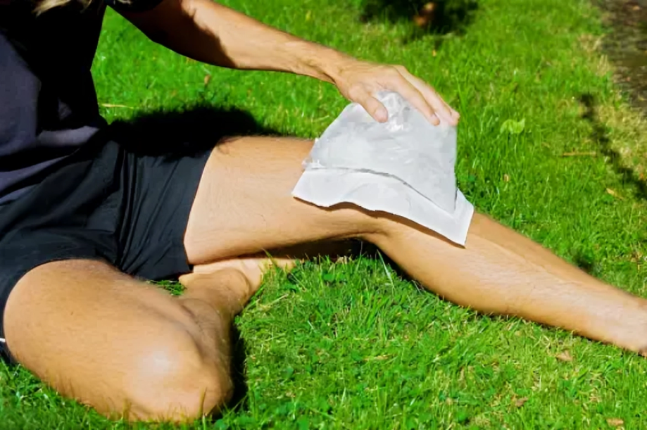
Ice can help reduce the severity of pain. Apply ice wrapped in a clean cloth to your skin as long as the pain persists. You can also repeat the procedure whenever the pain becomes unbearable. Ice also helps to reduce the swelling associated with medial tibial stress syndrome.

Compression:
Wearing compression bandages on the injured bone greatly reduces the pain and swelling. Moreover, compression bandages help you do the exercise smoothly by providing support to your injured bones, muscles, and ligaments.
Elevation:
Elevating the affected limb is a reasonable option to consider if you have swelling at the affected site along with pain.7Galbraith, R. M., & Lavallee, M. E. (2009). Medial tibial stress syndrome: conservative treatment options. Current reviews in musculoskeletal medicine, 2(3), 127–133. https://doi.org/10.1007/s12178-009-9055-6
Footwear and Training Modifications:
Wearing properly fitted, supportive footwear and modifying training routines are essential to prevent recurrence. Gradually increasing the intensity of activity can also help avoid overuse injuries.
Advanced Interventions for Severe Cases:
Phonophoresis:
It involves the use of ultrasound, which increases the absorption of drugs in a percutaneous layer of your skin.
Periosteal Pecking:
Orthopedics use large needles to reach the affected bone and peck (stroke) in periosteal pecking. As a result, certain neuroendocrine responses start in your body, which reduces the harshness of pain.
Ultrasound Therapy:
In medial tibial stress syndrome, ultrasound therapy is used. It increases blood flow in the affected area of the bone, thereby reducing the inflammation and chronic pain associated with it.
Fasciotomy:
Resistant and severe pain can be treated by a surgery called fasciotomy, in which tissues surrounding your injured bone are cut, leading to relief of pain.8Winters, M., Eskes, M., Weir, A., Moen, M. H., Backx, F. J., & Bakker, E. W. (2013). Treatment of medial tibial stress syndrome: a systematic review. Sports medicine (Auckland, N.Z.), 43(12), 1315–1333. https://doi.org/10.1007/s40279-013-0087-0
Calcium & Vitamin D Supplements:
If your pain doesn’t subside even with rest and activity modifications, check your Vitamin D and calcium levels. Deficiencies in these nutrients may contribute to persistent pain. Taking Vitamin D and calcium supplements can reduce pain and slow the progression of medial tibial stress syndrome.9Wesner M. L. (2012). Nutrient effects on stress reaction to bone. Canadian family physician Medecin de famille canadien, 58(11), 1226–1230.
Muscle associated with Medial Tibial Stress Syndrome
The tibialis posterior muscle, located in the posterior compartment of the lower leg, is the most commonly involved in this syndrome. Repetitive movements place significant traction on this muscle. The condition can also affect other muscles, such as the flexor digitorum longus and the soleus.
Can Exercise Help Relieve Medial Tibial Stress Syndrome Pain?
Yes, some exercises are quite beneficial in relieving the pain.10Moen, M. H., Holtslag, L., Bakker, E., Barten, C., Weir, A., Tol, J. L., & Backx, F. (2012). The treatment of medial tibial stress syndrome in athletes; a randomized clinical trial. Sports medicine, arthroscopy, rehabilitation, therapy & technology: SMARTT, 4, 12. https://doi.org/10.1186/1758-2555-4-12 These include:
- Toe walk
- Single leg raises while lying on the floor
- Toe curls
- Calf stretching exercises

Risk Factors
This syndrome can occur in anyone, but its incidence and severity increase with the following risk factors:11Winkelmann, Z. K., Anderson, D., Games, K. E., & Eberman, L. E. (2016). Risk Factors for Medial Tibial Stress Syndrome in Active Individuals: An Evidence-Based Review. Journal of Athletic Training, 51(12), 1049–1052. https://doi.org/10.4085/1062-6050-51.12.13
- Female gender
- Previous history of medial tibial stress syndrome or arthritis
- High BMI (obesity)
- Navicular bone drop
- Defects in ankle and foot alignment (flat feet)
- Abnormality in the lower extremity range of motions
- Wearing ill-fitted shoes during walking or running
- Doing high-impact exercises with increasing load
Prevention of MTSS
You can prevent medial tibial stress syndrome by taking proper rest and avoiding strenuous activities. Rest helps to resolve pain and inflammation. Engage in balanced exercises under a trainer’s supervision and use compression bandages on the injured area. Always work out on a soft surface while wearing supportive shoes with balanced soles. Stretching exercises are particularly effective in relieving symptoms. Maintaining a healthy weight is also crucial, as excess fat can exacerbate the condition.12Craig D. I. (2008). Medial tibial stress syndrome: evidence-based prevention. Journal of Athletic Training, 43(3), 316–318. https://doi.org/10.4085/1062-6050-43.3.316
Medial Tibial Stress Syndrome Vs. Stress Fracture:
Medial tibial stress syndrome develops when you overuse the tibia bone and its muscles through specific repetitive movements. On the other hand, stress fractures are small cracks in the bone caused by forceful actions. Stress fractures can also result from underlying conditions like osteoporosis, which weakens bones.
Pain associated with shin splints starts with movement and gradually decreases as the activity continues. However, pain from stress fractures worsens the longer you exercise. Orthopedics use a specific test called the “one-leg hop test” to differentiate between the two. In shin splints, patients can bear the jumping without significant pain, whereas with a stress fracture, the severity of pain prevents them from hopping more than once.
Healing Time for Medial Tibial Stress Syndrome
Almost every patient recovers from this syndrome, with the healing duration largely depending on the care given to the injured limb. With proper rest and treatment, recovery typically takes 4 to 12 weeks.
Prognosis
The prognosis is promising with proper care, as appropriate measures can heal the injury within weeks. However, neglecting sufficient rest and care can lead to complications, including stress fractures of the tibia.13Moen, M. H., Bongers, T., Bakker, E. W., Zimmermann, W. O., Weir, A., Tol, J. L., & Backx, F. J. (2012). Risk factors and prognostic indicators for medial tibial stress syndrome. Scandinavian journal of medicine & science in sports, 22(1), 34–39. https://doi.org/10.1111/j.1600-0838.2010.01144.x
Conclusion
In conclusion, excessive use of your lower limbs in strenuous exercises and running leads to this condition. Overuse of muscles and bone causes inflammation and swelling along with the pain. Pain is mostly experienced on your anterior shin when you start an activity, and it is a typical feature used to diagnose shin splints. MRI also helps to diagnose complicated cases. Resting and avoiding movement are the main treatment modalities. You can also massage ice on the painful site and wear compression bandages while walking or moving. Proper care promises a good prognosis, with recovery in almost 12 weeks.
Refrences
- 1Deshmukh, N. S., & Phansopkar, P. (2022). Medial Tibial Stress Syndrome: A Review Article. Cureus, 14(7). https://doi.org/10.7759/cureus.26641
- 2Franklyn, M., & Oakes, B. (2015). Aetiology and mechanisms of injury in medial tibial stress syndrome: Current and future developments. World journal of orthopedics, 6(8), 577–589. https://doi.org/10.5312/wjo.v6.i8.577
- 3Bliekendaal, S., Moen, M., Fokker, Y., Stubbe, J. H., Twisk, J., & Verhagen, E. (2018). Incidence and risk factors: a prospective study in Physical Education Teacher Education students. BMJ open sport & exercise medicine, 4(1), e000421. https://doi.org/10.1136/bmjsem-2018-000421
- 4Bhusari, N., & Deshmukh, M. (2023). Shin Splint: A Review. Cureus, 15(1), e33905. https://doi.org/10.7759/cureus.33905
- 5Nozawa, T., Bell-Peter, A., Doria, A. S., Marcuz, J. A., Stimec, J., Whitney, K., & Feldman, B. M. (2021). Tibia stress injury and the imaging appearance of a stress fracture in juvenile dermatomyositis: six patients’ experiences. Pediatric rheumatology online journal, 19(1), 17. https://doi.org/10.1186/s12969-021-00501-9
- 6Winters M. (2020). The diagnosis and management of medial tibial stress syndrome: An evidence update. Diagnostik und Therapie des Schienbeinkantensyndroms: Update zur Studienlage. Der Unfallchirurg, 123(Suppl 1), 15–19. https://doi.org/10.1007/s00113-019-0667-z
- 7Galbraith, R. M., & Lavallee, M. E. (2009). Medial tibial stress syndrome: conservative treatment options. Current reviews in musculoskeletal medicine, 2(3), 127–133. https://doi.org/10.1007/s12178-009-9055-6
- 8Winters, M., Eskes, M., Weir, A., Moen, M. H., Backx, F. J., & Bakker, E. W. (2013). Treatment of medial tibial stress syndrome: a systematic review. Sports medicine (Auckland, N.Z.), 43(12), 1315–1333. https://doi.org/10.1007/s40279-013-0087-0
- 9Wesner M. L. (2012). Nutrient effects on stress reaction to bone. Canadian family physician Medecin de famille canadien, 58(11), 1226–1230.
- 10Moen, M. H., Holtslag, L., Bakker, E., Barten, C., Weir, A., Tol, J. L., & Backx, F. (2012). The treatment of medial tibial stress syndrome in athletes; a randomized clinical trial. Sports medicine, arthroscopy, rehabilitation, therapy & technology: SMARTT, 4, 12. https://doi.org/10.1186/1758-2555-4-12
- 11Winkelmann, Z. K., Anderson, D., Games, K. E., & Eberman, L. E. (2016). Risk Factors for Medial Tibial Stress Syndrome in Active Individuals: An Evidence-Based Review. Journal of Athletic Training, 51(12), 1049–1052. https://doi.org/10.4085/1062-6050-51.12.13
- 12Craig D. I. (2008). Medial tibial stress syndrome: evidence-based prevention. Journal of Athletic Training, 43(3), 316–318. https://doi.org/10.4085/1062-6050-43.3.316
- 13Moen, M. H., Bongers, T., Bakker, E. W., Zimmermann, W. O., Weir, A., Tol, J. L., & Backx, F. J. (2012). Risk factors and prognostic indicators for medial tibial stress syndrome. Scandinavian journal of medicine & science in sports, 22(1), 34–39. https://doi.org/10.1111/j.1600-0838.2010.01144.x

