What are Reticular Veins?
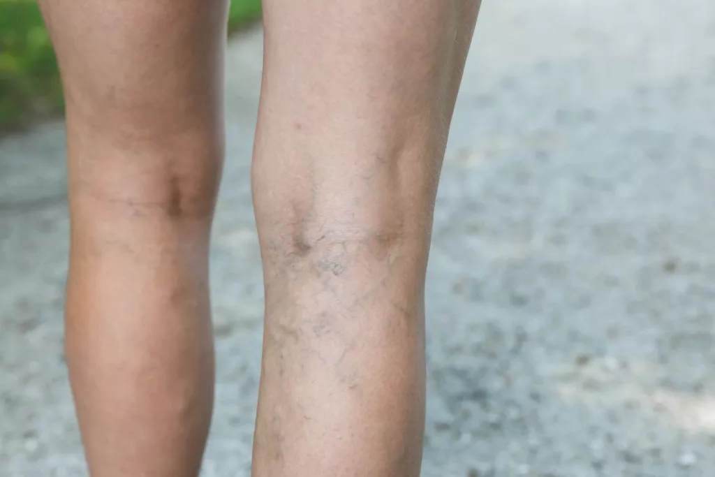
Reticular veins, sometimes called feeder veins or blue veins, are dilated veins with a diameter of less than 3.0 millimeters. Unlike larger veins, they often form a network of blue or greenish lines and can be found on the thighs, calves, and ankles.1Eklof B, Rutherford RB, Bergan JJ, Carpentier PH, Gloviczki P, Kistner RL, et al. Revision of the CEAP classification for chronic venous disorders: consensus statement. Journal of Vascular Surgery 2004;40(6):1248-52.
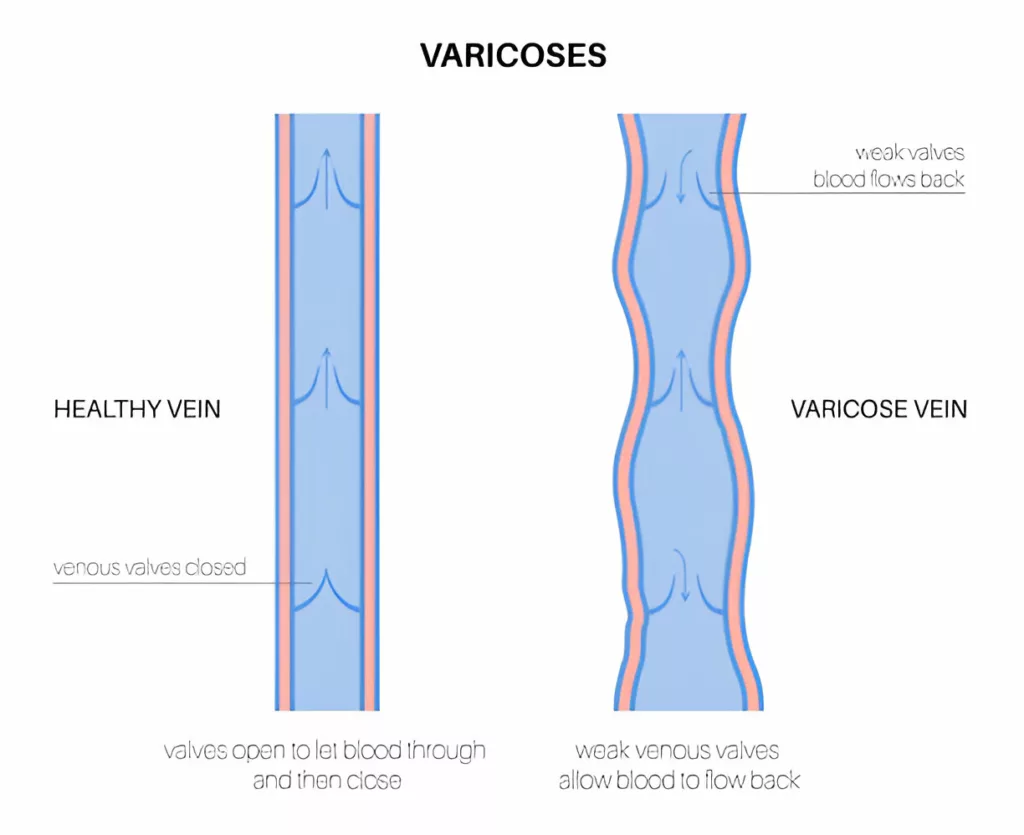
While typically not as prominent as varicose veins, reticular veins can indicate underlying venous insufficiency, a condition where blood flow through the veins is compromised, leading to increased pressure and the potential for blood to pool in the affected area. They are often tortuous and located in the body’s subdermal or subcutaneous tissue. The most common site is the lower limbs, where blood can pool in dilated veins due to gravity. These veins are generally considered a cosmetic concern; however, they can sometimes lead to symptoms like itching, aching, or a burning sensation in the affected area. Treatments for reticular veins include sclerotherapy and laser therapy, which aim to reduce their appearance and alleviate any associated symptoms.
Characteristics of Reticular Veins
The most common characteristics include the following:
- They are usually blue or purplish in appearance.
- The most common sites of the lower legs include the back of thighs and knees, inner thighs, and near ankles.
- Reticular veins are sometimes called “feeder veins” because they can supply blood to smaller spider veins, contributing to their development.
Symptoms of Reticular Veins
These veins do not always cause symptoms. Most commonly, this condition cause cosmetic concerns for individuals. When a group of reticular veins is present, they can cause varicose vein-like symptoms. If reticular veins are symptomatic, the following symptoms are present.
- Discomfort
- Heaviness
- Aching
- Fatigue
- Rarely burning, itching, or tenderness in the surrounding areas.
If reticular veins are left untreated for a long time, spider veins can develop.
Risk Factors & Causes
The prevalence of this condition is high, up to 60 percent in some populations. It is more common in women, and the risk of development increases with age.2Bertanha M, Jaldin RG, Moura R, Pimenta REF, Mariúba JVO, Lúcio Filho CEP, Alcantara GP, Padovani CR, Yoshida WB, Sobreira ML. Sclerotherapy for Reticular Veins in the Lower Limbs: A Triple-Blind Randomized Clinical Trial. JAMA Dermatol. 2017 Dec 1;153(12):1249-1255. doi: 10.1001/jamadermatol.2017.3426. PMID: 28973414; PMCID: PMC5817453. The risk factors are as follows:
Family History
People with family members who have venous concerns are more likely to develop reticular veins and other venous diseases.
Obesity
Obesity causes an increase in abdominal pressure, which places extra pressure on the venous system, resulting in increased blood backflow in veins. Elevated pressure can lead to impaired blood flow and venous insufficiency, where the veins struggle to return blood to the heart efficiently. As a result, veins may become dilated, contributing to the development of feeder veins.3Scholl L, Dörler M, Stücker M. Ulkus bei Adipositas-assoziierter chronischer Veneninsuffizienz [Ulcers in obesity-associated chronic venous insufficiency]. Hautarzt. 2017 Jul;68(7):560-565. German. doi: 10.1007/s00105-017-3971-y. PMID: 28357467.
Pregnancy
A growing uterus in pregnancy can affect blood flow, resulting in the development of dilated veins.4Skudder PA, Farrington DT. Venous conditions associated with pregnancy. Semin Dermatol. 1993 Jun;12(2):72-7. PMID: 8512797.
Aging
The risk of venous insufficiency increases with increasing age.
Occupations
Certain occupations have a higher risk of developing this condition. People who stand more, such as shopkeepers, teachers, and factory workers, are more likely to develop feeder veins.5Bass A. [The effect of standing in the workplace and developing chronic venous insufficiency]. Harefuah. 2007 Sep;146(9):675-6, 734-5. Hebrew. PMID: 17969303. Similarly, those sitting at a computer for prolonged periods are at risk of developing reticular veins behind the knees.6Restaino RM, Holwerda SW, Credeur DP, Fadel PJ, Padilla J. Impact of prolonged sitting on lower and upper limb micro- and macrovascular dilator function. Exp Physiol. 2015 Jul 1;100(7):829-38. doi: 10.1113/EP085238. Epub 2015 Jun 10. PMID: 25929229; PMCID: PMC4956484.
Oral Contraceptive Pills
OCPs can cause venous stasis and venous wall changes that can develop reticular veins.7Cohen J. Insuffisance veineuse et contraception orale [Venous insufficiency and oral contraception]. Rev Fr Gynecol Obstet. 1991 Feb 25;86(2 Pt 2):187-9. French. PMID: 1767172.
Reticular Veins Vs. Spider Veins or Telangiectasias
Spider veins, also known as telangiectasias or thread veins, are prominent clusters of damaged vessels in the skin. They are usually 1 millimeter in diameter.8Eklof B, Rutherford RB, Bergan JJ, Carpentier PH, Gloviczki P, Kistner RL, et al. Revision of the CEAP classification for chronic venous disorders: consensus statement. Journal of Vascular Surgery 2004;40(6):1248-52. Spider veins appear as blue or purple lines coming out of a central focus, which gives the appearance of a spider.9Sandean DP, Winters R. Spider Veins. [Updated 2023 Jul 4]. In: StatPearls [Internet]. Treasure Island (FL): StatPearls Publishing; 2024 Jan-. Available from: https://www.ncbi.nlm.nih.gov/books/NBK563218/
The significant difference between spider veins and reticular veins is their size. Reticular veins are usually more extensive and prominent, with a diameter of up to 3mm, compared to spider veins that are 1mm in diameter. Reticular veins sometimes serve as feeder veins to the spider veins. You might see a reticular vein from which several spider veins are extending.
Reticular Veins vs. Varicose Veins
Reticular veins are smaller than varicose veins. While varicose veins are typically larger, twisted, and bulging, reticular veins appear as flat, blue, or greenish lines beneath the skin. These veins are usually not palpable to the touch due to their flat nature, whereas varicose veins protrude from the skin and can be easily felt. Furthermore, discomfort, aching, and fatigue are more common with varicose veins. Feeder veins are usually asymptomatic.
Are Reticular Veins Dangerous?
These veins are cosmetically unattractive and can cause some discomfort and pain, but they are not dangerous. They show that the blood in the veins is pooling due to venous insufficiency. Investigating the cause of reticular veins and eliminating controllable risk factors is essential. If there are signs of the development of varicose veins, then it can be dangerous. If this condition become painful, a visit to the doctor is necessary.
Treatment Options for Reticular Veins
Reticular veins can be treated using various methods, depending on their severity and associated symptoms. The following options are available:
Sclerotherapy
The most effective treatment for reticular veins is sclerotherapy, especially when reticular veins are enlarged and painful.10Smith PC. Management of reticular veins and telangiectases. Phlebology. 2015 Nov;30(2 Suppl):46-52. doi: 10.1177/0268355515592770. PMID: 26556703. This procedure involves injecting a sclerosing agent directly into the affected veins, causing damage to the vein walls. As a result, the veins collapse and are gradually absorbed by the body. An ultrasound device may be used to guide the injection and ensure accurate placement of the sclerosing agent in the targeted veins.
Intense Pulsed Light (IPL) Treatment
Also known as IPL, this therapy is mainly used to treat spider veins and can sometimes help with slightly more prominent veins. It uses bursts of light to target and destroy the affected veins, similar to how laser treatments work.
Laser Therapy
Laser therapy is a treatment method that uses focused light to target and destroy reticular veins. Unlike sclerotherapy, which involves injecting a solution into the veins, laser therapy does not require any injections. Instead, it uses concentrated light beams to heat and damage the vein, causing it to collapse and be absorbed by the body. Laser therapy is often less invasive and may be less painful than sclerotherapy for some patients. However, it can lead to side effects such as skin discoloration or irritation and is generally more expensive.
Microphlebectomy
This technique involves removing veins through minor cuts using special tools. It’s a common approach for getting rid of varicose veins.
Thermocoagulation
This treatment uses radiofrequency energy to heat and damage the veins. A small needle delivers the radiofrequency, which destroys the veins through heat.
Reticular veins are usually more of a cosmetic issue than a serious health concern. Still, they can be uncomfortable and may indicate deeper vein problems. Knowing what causes them, where they appear, and what symptoms they might bring can help you manage them better. Simple steps like elevating your legs and wearing compression stockings can be practical. Treatments like sclerotherapy, IPL, laser therapy, microphlebectomy, and thermocoagulation can relieve more severe cases. Suppose you find the veins painful or notice worsening symptoms. In that case, you should check in with a healthcare provider to keep your legs feeling their best and address any underlying issues.
Prevention of Reticular Veins
If you notice the appearance of reticular veins, increasing blood flow through your legs can help manage and prevent further development. It can be done in the following ways:
Leg Elevation
Elevation of the legs improves the blood flow back to the heart and prevents blood pooling in the legs. Thus, it relieves reticular veins.
Avoid Leg Crossing
It is important not to cross your legs while sitting. Crossing the legs interferes with blood flow, leading to stasis and pooling.
Compression Stockings
People in occupations where they stand a lot should wear compression stockings. These stockings help maintain the blood flow in prolonged standing. Compression stockings are standard therapy for all venous and lymphatic disorders.11Rabe E, Partsch H, Hafner J, Lattimer C, Mosti G, Neumann M, Urbanek T, Huebner M, Gaillard S, Carpentier P. Indications for medical compression stockings in venous and lymphatic disorders: An evidence-based consensus statement. Phlebology. 2018 Apr;33(3):163-184. doi: 10.1177/0268355516689631. Epub 2017 Feb 22. PMID: 28549402; PMCID: PMC5846867.
Exercise
Exercising regularly helps maintain weight and improve blood flow.
Conclusion
In conclusion, reticular veins, often seen as a milder form of venous insufficiency, can significantly impact a person’s quality of life despite their less prominent appearance compared to varicose veins. Understanding their causes, symptoms, and treatment options is crucial for effective management and prevention. Early intervention, including lifestyle changes, medical treatments, and possibly minimally invasive procedures, can help alleviate symptoms and improve overall venous health. By addressing this condition proactively, individuals can maintain better leg health and enhance their comfort and well-being.
Refrences
- 1Eklof B, Rutherford RB, Bergan JJ, Carpentier PH, Gloviczki P, Kistner RL, et al. Revision of the CEAP classification for chronic venous disorders: consensus statement. Journal of Vascular Surgery 2004;40(6):1248-52.
- 2Bertanha M, Jaldin RG, Moura R, Pimenta REF, Mariúba JVO, Lúcio Filho CEP, Alcantara GP, Padovani CR, Yoshida WB, Sobreira ML. Sclerotherapy for Reticular Veins in the Lower Limbs: A Triple-Blind Randomized Clinical Trial. JAMA Dermatol. 2017 Dec 1;153(12):1249-1255. doi: 10.1001/jamadermatol.2017.3426. PMID: 28973414; PMCID: PMC5817453.
- 3Scholl L, Dörler M, Stücker M. Ulkus bei Adipositas-assoziierter chronischer Veneninsuffizienz [Ulcers in obesity-associated chronic venous insufficiency]. Hautarzt. 2017 Jul;68(7):560-565. German. doi: 10.1007/s00105-017-3971-y. PMID: 28357467.
- 4Skudder PA, Farrington DT. Venous conditions associated with pregnancy. Semin Dermatol. 1993 Jun;12(2):72-7. PMID: 8512797.
- 5Bass A. [The effect of standing in the workplace and developing chronic venous insufficiency]. Harefuah. 2007 Sep;146(9):675-6, 734-5. Hebrew. PMID: 17969303.
- 6Restaino RM, Holwerda SW, Credeur DP, Fadel PJ, Padilla J. Impact of prolonged sitting on lower and upper limb micro- and macrovascular dilator function. Exp Physiol. 2015 Jul 1;100(7):829-38. doi: 10.1113/EP085238. Epub 2015 Jun 10. PMID: 25929229; PMCID: PMC4956484.
- 7Cohen J. Insuffisance veineuse et contraception orale [Venous insufficiency and oral contraception]. Rev Fr Gynecol Obstet. 1991 Feb 25;86(2 Pt 2):187-9. French. PMID: 1767172.
- 8Eklof B, Rutherford RB, Bergan JJ, Carpentier PH, Gloviczki P, Kistner RL, et al. Revision of the CEAP classification for chronic venous disorders: consensus statement. Journal of Vascular Surgery 2004;40(6):1248-52.
- 9Sandean DP, Winters R. Spider Veins. [Updated 2023 Jul 4]. In: StatPearls [Internet]. Treasure Island (FL): StatPearls Publishing; 2024 Jan-. Available from: https://www.ncbi.nlm.nih.gov/books/NBK563218/
- 10Smith PC. Management of reticular veins and telangiectases. Phlebology. 2015 Nov;30(2 Suppl):46-52. doi: 10.1177/0268355515592770. PMID: 26556703.
- 11Rabe E, Partsch H, Hafner J, Lattimer C, Mosti G, Neumann M, Urbanek T, Huebner M, Gaillard S, Carpentier P. Indications for medical compression stockings in venous and lymphatic disorders: An evidence-based consensus statement. Phlebology. 2018 Apr;33(3):163-184. doi: 10.1177/0268355516689631. Epub 2017 Feb 22. PMID: 28549402; PMCID: PMC5846867.
