What is Cholesteatoma?
Types of Cholesteatoma
It is classified into two major types.1Kennedy, K. L. (2023, July 4). Middle ear cholesteatoma. StatPearls – NCBI Bookshelf. https://www.ncbi.nlm.nih.gov/books/NBK448108/
Congenital Cholesteatoma:
It occurs rarely when the remnants of ear epithelium in the tympanic membrane are present during the development.
Acquired Cholesteatoma:
It most commonly occurs due to an infection that causes abnormal growth of the ear epithelium (squamous keratinized).
Epidemiology
The incidence and prevalence of this condition are not well understood. However, some studies provide help with complete demographic statistics about cholesteatoma. According to studies, In the United States of America (USA), there are about 6 cases of cholesteatoma per 10,000 people. Most of the time, it remains asymptomatic, so it remains neglected. Besides this, the diagnostic age of this tumor is about 9-10 years of age.2Patel, V. A., MD. (n.d.). Cholesteatoma medication. https://emedicine.medscape.com/article/860080-medication?form=fpf&scode=msp&st=fpf&socialSite=google&icd=login_success_gg_match_fpf#1
According to another study, it was noted that the incidence of acquiring it increases in people with poor socioeconomic status.3Khalid-Raja M, Tikka T, Coulson C. Cholesteatoma: a disease of the poor (socially deprived)? Eur Arch Otorhinolaryngol. 2015 Oct;272(10):2799-805. doi: 10.1007/s00405-014-3285-y. Epub 2014 Sep 18. PMID: 25231708.
Understanding the Mechanism: Cholesteatoma Pathogenesis
The presence of abnormal skin growth within the ear can give rise to a myriad of issues, including hearing impairment and erosion of neighboring structures. This condition triggers an inflammatory response, releasing various factors that can damage the ear canal. Among these factors, the culprits include:
- Growth factors
- Lytic enzymes
- Cytokines
These factors recruit osteoclasts (bone-dissolving cells) that start bone destruction. If cholesteatoma remains untreated, it can erode adjacent bony structures and enter the brain, nose, face, and neck. However, if it becomes infected, it grows more quickly than non-infected.4Kennedy, K. L. (2023, July 4). Middle ear cholesteatoma. StatPearls – NCBI Bookshelf. https://www.ncbi.nlm.nih.gov/books/NBK448108/
Causes of Cholesteatoma
There can be multiple causes of this disease depending upon the type.
Congenital Cholesteatoma
It results when the remnants of the squamous epithelium are trapped inside the temporal bone during embryogenesis (embryo formation). Cyst formation is located in the anterior superior quadrant and is generally diagnosed in childhood.
The cyst formation in congenital cholesteatoma blocks the eustachian tube, accumulating the middle ear fluid that leads to conductive hearing loss. Unlike other cholesteatomas, this type is identified by having an intact tympanic membrane with no history of infection, ontological surgeries, and tympanic membrane perforation.
Congenital cholesteatoma commonly develops unilaterally, but it can occur bilaterally in rare cases. According to a study performed on 604 patients by Lee et al., it was noted that about 1.6% of the patients have bilateral congenital cholesteatoma.5Centers for Disease Control and Prevention. (2022). Chronic Diseases and Health Promotion. Retrieved from https://emedicine.medscape.com/article/860080-overview?form=fpf
Primary Acquired Cholesteatoma
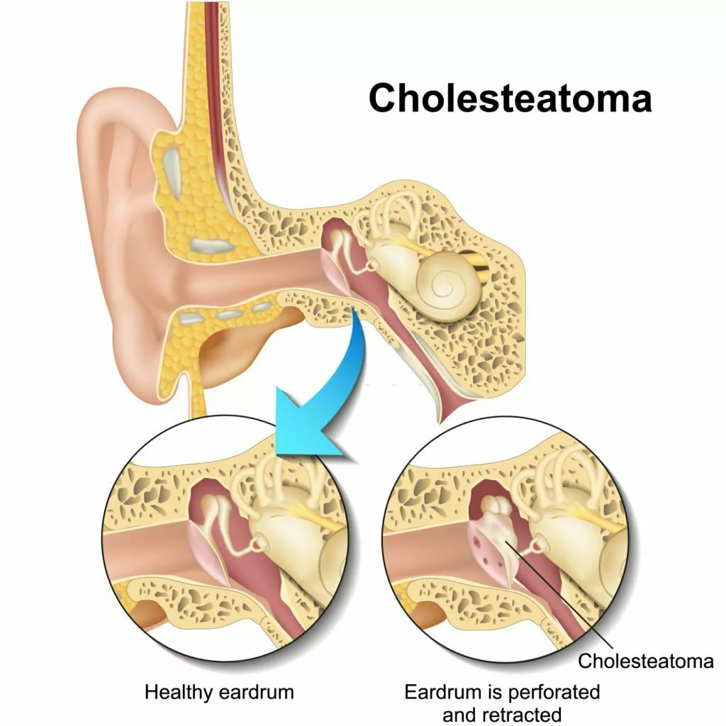
This type develops due to retraction of the tympanic membrane (eardrum). Moreover, continuous tympanic membrane retraction destroys ossicles, resulting in vertigo and deafness.
Secondary Acquired Cholesteatoma
This type of cholesteatoma results from injury to the tympanic membrane. Injury to the tympanic membrane is due to the following reasons:
- Perforation
- Chronic otitis media (middle ear infection)
- Surgical procedures
Some other risk factors include:
Craniofacial Abnormalities
According to a research study, it was noted that patients with craniofacial abnormalities such as Down syndrome are 20 times more prone to have cholesteatoma than others.6Patel, V. A., MD. (n.d.). Cholesteatoma medication. https://emedicine.medscape.com/article/860080-medication?form=fpf&scode=msp&st=fpf&socialSite=google&icd=login_success_gg_match_fpf#1
Symptoms of Cholesteatoma
Sometimes, it remains asymptomatic and undiagnosed.
It is a benign lesion that can cause complications because it can erode into the central nervous system (CNS) if neglected. This is why it can show a few symptoms related to CNS dysfunction.7Website, N. (2023, June 26). Cholesteatoma. nhs.uk. https://www.nhs.uk/conditions/cholesteatoma/#:~:text=A%20cholesteatoma%20is%20an%20abnormal,essential%20for%20hearing%20and%20balance.The most common symptoms of the cholesteatoma are:
Painless Otorrhea (Ear Discharge)
The hallmark of this disease is recurrent painless, foul-smelling ear discharge. There is no blood supply for cholesteatoma, so systemic antibiotics can not eradicate it. Topical antibiotics can only superficially control the infection, while large cholesteatomas are difficult to treat. Due to this reason, recurrent otorrhea develops despite aggressive antibiotic therapy.
Hearing loss
It is the most common symptom of cholesteatoma. Large cholesteatomas fill the ear space and interfere with sound transmission, impairing hearing. Additionally, cholesteatoma-induced ossicular damage (damage to the ossicles of your ear) also causes conductive hearing loss.
Several research studies demonstrate that there is a potential association between cholesteatoma and sensorineural hearing loss.
Vertigo
It is a progressive symptom of cholesteatoma that occurs due to a lack of early diagnosis and treatment. It develops due to erosion of the middle ear ossicles and when the cholesteatoma involves the stapes footplate.
Tinnitus
Hearing of sound coming from inside of the body rather than outside.
Anxiety
Dr chin-lung Ku performed some population-based studies that show the link between cholesteatoma and depression. As discussed above, cholesteatoma is an abnormal growth of skin epithelial cells in the temporal bone, most commonly caused by the middle ear infection. Even though it is a benign lesion, it causes damage to the middle ear, resulting in foul-smelling discharge, hearing impairment, and tinnitus. In addition, these impairments result in psychological manifestations like the affected person becoming socially withdrawn, which leads to depressive disorder.8Relationship between Depression and Cholesteatoma. (n.d.). https://scitemed.com/Press/1206/Relationship-between-Depression-and-Cholesteatoma
When to see your Doctor?
Whenever you feel some foul-smelling discharge coming out from your ear associated with hearing loss, immediately book an appointment with your doctor. Early diagnosis and treatment can prevent you from serious complications like meningitis or epidural abscess.
What are the Complications of Cholesteatoma?
It can cause serious complications if it remains untreated. These complications are 9Kennedy, K. L. (2023, July 4). Middle ear cholesteatoma. StatPearls – NCBI Bookshelf. https://www.ncbi.nlm.nih.gov/books/NBK448108/
- Epidural abscess
- Meningitis
- Facial nerve involvement
- Sigmoid sinus thrombosis
How to Diagnose Cholesteatoma?
There is no need for laboratory investigations and biopsy to diagnose the cholesteatoma. Moreover, the diagnosis is mainly based on history, physical examination, laboratory investigations, and radiological investigations.
Here are the steps to the detailed diagnosis of cholesteatoma:
History:
A detailed history is always a key point to reaching the diagnosis. Your doctor may ask multiple questions related to it. These can be:
- Your biodata (Name, age, weight)
- Do you have an impairment? If yes, then what is the onset of hearing impairment? Was it unilateral or bilateral?
- Do you have pain in the ear?
- Do you have vertigo?
- Do you have a headache?
- Do you have a fever?
- Any ear discharge? If yes, is it foul-smelling or not?
- Have you taken any antibiotics or ear drops? If yes, then which type of antibiotics or drops?
- Any previous medical illness?
- Do you feel anxious or not?
- Have you seen other doctors for this problem or not?
- Have you gone through any radiological or laboratory investigations?
- Family history
- Socioeconomic status
Physical Examination:
After a detailed history, a physical examination is crucial for the timely diagnosis of cholesteatoma. Physical examination includes;
Inspection
In this phase, your doctor will inspect you properly. Inspection includes:
- Structure of the ear
- Any swelling or deformity
- Any discharge coming out of it
- Color of discharge
- Consistency of discharge
- Inspection of nose
- Inspection of face
- Inspection of head and neck
Palpation
After inspection, your doctor will palate your ear to see:
- Examination ear
- Any tenderness
- Warm or cold
- Palpation of cervical lymph node
Otoscopy
An otoscope is an instrument used to visualize the interior of the ear. It is larger and is easily visible on otoscopic examination. In most cases, the tympanic membrane perforation is also present.
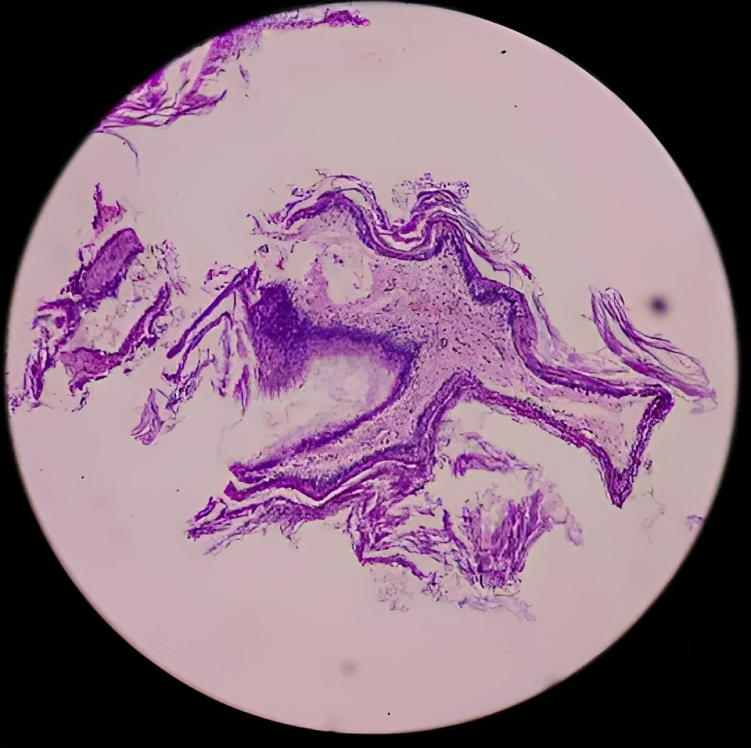
Microscopic Examination:
A microscopic examination is also useful in diagnosing cholesteatoma. In microscopic examination, white keratinized epithelial debris collections are seen posterosuperior to the tympanic membrane.
Audiometry:
This test should be performed preoperatively to assess the normal hearing baseline.10Patel, V. A., MD. (n.d.). Cholesteatoma medication. https://emedicine.medscape.com/article/860080-medication?form=fpf&scode=msp&st=fpf&socialSite=google&icd=login_success_gg_match_fpf#1
Laboratory Investigations:
Your doctor may suggest laboratory investigations to rule out another disease in suspicious cases. Laboratory investigations include:
- Complete blood count (CBC) to check anemia
- Erythrocyte sedimentation rate (ESR)
- C reactive protein (CRP) to check for active infection
- White blood cell count to check for infection
- Platelets count
Radiological Investigations:
Radiological investigations are the prime one to diagnose cholesteatoma. It includes
High Resonance Computed Tomography Scan (HRCT)
It is the gold standard investigation of choice for cholesteatoma. Because a CT scan can diagnose bony abnormalities in the ear. However, CT scans cannot differentiate between the cholesteatoma and the granulation of tissues. Bony defects that can be seen on CT scans are:
- Labyrinth fistula formation
- Tegmen defect
- Erosion of the ossicles
- Scutal erosion
Magnetic Resonance Imaging (MRI)
MRI also helps to diagnose cholesteatoma. It shows isointense on T1 and hyperintense on T2. MRI is indicated when there are the following clinical concerns:
- Dural invasion
- Epidural Abscess
- Herniation of the brain into the mastoid cavity
- Facial nerve involvement
- Meningitis
- Thrombus formation in sigmoid sinus.
How to treat Cholesteatoma?
Surgical removal of the cholesteatoma remains the treatment of choice. Surgery is contraindicated in patients with serious comorbidities. A close follow-up is required to avoid recurrence. The surgical procedures involved in treating cholesteatoma are:
1) Mastoidectomy:
It is a surgical procedure in which the part or the whole mastoid bone is removed. In certain circumstances, canal wall-up and canal wall-down techniques can be used.
Canal Wall Down Mastoidectomy
The canal wall-down procedure is the most definitive procedure with a high success rate in treating cholesteatoma. It is a surgical procedure in which the superior and posterior bony external auditory canals are removed, forming a continuous opening. Moreover, this opening consists of the external auditory lumen, epitympanum, and open mastoid cavity. There are about three types of canal wall-down mastoidectomy.
Radical Mastoidectomy:
It is a surgical procedure in which the entire tympanomastoid is removed. Removal of the whole bone results in poor hearing after surgery.
Modified Radical Mastoidectomy:
It is a surgical procedure in which only the epitympanum and mastoid cavity are removed.
The Bondy-Modified Radical Mastoidectomy:
This procedure is suitable when there is a small lateral epitympanic cholesteatoma with the following features:
- Mesotympanum is not involved
- The ossicular chain is intact
- The canal wall is drilled down
Advantages & Disadvantages of Canal Wall-Down Mastoidectomy
This procedure has a high success rate and a better response than canal wall-up. The advantages and disadvantages of this procedure are:
- Meatus is enlarged
- There is difficulty in fitting hearing aids
- After six months or a year, canal cleaning is required
- Water tolerance is not good
- It is a single procedure
- Recurrence rate is low
Canal Wall-Up Mastoidectomy
During mastoidectomy, surgeons remove mastoid air cells, creating a mastoid cavity while preserving the posterior wall and external auditory canal. In canal wall-up mastoidectomy, surgeons maintain the external auditory canal for a normal appearance, but it entails a higher risk of recurrence. Due to this reason, a second look at tympanomastoidectomy is advised after a few years of canal wall-up mastoidectomy.11Prasad SC, La Melia C, Medina M, Vincenti V, Bacciu A, Bacciu S, Pasanisi E. Long-term surgical and functional outcomes of the intact canal wall technique for middle ear cholesteatoma in the pediatric population. Acta Otorhinolaryngol Ital. 2014 Oct;34(5):354-61. PMID: 25709151; PMCID: PMC4299157.
Advantages & Disadvantages of Canal Wall-Up Procedure
Preoperatively, your doctor will explain the advantages and disadvantages of the canal wall-up procedure. However, these advantages and disadvantages are:
- Auricular appearance is normal
- Hearing aids are easy to fit
- Routine ear cleaning is not necessary
- Tolerance against water exposure
- Have a high rate of recurrence
- It is a staged procedure
2) Tympanoplasty
It is a surgical procedure in which the surgeon repairs the ruptured tympanic membrane with the help of a graft.
Goals of surgical procedure
The goal of the operation should be kept in mind for successful surgery. The goal of mastoidectomy is:
- To ensure the safety of the ear by removing infection and cholesteatoma
- To make ears functional for daily activities of living
- To conserve hearing impairment
What are the Postoperative Complications of Cholesteatoma?
The postoperative complications are as follows:
- Stenosis
- Injury to facial nerve
- Permanent hearing loss
- Graft failure
- Vertigo
- Otorrhea
- Dysgeusia (alteration of taste)
What is the Prognosis of Cholesteatoma?
Most of the time, it needs removal, but sometimes it requires multiple surgeries. Nowadays, complications of cholesteatoma are rare. However, 5% of the patients need re-operation for canal wall-down mastoidectomy. Additionally, The recurrence rate is much higher in patients with canal wall-up surgery. Unfortunately, hearing loss associated with cholesteatoma is permanent, but death from cholesteatoma rarely occurs.
Conclusion
To conclude, it is a benign lesion that remains asymptomatic for a long period. Over time, a foul-smelling discharge with progressive hearing loss can be the major symptom of cholesteatoma. Moreover, it can cause complications because it can erode into the central nervous system (CNS) if neglected. Additionally, it is an avascular (having no blood supply) tumor, so systemic antibiotics do not affect it, while topical antibiotics can only treat superficial infections. The possible treatment of cholesteatoma is surgery. Whenever you have a foul-smelling discharge from your ears, immediately consult your doctor.
Refrences
- 1Kennedy, K. L. (2023, July 4). Middle ear cholesteatoma. StatPearls – NCBI Bookshelf. https://www.ncbi.nlm.nih.gov/books/NBK448108/
- 2Patel, V. A., MD. (n.d.). Cholesteatoma medication. https://emedicine.medscape.com/article/860080-medication?form=fpf&scode=msp&st=fpf&socialSite=google&icd=login_success_gg_match_fpf#1
- 3Khalid-Raja M, Tikka T, Coulson C. Cholesteatoma: a disease of the poor (socially deprived)? Eur Arch Otorhinolaryngol. 2015 Oct;272(10):2799-805. doi: 10.1007/s00405-014-3285-y. Epub 2014 Sep 18. PMID: 25231708.
- 4Kennedy, K. L. (2023, July 4). Middle ear cholesteatoma. StatPearls – NCBI Bookshelf. https://www.ncbi.nlm.nih.gov/books/NBK448108/
- 5Centers for Disease Control and Prevention. (2022). Chronic Diseases and Health Promotion. Retrieved from https://emedicine.medscape.com/article/860080-overview?form=fpf
- 6Patel, V. A., MD. (n.d.). Cholesteatoma medication. https://emedicine.medscape.com/article/860080-medication?form=fpf&scode=msp&st=fpf&socialSite=google&icd=login_success_gg_match_fpf#1
- 7Website, N. (2023, June 26). Cholesteatoma. nhs.uk. https://www.nhs.uk/conditions/cholesteatoma/#:~:text=A%20cholesteatoma%20is%20an%20abnormal,essential%20for%20hearing%20and%20balance.
- 8Relationship between Depression and Cholesteatoma. (n.d.). https://scitemed.com/Press/1206/Relationship-between-Depression-and-Cholesteatoma
- 9Kennedy, K. L. (2023, July 4). Middle ear cholesteatoma. StatPearls – NCBI Bookshelf. https://www.ncbi.nlm.nih.gov/books/NBK448108/
- 10Patel, V. A., MD. (n.d.). Cholesteatoma medication. https://emedicine.medscape.com/article/860080-medication?form=fpf&scode=msp&st=fpf&socialSite=google&icd=login_success_gg_match_fpf#1
- 11Prasad SC, La Melia C, Medina M, Vincenti V, Bacciu A, Bacciu S, Pasanisi E. Long-term surgical and functional outcomes of the intact canal wall technique for middle ear cholesteatoma in the pediatric population. Acta Otorhinolaryngol Ital. 2014 Oct;34(5):354-61. PMID: 25709151; PMCID: PMC4299157.
