Angiomyolipomas are benign tumors composed of an abnormal mixture of blood vessels, smooth muscle tissue, and fat cells. In the human body, cells divide and multiply in a highly regulated manner to support normal growth and function. However, when this process becomes uncontrolled, it can lead to the development of tumors—one such example being angiomyolipoma. Although it can occur in different organs, angiomyolipoma most commonly arises in the kidney. It is medically referred to as renal angiomyolipoma.1Çalışkan, S., Gümrükçü, G., Özsoy, E., Topaktas, R., & Öztürk, M. İ. (2019). Renal angiomyolipoma. Revista da Associacao Medica Brasileira (1992), 65(7), 977–981. https://doi.org/10.1590/1806-9282.65.7.9772Eble J. N. (1998). Angiomyolipoma of kidney. Seminars in diagnostic pathology, 15(1), 21–40.
What is Angiomyolipoma?
Angiomyolipoma (AML) is a rare, typically benign tumor that most commonly affects the kidneys, although it can also arise in other organs. AML is composed of three types of tissues: blood vessels (angio), smooth muscle cells (myo), and fat cells (lipo), which gives it a characteristic appearance in imaging studies such as CT or MRI scans.3Lienert, A. R., & Nicol, D. (2012). Renal angiomyolipoma. BJU international, 110 Suppl 4, 25–27. https://doi.org/10.1111/j.1464-410X.2012.11618.x
Renal angiomyolipoma usually occurs sporadically, but about 20–30% of cases are associated with tuberous sclerosis complex (TSC), a genetic disorder. Most AMLs are discovered incidentally during imaging for unrelated issues, as they often remain asymptomatic, particularly when small. Despite its benign nature, complications may arise—especially if the tumor grows larger than 4 cm, at which point the risk of spontaneous hemorrhage increases significantly. This can lead to symptoms such as flank pain, abdominal mass, gross hematuria (visible blood in urine), or in rare cases, life-threatening retroperitoneal bleeding. Larger tumors may also compress nearby structures and cause secondary symptoms like hypertension or anemia due to chronic blood loss.4Eble, J. N. (1998). Angiomyolipoma of the kidney. Seminars in Diagnostic Pathology, 15(1), 21–40.
Growth Rate & Prevalence
In most cases, the growth rate of angiomyolipoma remains negligible for years; however, in some instances, it progresses slowly at a rate of approximately 0.19 cm per year. This growth can accelerate significantly in pregnant women, increasing the risk of complications due to hormonal changes.5 Courtney, M., Mulholland, D., O’Neill, D., Redmond, C., Ryan, J., Geoghegan, T., Torreggiani, W., & Lee, M. (2021). The natural growth pattern of sporadic renal angiomyolipoma. Acta radiologica (Stockholm, Sweden : 1987), 62(2), 276–280. https://doi.org/10.1177/0284185120918372 That’s why it is advisable for women diagnosed with this tumor to receive proper treatment before planning a pregnancy. Angiomyolipoma is more prevalent in females, especially those between the ages of 40 and 60.6Nelson CP, Sanda MG. Contemporary diagnosis and management of renal angiomyolipoma. J Urol. 2002 Oct;168(4 Pt 1):1315-25. doi: 10.1016/S0022-5347(05)64440-0. PMID: 12352384.
Certain medical conditions like tuberous sclerosis complex (TSC) and lymphangioleiomyomatosis (LAM)significantly increase the risk of developing this tumor. In fact, more than 50% of patients diagnosed with TSC also present with renal angiomyolipomas.7Maulaz, P., Malinge, M. C., Farges, D., Ingster, O., Azzouzi, A. R., & Bigot, P. (2020). Prévalence de la sclérose tubéreuse de Bourneville chez des patients pris en charge pour un angiomyolipome rénal [Prevalence of the tuberous sclerosis complex at patients taken care for a renal angiomyolipoma]. Progres en urologie : journal de l’Association francaise d’urologie et de la Societe francaise d’urologie, 30(10), 500–506. https://doi.org/10.1016/j.purol.2020.05.011.
Causes of Angiomyolipoma
The primary cause of renal angiomyolipoma is the uncontrolled proliferation of adipocytes, smooth muscle cells, and vascular components. Although the exact trigger remains uncertain, gene mutations—particularly in TSC1 and TSC2 genes—are believed to play a central role. Environmental exposure to radiation or certain chemicals, as well as inherited genetic disorders, can contribute to these mutations. These genetic changes are commonly seen in patients with tuberous sclerosis complex (TSC)—a condition strongly linked to the development of angiomyolipoma.
Another rare but important association is with lymphangioleiomyomatosis (LAM), a disease that affects the lungs and shares similar cellular abnormalities. Both TSC and LAM increase the risk of developing this tumor. While the exact etiology remains under investigation, gene-level disturbances leading to abnormal cell growth are widely recognized as the main culprit in angiomyolipoma formation.8Wang, M. X., Segaran, N., Bhalla, S., Pickhardt, P. J., Lubner, M. G., Katabathina, V. S., & Ganeshan, D. (2021). Tuberous Sclerosis: Current Update. Radiographics: a review publication of the Radiological Society of North America, Inc, 41(7), 1992–2010. https://doi.org/10.1148/rg.2021210103

Signs & Symptoms of Angiomyolipoma
Most renal angiomyolipomas remain asymptomatic, and many patients discover them incidentally during imaging for unrelated issues. Nevertheless, in certain cases, especially when the tumor is large or actively bleeding, symptoms do appear and prompt medical attention.
The major symptoms appearing in patients of renal angiomyolipoma include:
- Flank pain: Patients experience continuous mild pain in the lower backside. It usually occurs on the side where the kidney is affected
- Fever: This may occur as a result of internal bleeding or inflammation around the tumor.
- Hematuria: This is a condition in which patients have blood in the urine.
- Urinary difficulties: Including urgency, frequency, or discomfort.
- Bleeding: Spontaneous rupture of the tumor can lead to retroperitoneal hemorrhage, a potentially life-threatening condition.
- Anemia: Continuous bleeding can cause blood deficiency in the body.
- High blood pressure: Kidneys are the major body organ responsible for maintaining homeostasis in the body. Damage to the kidney disrupts its functioning which results in high blood pressure.9Flum, A. S., Hamoui, N., Said, M. A., Yang, X. J., Casalino, D. D., McGuire, B. B., Perry, K. T., & Nadler, R. B. (2016). Update on the Diagnosis and Management of Renal Angiomyolipoma. The Journal of Urology, 195(4 Pt 1), 834–846. https://doi.org/10.1016/j.juro.2015.07.126
Diagnosis of Angiomyolipoma
Doctors often diagnose angiomyolipoma incidentally during imaging for other medical conditions or during routine health checkups. The diagnosis involves the following steps:
- A complete history of the patient with a special focus on past medical illness gives information about the risk factors
- A physical examination may reveal a palpable mass or tenderness in the kidney area.
- Diagnosis is confirmed via radiological Imaging.
- Percutaneous biopsy may also be needed in some cases.
Radiology & Angiomyolipoma:
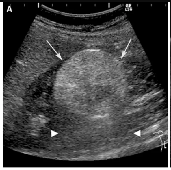
Major Imaging Modalities include CT, MRI, ultrasound, and Digital subtraction angiography (DSA). A CT scan is typically the first choice and can show the fat content within the tumor, which is characteristic of angiomyolipoma. However, in fat-poor variants, CT might not be sufficient. In such cases, MRI provides better detail and helps differentiate the lesion from malignancy. Ultrasound and MRI are also helpful in assessing the extent of the soft tissue component. DSA is particularly useful when planning embolization, as it reveals the vascular supply of the tumor.10Halpenny, D., Snow, A., McNeill, G., & Torreggiani, W. C. (2010). The radiological diagnosis and treatment of renal angiomyolipoma-current status. Clinical radiology, 65(2), 99–108. https://doi.org/10.1016/j.crad.2009.09.014
Treatment of Angiomyolipoma
If the tumor is small and grows slowly, doctors usually recommend active surveillance. This involves regular imaging and monitoring of kidney function to ensure the tumor is not progressing or causing complications. However, if the tumor is larger than 4 cm, growing rapidly, or causing symptoms such as pain or bleeding, treatment becomes necessary. Depending on the individual case, the following options are available:
Medical Therapy:
mTOR inhibitors (e.g., everolimus) are used to reduce tumor size, especially in patients with tuberous sclerosis complex or those who are not surgical candidates.
Surgical Options:
The surgical removal options include:
Ablation Therapy:
In this surgical procedure, the use of radiofrequency with a precise target is required to destroy the diseased portion.11Strunk H. M. (2002). Renale Angiomyolipome [Renal angiomyolipoma]. Ultraschall in der Medizin (Stuttgart, Germany : 1980), 23(6), 367–372. https://doi.org/10.1055/s-2002-36174
Cryotherapy:
Cryoablation or Cryotherapy is a modern treatment methodology that aims to remove the tumorous mass without damaging the nearby structures. This therapy involves damaging the tumor mass with the help of extremely low temperatures.
Embolization:
In this surgical procedure, the medical team restricts the blood supply to the tumor mass, leading to a decrease in the chances of bleeding.
Nephrectomy:
In this surgical procedure, the medical team conducts a partial nephrectomy to remove a portion of the affected kidney. Alternatively, they perform a complete nephrectomy to remove the entire kidney.
Emergency Management in Case of Bleeding:
All patients with angiomyolipoma who develop symptoms or signs suggestive of active bleeding should undergo prompt imaging—typically with CT or MRI—to assess the extent and source of hemorrhage.
Bleeding from renal angiomyolipomas can range from mild to life-threatening. In severe cases, it may lead to hemorrhagic shock, irreversible kidney damage, or even death.
Patients presenting with active bleeding require immediate resuscitation if they are hemodynamically unstable. If feasible, prompt angiography followed by selective arterial embolization (SAE) is the preferred approach to control the hemorrhage and preserve kidney function.
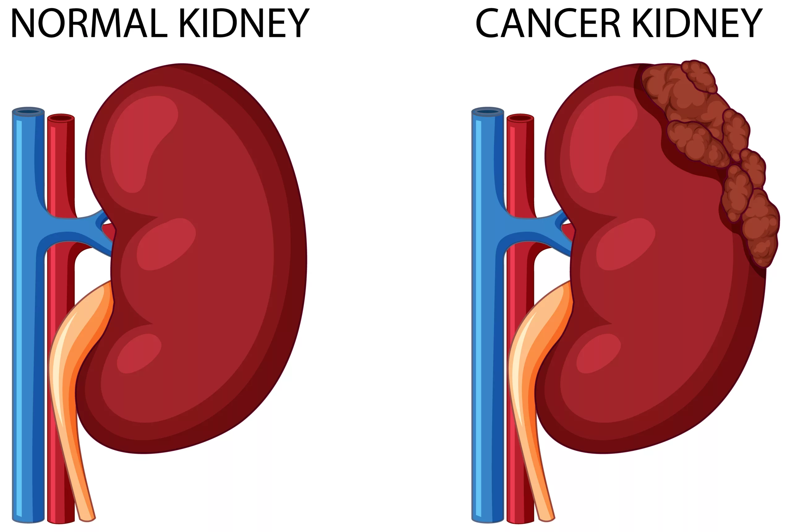
Difference Between Renal Angiomyolipoma & Renal Cell Carcinoma
While both renal angiomyolipoma and renal cell carcinoma are tumor-related conditions of the kidney, they differ in origin, behavior, and clinical implications. Angiomyolipoma arises from the unchecked growth of fat, smooth muscle, and blood vessel cells, whereas renal cell carcinoma develops due to abnormal proliferation of epithelial cells within the renal tubules.
Angiomyolipoma is generally benign and non-invasive, whereas renal cell carcinoma is malignant and has a tendency to metastasize to distant organs such as the lungs, liver, or bones. Symptoms in angiomyolipoma are often absent, while renal cell carcinoma typically presents with significant warning signs such as weight loss, persistent flank pain, blood in the urine, and night sweats. Due to its aggressive nature, renal cell carcinoma is treated with targeted therapies, immunotherapy, or radiotherapy. In contrast, angiomyolipoma is usually managed with conservative monitoring or surgical excision if necessary.12Xu, Z. F., Xu, H. X., Xie, X. Y., Liu, G. J., Zheng, Y. L., & Lu, M. D. (2010). Renal cell carcinoma and renal angiomyolipoma: differential diagnosis with real-time contrast-enhanced ultrasonography. Journal of ultrasound in medicine: official journal of the American Institute of Ultrasound in Medicine, 29(5), 709–717. https://doi.org/10.7863/jum.2010.29.5.709
Wrap Up
To sum it up, Angiomyolipoma is a rare benign tumor of the kidney which affects the normal functioning of the kidney. It appears to be a symptomless disease, but sometimes it creates potential complications like severe bleeding which is life-threatening for the patients.
Refrences
- 1Çalışkan, S., Gümrükçü, G., Özsoy, E., Topaktas, R., & Öztürk, M. İ. (2019). Renal angiomyolipoma. Revista da Associacao Medica Brasileira (1992), 65(7), 977–981. https://doi.org/10.1590/1806-9282.65.7.977
- 2Eble J. N. (1998). Angiomyolipoma of kidney. Seminars in diagnostic pathology, 15(1), 21–40.
- 3Lienert, A. R., & Nicol, D. (2012). Renal angiomyolipoma. BJU international, 110 Suppl 4, 25–27. https://doi.org/10.1111/j.1464-410X.2012.11618.x
- 4Eble, J. N. (1998). Angiomyolipoma of the kidney. Seminars in Diagnostic Pathology, 15(1), 21–40.
- 5Courtney, M., Mulholland, D., O’Neill, D., Redmond, C., Ryan, J., Geoghegan, T., Torreggiani, W., & Lee, M. (2021). The natural growth pattern of sporadic renal angiomyolipoma. Acta radiologica (Stockholm, Sweden : 1987), 62(2), 276–280. https://doi.org/10.1177/0284185120918372
- 6Nelson CP, Sanda MG. Contemporary diagnosis and management of renal angiomyolipoma. J Urol. 2002 Oct;168(4 Pt 1):1315-25. doi: 10.1016/S0022-5347(05)64440-0. PMID: 12352384.
- 7Maulaz, P., Malinge, M. C., Farges, D., Ingster, O., Azzouzi, A. R., & Bigot, P. (2020). Prévalence de la sclérose tubéreuse de Bourneville chez des patients pris en charge pour un angiomyolipome rénal [Prevalence of the tuberous sclerosis complex at patients taken care for a renal angiomyolipoma]. Progres en urologie : journal de l’Association francaise d’urologie et de la Societe francaise d’urologie, 30(10), 500–506. https://doi.org/10.1016/j.purol.2020.05.011.
- 8Wang, M. X., Segaran, N., Bhalla, S., Pickhardt, P. J., Lubner, M. G., Katabathina, V. S., & Ganeshan, D. (2021). Tuberous Sclerosis: Current Update. Radiographics: a review publication of the Radiological Society of North America, Inc, 41(7), 1992–2010. https://doi.org/10.1148/rg.2021210103
- 9Flum, A. S., Hamoui, N., Said, M. A., Yang, X. J., Casalino, D. D., McGuire, B. B., Perry, K. T., & Nadler, R. B. (2016). Update on the Diagnosis and Management of Renal Angiomyolipoma. The Journal of Urology, 195(4 Pt 1), 834–846. https://doi.org/10.1016/j.juro.2015.07.126
- 10Halpenny, D., Snow, A., McNeill, G., & Torreggiani, W. C. (2010). The radiological diagnosis and treatment of renal angiomyolipoma-current status. Clinical radiology, 65(2), 99–108. https://doi.org/10.1016/j.crad.2009.09.014
- 11Strunk H. M. (2002). Renale Angiomyolipome [Renal angiomyolipoma]. Ultraschall in der Medizin (Stuttgart, Germany : 1980), 23(6), 367–372. https://doi.org/10.1055/s-2002-36174
- 12Xu, Z. F., Xu, H. X., Xie, X. Y., Liu, G. J., Zheng, Y. L., & Lu, M. D. (2010). Renal cell carcinoma and renal angiomyolipoma: differential diagnosis with real-time contrast-enhanced ultrasonography. Journal of ultrasound in medicine: official journal of the American Institute of Ultrasound in Medicine, 29(5), 709–717. https://doi.org/10.7863/jum.2010.29.5.709
