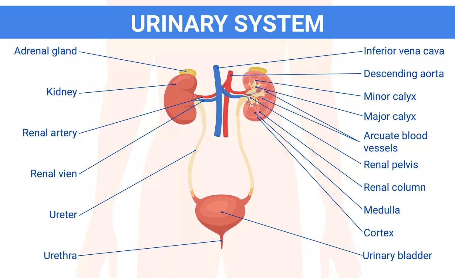
Urolithiasis, commonly known as kidney stones, refers to the formation of hard mineral and salt deposits within the urinary tract, including the kidneys, ureters, bladder, or urethra. These stones can vary in size and may cause significant pain, especially if they obstruct the flow of urine. Urolithiasis affects millions of people worldwide and often requires medical intervention due to the risk of complications such as severe renal colic, hematuria, or infection.1Coe FL, Evan AP, Worcester EM, Lingeman JE. The pathogenesis and treatment of kidney stones. N Engl J Med. 2005; 393(10): 1141–1151.
Urolithiasis vs. Nephrolithiasis
The terms urolithiasis and nephrolithiasis share the same meaning but have distinct definitions. Urolithiasis refers to stones located in the complete urinary tract system, which includes the kidneys and ureters, and bladder. The term nephrolithiasis indicates stone development specifically in the kidneys. The distinction between these terms matters because it determines how doctors will diagnose and treat patients
The prevalence of kidney stones in American adults reaches 10% throughout their lifetimes.2Scales CD Jr, Smith AC, Hanley JM, Saigal CS. Prevalence of kidney stones in the United States. Eur Urol. 2012;62(1):160–165. The incidence of urolithiasis has continued to rise over the past few decades, especially among groups with elevated risks stemming from diet, hydration patterns, and genetic susceptibility. These stones require proper treatment because improper management will lead to pain as well as possible complications, including urinary tract infections or damage to the kidneys.
Kidney Stones Formation
Kidney stones are formed on a step-by-step basis in the urinary tract. Different factors, such as the concentration of minerals, pH levels, and the presence of substances that can favour or inhibit the formation of stones, are responsible for the formation of these stones.3Evan AP. Physiopathology and etiology of stone formation in the kidney and the urinary tract. Pediatr Nephrol. 2010;25(5):831–841.
Kidney stones develop when there is an imbalance between the amounts of calcium, oxalate, and uric acid in urine. Such an imbalance can cause these substances to crystallize, and these will eventually aggregate in stone form.
Common Types of Kidney Stones
The most common types of kidney stones include:
- Calcium Stones: The most prevalent type is usually in the form of calcium oxalate.4Khan SR. Kidney stones of calcium oxalate. World J Urol. 1997;15(3):165–171.
- Uric Acid Stones: These form in people who lose too much fluid (e.g., through chronic diarrhea), consume diets high in purines (found in red meat, seafood, and organ meats), or have gout. They tend to form in acidic urine.
- Struvite Stones: These occur in women with urinary tract infections (UTIs). These stones can grow quickly and may become large enough to obstruct parts of the urinary tract.
- Cystine Stones: A rare form of kidney stone that occurs in individuals with cystinuria, a genetic disorder in which the kidneys excrete too much cystine into the urine.
Common Causes of Urolithiasis
The causes of the kidney stones are diet, hydration, and health conditions.
Dietary Factors:
Kidney stone prevention starts with what you eat. Eating too much salt, red meat, or foods high in oxalate (like spinach or nuts) can raise your chances of forming stones. Instead, focus on a diet full of fruits, vegetables, and whole grains. These foods help keep your body balanced and your urine less likely to form crystals. Staying well-hydrated is just as important—water helps flush out substances that could otherwise form stones.
Dehydration:
The risk of urolithiasis increases when people fail to drink enough fluids, which results in dehydration. The body needs proper fluids to maintain adequate urine concentration, which prevents stone development. Drinking sufficient water prevents dehydration.
Other Diseases:
Some conditions, such as hyperparathyroidism, Urinary tract infections, and chronic inflammation, increase the chances of this disease. The effective management of these conditions helps decrease the probability of urolithiasis development.
Genetic Factors:
The development of kidney stones heavily depends on genetic factors, among other factors. People who have family members who developed urolithiasis face an increased chance of forming kidney stones. Sharing your family medical history with your doctor helps them prevent and manage your condition at an early stage.5Goldfarb DS, Arowojolu O. Metabolic evaluation of first-time and recurrent stone formers. Urol Clin North Am. 2013;40(1):13–2
Urolithiasis Risk Factors
Urolithiasis, or the formation of urinary tract stones, is influenced by a range of demographic, environmental, dietary, and medical factors, although these do not all act directly on stone formation mechanisms.
Age, Gender & Family History:
The risk of kidney stones peaks between 20 to 49 years of age. Men are generally at a higher risk than women, although the gender gap has narrowed in recent years. A family history of kidney stones significantly increases the likelihood of recurrence in relatives, suggesting a genetic or shared lifestyle influence.
Geographic & Climate Considerations:
Living in hot, arid regions increases the risk due to dehydration, which reduces urine volume and concentrates stone-forming substances. Local dietary habits and water composition also play a role in stone prevalence in different regions.
Lifestyle Factors:
High intake of animal protein, salt, and added sugars—especially fructose—can increase the risk. Low fluid intake is one of the strongest modifiable risk factors. Conversely, a balanced diet rich in fruits, vegetables, and adequate calcium may offer protective benefits.6Curhan GC, Willett WC, Rimm EB, Stampfer MJ. A prospective study of dietary calcium and other nutrients and the risk of symptomatic kidney stones. N Engl J Med. 1993;328(12):833–838
Medical Conditions that Increase Risk:
Medical diseases have the potential to cause urolithiasis. The development of urolithiasis results from diseases that include urinary tract infections, as well as renal tubular acidosis and hyperparathyroidism-related conditions, and cystinuria. The knowledge and management of these conditions decreases the likelihood of developing kidney stones.
Urolithiasis Symptoms
The early detection of urolithiasis symptoms stands as an essential diagnostic requirement. The symptoms of kidney stones extend across multiple levels of severity. The medical condition creates significant interruptions to your daily routine. The hallmark symptom is sudden, severe flank pain that may radiate to the lower abdomen or groin.
Symptoms Based on Stone Location:
The position where the stone exists determines what symptoms will appear to the patient.
- Renal Stones: Pain in the flank or back, just below the ribs.
- Ureteral Stones: Pain radiates from the lower abdomen to the groin, often accompanied by nausea or vomiting.
- Bladder Stones: May cause painful urination (dysuria), urinary frequency, and urgency.
Additional symptoms may include hematuria (blood in urine), cloudy or foul-smelling urine, and in some cases, fever or chills if infection is present.
When to Seek Medical Attention:
The following signs require immediate medical help when you have severe pain with nausea and vomiting and stomach discomfort, and increased urination frequency.
- Nausea, vomiting, stomach upset.
- The need to urinate more frequently than normal.
- Urine color and smell are changed.
- The presence of fever along with infection symptoms indicates a medical issue.
These could be signs of a more serious condition.
Diagnosis of Urolithiasis
The diagnosis of urolithiasis requires medical history information and physical examination results along with modern diagnostic tests. Medical professionals use this complete method to properly diagnose kidney stones while selecting the most suitable therapeutic approach.
Medical History & Physical Examination
To detect urolithiasis, healthcare providers begin with a physical examination combined with a review of the patient’s medical history. They assess symptoms along with the patient’s background and lifestyle to identify possible causes and risk factors for kidney stones.
Imaging Tests
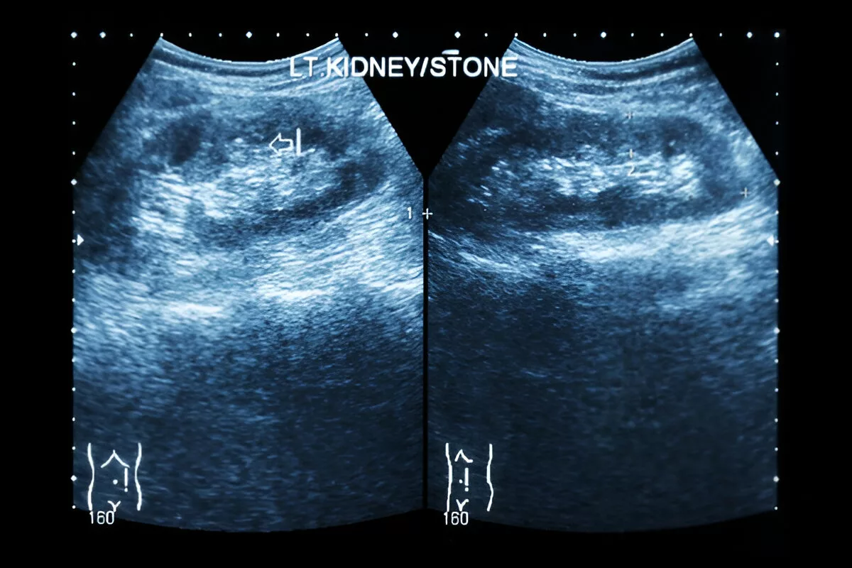
The visualization of kidney stones in urolithiasis diagnosis relies heavily on imaging tests. The standard diagnostic tests used in medical practice are:
- CT Scans: It enable them to detect tiny stones effectively thus making them the preferred diagnostic method.7Smith RC, Verga M, McCarthy S, Rosenfield AT. Diagnosis of acute flank pain: value of unenhanced helical CT. AJR Am J Roentgenol. 1996;166(1):97–101.
- Ultrasound: The ultrasound method uses sound waves to detect kidney stones and serves as a safe non-invasive tool particularly useful for pregnant women and children.
- X-rays: They serve as a diagnostic tool for particular kidney stone types, yet they fail to identify all stone variations.
Laboratory Tests
Medical tests help both diagnose and manage urolithiasis cases. These include:
- Urinalysis: The urinalysis test detects urine infections as well as blood presence. It can show if kidney have stones.
- Blood Tests: Healthcare providers achieve accurate urolithiasis diagnosis and create patient-specific treatment plans by integrating these diagnostic approaches.
- Stone Analysis: The analysis of passed stones reveals their chemical composition. This helps prevent more stones from forming.
These tests check how well the kidneys are working. The tests evaluate infections and other medical problems within the patient’s body.
Urolithiasis Treatment
The size and position, together with the type of stone, determine the necessary treatment approach for urolithiasis patients. The proper selection of a treatment plan requires healthcare providers to understand these important factors.
Conservative Management:
Doctors may allow small stones to pass through the body without treatment. The approach enables your body to naturally eliminate the stone.
- Pain Management is key. NSAIDs, alongside opioids, are among the available pain management medications that healthcare providers may use.
- The patient under watchful waiting receives ongoing monitoring of their stone. Doctors will conduct imaging tests to determine if the stone will pass independently.
Medication Options:
Some medicines can help with kidney stones. The treatment improves symptoms, together with stone dissolution.
- Alpha-blockers enable the muscles found in the ureter to become less tense. The relaxed muscles of the ureter enable stone passage.
- People suffering from kidney stones can find relief from pain by taking over-the-counter medication.
- Stone-dissolving medications dissolve particular types of stones, including those formed from uric acid.
Surgical Interventions
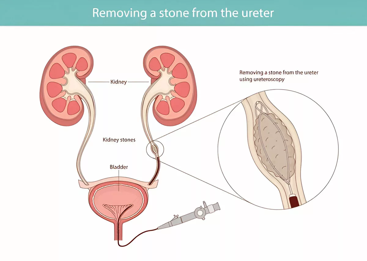
Surgical procedures become necessary for treating stones that are too large or produce intense symptoms.
- The non-invasive shock wave treatment known as Extracorporeal Shock Wave Lithotripsy (ESWL) breaks down stones into smaller pieces, which the body can then eliminate.8Lingeman JE. Shock wave lithotripsy: complications and outcomes. J Urol. 1997;158(1):21–25.
- Medical staff use small scopes for visualization of stones through ureteroscopy and cystoscopy to enable both stone removal and fragmentation procedures.
- The minimally invasive treatment, Percutaneous Nephrolithotomy, involves making a small cut to access the stone directly for removal.
The appropriate treatment for urolithiasis requires assessment of both stone characteristics along with patient health status. Patients who understand their treatment choices will be able to make informed decisions about their medical care.
Urolithiasis Prevention
A prevention plan for urolithiasis recurrence includes diet changes, together with sufficient hydration and specific medical treatments. The implementation of preventive strategies helps people minimize their chances of developing future kidney stones.
Dietary Modifications:
The prevention of urolithiasis recurrence depends heavily on dietary factors. A person who selects their food wisely will reduce their risk factors that contribute to kidney stone development.
Foods to Avoid
Some particular foods raise the chances of developing kidney stones. The risk of developing kidney stones increases with foods that contain high amounts of oxalate as well as sodium and animal proteins. Reducing consumption of these foods helps stop stone formation.
- Foods high in oxalate: Spinach, beets, rhubarb, and nuts
- High sodium foods: Processed and packaged foods
- Animal protein: Excessive consumption of meat, poultry, and fish
Beneficial Foods
Consuming specific foods as part of your diet helps prevent the formation of new kidney stones. The following foods have beneficial effects:
- Calcium-rich foods: Dairy products, leafy greens
- Potassium-rich foods: Bananas, citrus fruits
- Adequate fiber: Whole grains, fruits, and vegetables
Hydration Strategies:
The prevention of kidney stone recurrence requires proper hydration of the body. Drinking enough water helps dissolve minerals in urine, which decreases their chances of forming stones.
Tips for staying hydrated
- Each day, people should drink at least 8-10 glasses of water.
- Monitor urine color: It should be pale yellow
- Sugary drinks should be avoided since they increase the chances of developing kidney stones
Medication for Prevention:
Doctors may prescribe medicines to stop urolithiasis from recurring in specific circumstances. The selection of medication depends on the stone type and the underlying causes.
Common Medications used
- Thiazide diuretics for calcium stones.9Borghi L, Meschi T, Amato F, Briganti A, Novarini A, Giannini A. Urinary volume, water and recurrences in idiopathic calcium nephrolithiasis: a 5-year randomized prospective study. J Urol. 1996;155(3):839–843.
- Allopurinol for uric acid stones
- Potassium citrate for certain types of stones
Lifestyle Changes:
The prevention of recurrent kidney stones requires implementing dietary changes and hydration practices, and selected lifestyle modifications. A person’s risk of developing kidney stones decreases when they control their weight and handle medical issues while restricting vitamin and mineral overconsumption.
These preventive measures decrease the likelihood of urolithiasis recurrence and enhance kidney health overall.
Living with Urolithiasis: Managing Chronic Stone Disease
Patients who have urolithiasis for the long term must face the ongoing challenge of stone disease. A person must manage this condition with medical care combined with lifestyle changes and continuous supervision.
Long-term Monitoring
People with a history of urolithiasis need regular check-ups with their healthcare provider. Patients need regular imaging tests and urine analysis to detect early signs of new stone formation.
Recurrent Episodes
The occurrence of repeated kidney stones causes significant discomfort to the patient. People should keep stone diaries to record symptoms and triggers while consulting healthcare professionals or support groups for guidance.
Quality of Life With Disease
Patients who receive successful urolithiasis treatment achieve better quality of life. Medical treatment alongside dietary changes and hydration constitutes the basis for successful management of this condition.
Support Resources
Several support resources exist to benefit patients, including online forums together with nutritional counseling, and patient advocacy groups. These resources deliver important information together with emotional support to patients.
Special Populations & Urolithiasis
The medical condition of urolithiasis or kidney stones creates specific difficulties for three special groups, including elderly patients and pregnant women, and children.
Kidney Stones In Children
Urolithiasis occurs less frequently in children than in adults, but it can affect both renal development and future health complications. The majority of patients in this group have inherited and metabolic disorders.
Urolithiasis During Pregnancy
The physiological changes of pregnancy lead to urolithiasis through increased ureter dilation and calcium excretion. The medical treatment of renal stones during pregnancy needs multiple safety measures to protect both the fetus and the mother.
Elderly Patients with Kidney Stones
Older adults tend to develop urolithiasis because of their lower mobility levels and dietary modifications, as well as their age-related kidney changes. The treatment approach for these patients needs to account for their existing medical conditions, together with their medication interactions.
Patients with Comorbidities
The presence of inflammatory bowel disease, together with gout and additional health issues, elevates the risk of developing urolithiasis in patients. These conditions need treatment to prevent the development of kidney stones.10.Pak CY. Medical management of urinary stone disease. Nephron Clin Pract. 2004;98(2):c49–c53.
Healthcare providers must identify both the special risk factors and challenges of urolithiasis to achieve successful prevention of complications in specific populations.
Conclusion: Taking Control of Your Kidney Health
The study of urolithiasis serves as a basis for both the prevention and management of the condition. Knowledge of kidney stone causes, risk factors, and symptoms helps people adopt preventive measures to protect their kidneys from disease. A combination of proper diet management along with adequate hydration, and medical condition control enables urolithiasis prevention.
People who have kidney stones should take steps to prevent urolithiasis from creating new stones. The treatment options for urolithiasis depend on stone size and type and range between noninvasive approaches and surgical procedures.
The information in this guide enables people to protect their kidneys while reducing their chances of developing urolithiasis. Urolithiasis prevention and treatment demand a comprehensive approach that combines medical support with lifestyle adjustments and disease awareness programs.
Refrences
- 1Coe FL, Evan AP, Worcester EM, Lingeman JE. The pathogenesis and treatment of kidney stones. N Engl J Med. 2005; 393(10): 1141–1151.
- 2Scales CD Jr, Smith AC, Hanley JM, Saigal CS. Prevalence of kidney stones in the United States. Eur Urol. 2012;62(1):160–165.
- 3Evan AP. Physiopathology and etiology of stone formation in the kidney and the urinary tract. Pediatr Nephrol. 2010;25(5):831–841.
- 4Khan SR. Kidney stones of calcium oxalate. World J Urol. 1997;15(3):165–171.
- 5Goldfarb DS, Arowojolu O. Metabolic evaluation of first-time and recurrent stone formers. Urol Clin North Am. 2013;40(1):13–2
- 6Curhan GC, Willett WC, Rimm EB, Stampfer MJ. A prospective study of dietary calcium and other nutrients and the risk of symptomatic kidney stones. N Engl J Med. 1993;328(12):833–838
- 7Smith RC, Verga M, McCarthy S, Rosenfield AT. Diagnosis of acute flank pain: value of unenhanced helical CT. AJR Am J Roentgenol. 1996;166(1):97–101.
- 8Lingeman JE. Shock wave lithotripsy: complications and outcomes. J Urol. 1997;158(1):21–25.
- 9Borghi L, Meschi T, Amato F, Briganti A, Novarini A, Giannini A. Urinary volume, water and recurrences in idiopathic calcium nephrolithiasis: a 5-year randomized prospective study. J Urol. 1996;155(3):839–843.
- 10.Pak CY. Medical management of urinary stone disease. Nephron Clin Pract. 2004;98(2):c49–c53.
